. 2003 Jul 2;23(13):5496-502.
doi: 10.1523/JNEUROSCI.23-13-05496.2003.

Acid-sensing ion channel 1 is localized in brain regions with high synaptic density and contributes to fear conditioning

Affiliations

Acid-sensing ion channel 1 is localized in brain regions with high synaptic density and contributes to fear conditioning

John A Wemmie et al. J Neurosci. .

Abstract

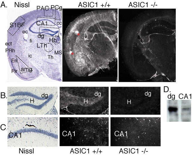
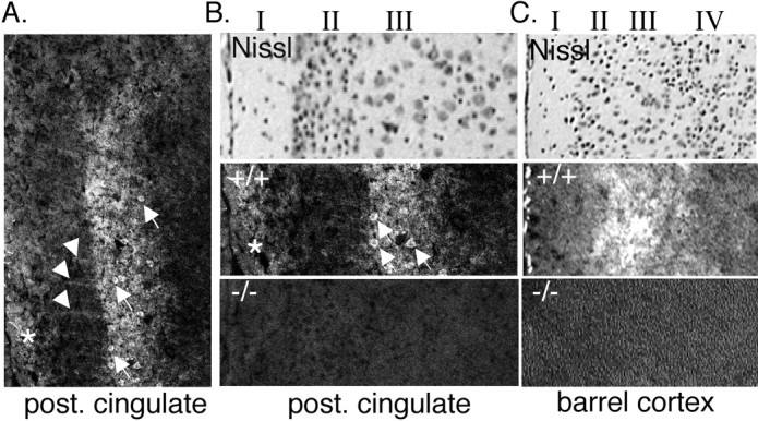
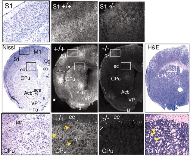
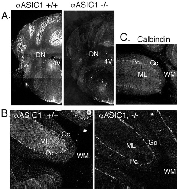
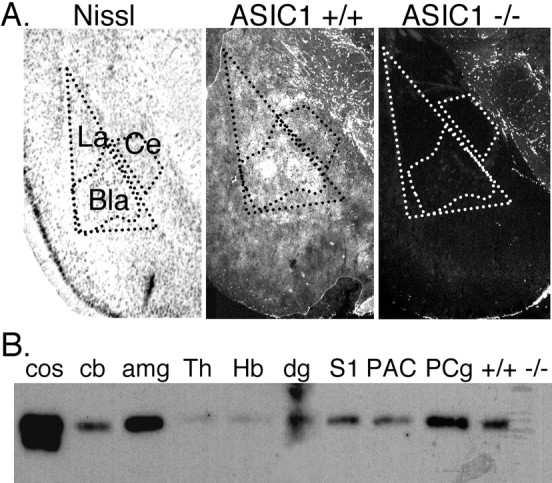
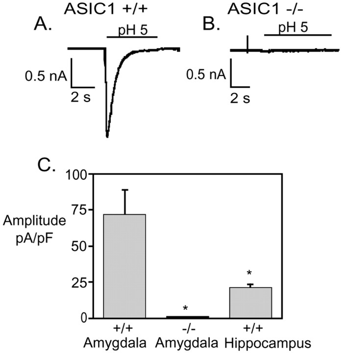
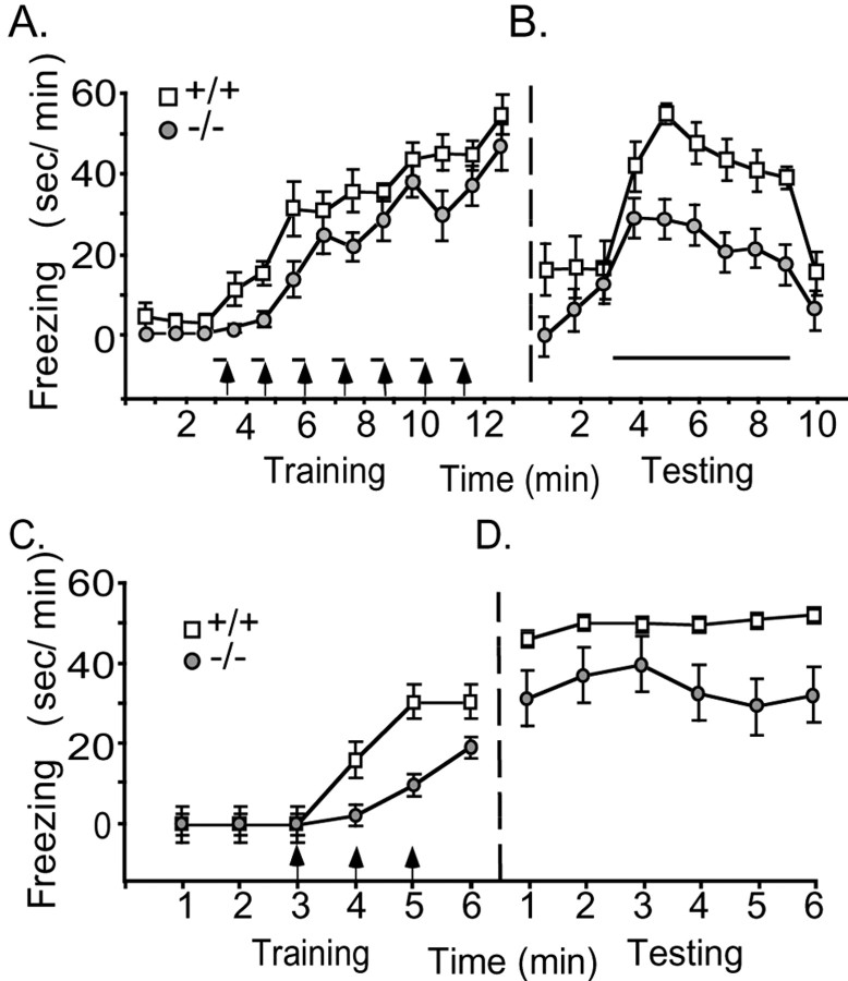
The acid-sensing ion channel, ASIC1, contributes to synaptic plasticity in the hippocampus and to hippocampus-dependent spatial memory. To explore the role of ASIC1 in brain, we examined the distribution of ASIC1 protein. Surprisingly, although ASIC1 was present in the hippocampal circuit, it was much more abundant in several areas outside the hippocampus. ASIC1 was enriched in areas with strong excitatory synaptic input such as the glomerulus of the olfactory bulb, whisker barrel cortex, cingulate cortex, striatum, nucleus accumbens, amygdala, and cerebellar cortex. Because ASIC1 levels were particularly high in the amygdala, we focused further on this area. We found that extracellular acidosis elicited a greater current density in amygdala neurons than hippocampal neurons and that disrupting the ASIC1 gene eliminated H+-evoked currents in the amygdala. We also tested the effect of ASIC1 on amygdala-dependent behavior; ASIC1-null mice displayed deficits in cue and context fear conditioning, yet baseline fear on the elevated plus maze was intact. These studies suggest that ASIC1 is distributed to regions supporting high levels of synaptic plasticity and contributes to the neural mechanisms of fear conditioning.


Figures

Figure 1.
Figure 1.
ASIC1 immunolocalization in forebrain. A, Coronal sections were stained for Nissl substance or immunolabeled for ASIC1 protein in +/+ and –/– mice. Areas marked by dashed lines in the Nissl-stained section are the areas dissected to prepare protein extracts for Western blotting in D and Figure 6 B. Asterisks in ASIC1 +/+ hemisphere denote areas of nonspecific staining that did not occur bilaterally or in multiple sections. B, C, Enlarged images of dentate gyrus and CA1 respectively. D, Western blot of ASIC1 protein in 100μg protein extract from dentate gyrus and CA1. amg, amygdala; cc, corpus callosum; dg, dentate gyrus; ec, external capsule; ect, ectorhinal cortex; En, endopiriform nuclei; fi, fimbria; Hb, habenula; H, hilus (polymorphic layer); ic, internal capsule; LTh, lateral thalamus; MS, medial septal nuclei; PAC, parietal association cortex; Pir, piriform cortex; PCg, posterior cingulate cortex; PRh, perirhinal cortex; S1BF, somatosensory barrel field; Th, thalamus.
Figure 2.
Figure 2.
ASIC1 immunolocalization in cortex. A, B, Immunolabeling in the posterior (post.) cingulate cortex. Stripes extending through layer II are labeled with an arrowhead. Positive-staining pyramidal cells in layer III are labeled with arrows. *ASIC1-specific staining in layer I. C, ASIC1 immunostaining is also elevated in layer III of barrel cortex.
Figure 3.
Figure 3.
Immunolocalization of ASIC1 in the sensorimotor cortex and striatum. Coronal sections through the forebrain were stained for Nissl substance, hematoxylin and eosin (H&E), or ASIC1 protein in ASIC1 +/+ or –/– mice. Center row, staining of representative coronal slices. Top row, insets of somatosensory cortex at higher magnification. Bottom row, insets of external capsule/corpus callosum and striatum at higher magnification. White matter tracts are labeled with arrows. ASIC1 immunolabeling was noticeably reduced in the white matter tracts. Areas of staining that were not present bilaterally and not present in multiple slices, suggesting nonspecific staining, are marked with an asterisk. aca, anterior commissure; Acb, accumbens nucleus; cc, corpus callosum; Cg, cingulate cortex; CPu, caudate/putamen (striatum); ec, external capsule; M1, primary motor cortex; Pir, piriform cortex; S1, somatosensory cortex; VP, ventral pallidum; Tu, olfactory tubercle.
Figure 4.
Figure 4.
Immunolocalization of ASIC1 in the olfactory bulb. Coronal sections through the olfactory bulb were stained for Nissl substance or immunolabeled for ASIC1 protein in ASIC1 +/+ and –/– mice. Higher magnifications at bottom demonstrate ASIC1 immunostaining in glomeruli (arrowheads). E/OV, ependymal and subendymal layer/olfactory ventricle; EPl, external plexiform layer; Gl, glomerular layer; Gr, granule cell layer; IPl, internal plexiform layer; Mi, mitral cell layer; ON, olfactory nerve layer.
Figure 5.
Figure 5.
Immunolocalization of ASIC1 in the cerebellum. ASIC1 immunohistochemistry in coronal (A) and parasagittal (B) sections of the cerebellum. C, Immunostaining with anti-calbindin D-28K antibody in fresh-frozen tissue. 4V, fourth ventricle; DN, deep cerebellar nuclei; Gc, granule cell layer; ML, molecular layer; Pc, pyramidal cell layer; WM, white matter.
Figure 6.
Figure 6.
A, ASIC1 immunolocalization in the amygdala complex. Bla, basolateral nucleus; Ce, central nucleus; La, lateral nucleus. B, Western blotting of ASIC1 protein in 100 μg of protein extract per lane isolated from indicated brain region. Cos-7 cells transfected with mASIC1, cos. Because the entire cerebellum was used to generate the cb extract, the subcortical structures with little ASIC1 may have diluted out the high expression level seen by immunohistological staining in the cerebellar cortex (Fig. 5). +/+ and –/–, whole-brain extract from ASIC1 +/+ or –/– mouse; amg, amygdala; cb, cerebellum; dg, dentate gyrus; Hb, habenula; S1, somatosensory barrel field; Th, thalamus; PAC, parietal association cortex; PCg, posterior cingulate cortex.
Figure 7.
Figure 7.
Proton-gated currents in amygdala neurons. A, B, Representative recordings of pH 5 evoked response in amygdala neurons from ASIC1 +/+ and –/– mice. C, Average current density of peak pH 5-evoked response in amygdala neurons from ASIC1 +/+ (n = 14) and –/– (n = 18) mice and hippocampal neurons from ASIC1 +/+ mice (n = 67; *p < 0.01).
Figure 8.
Figure 8.
Behavioral analysis of learned fear. A, B, Cued fear conditioning. The amount of freezing in 1 min intervals was determined during training (A) and testing (B). During testing, the ASIC1 –/– mice froze significantly less than +/+ controls with the presentation of the conditioned stimulus (intervals 4–9) (p = 0.02) (+/+, n = 5; –/–, n = 9). C, D, Context fear conditioning. The difference in freezing between +/+ and –/– mice was significant during training (intervals 4–6; p = 0.002) and during testing (p = 0.03) (+/+, n = 7; –/–,n=8). Footshock, arrows; tone, bars. Statistical significance was tested by ANOVA with repeated measures.

References

    1. Allen NJ, Attwell D ( 2002) Modulation of ASIC channels in rat cerebellar Purkinje neurons by ischaemia-related signals. J Physiol (Lond) 543: 521–529. - PMC - PubMed
    1. Alvarez de la Rosa D, Krueger SR, Kolar A, Shao D, Fitzsimonds RM, Canessa CM ( 2003) Distribution, subcellular localization and ontogeny of ASIC1 in the mammalian central nervous system. J Physiol (Lond) 546: 77–87. - PMC - PubMed
    1. Benson CJ, Xie J, Wemmie JA, Price MP, Henss JM, Welsh MJ, Snyder PM ( 2002) Heteromultimerics of DEG/ENaC subunits form H +-gated channels in mouse sensory neurons. Proc Natl Acad Sci USA 99: 2338–2343. - PMC - PubMed
    1. Bianchi L, Driscoll M ( 2002) Protons at the gate: DEG/ENaC ion channels help us feel and remember. Neuron 34: 337–340. - PubMed
    1. Bolshakov KV, Essin KV, Buldakova SL, Dorofeeva NA, Skatchkov SN, Eaton MJ, Tikhonov DB, Magazanik LG ( 2002) Characterization of acid-sensitive ion channels in freshly isolated rat brain neurons. Neuroscience 110: 723–730. - PubMed

Publication types

MeSH terms