. 2003 Jul 2;23(13):5507-19.
doi: 10.1523/JNEUROSCI.23-13-05507.2003.

Genetic analysis of the roles of Hh, FGF8, and nodal signaling during catecholaminergic system development in the zebrafish brain

Affiliations

Genetic analysis of the roles of Hh, FGF8, and nodal signaling during catecholaminergic system development in the zebrafish brain

Jochen Holzschuh et al. J Neurosci. .

Abstract

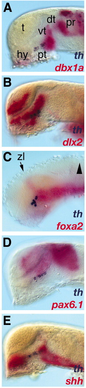
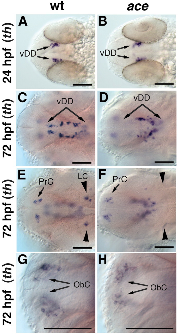
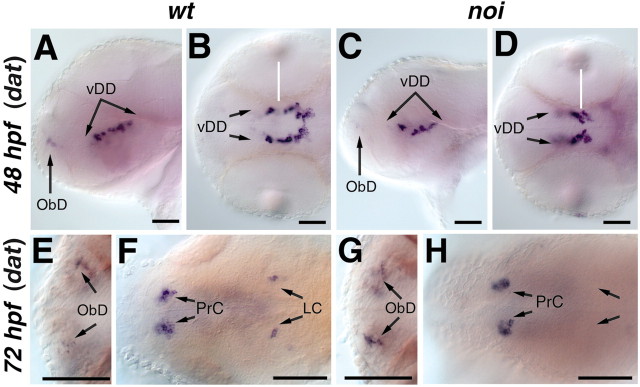
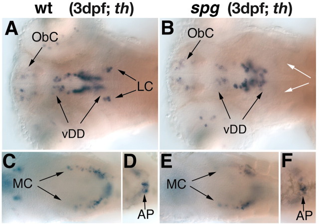
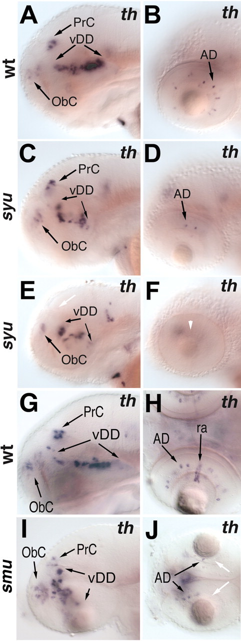
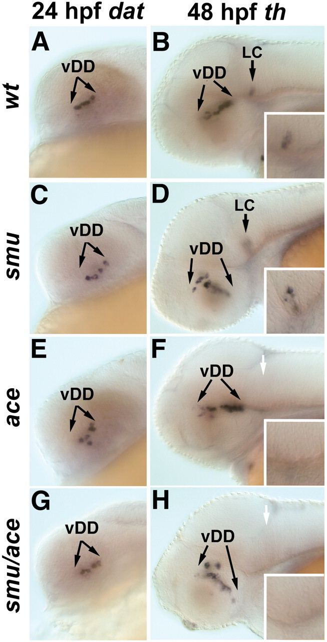
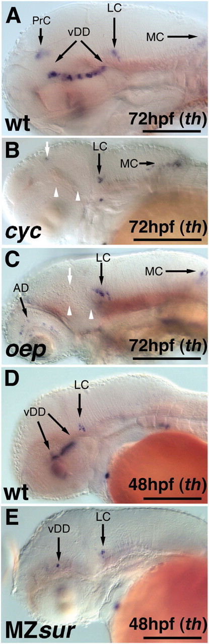
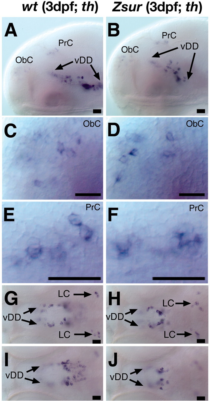
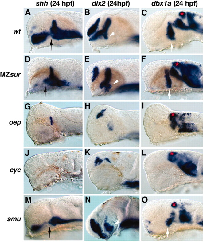
CNS catecholaminergic neurons can be distinguished by their neurotransmitters as dopaminergic or noradrenergic and form in distinct regions at characteristic embryonic stages. This raises the question of whether all catecholaminergic neurons of one transmitter type are specified by the same set of factors. Therefore, we performed genetic analyses to define signaling requirements for the specification of distinct clusters of catecholaminergic neurons in zebrafish. In mutants affecting midbrain- hindbrain boundary (MHB) organizer formation, the earliest ventral diencephalic dopaminergic neurons appear normal. However, after 2 d of development, we observed fewer cells than in wild types, which suggests that the MHB provides proliferation or survival factors rather than specifying ventral diencephalic dopaminergic clusters. In hedgehog (Hh) pathway mutants, the formation of catecholaminergic neurons is affected only in the pretectal cluster. Surprisingly, neither fibroblast growth factor 8 (FGF8) alone nor in combination with Hh signaling is required for specification of early developing dopaminergic neurons. We analyzed the formation of prosomeric territories in the forebrain of Hh and Nodal pathway mutants to determine whether the absence of specific dopaminergic clusters may be caused by early patterning defects ablating corresponding parts of the CNS. In Nodal pathway mutants, ventral diencephalic and pretectal catecholaminergic neurons fail to develop, whereas both anatomical structures form at least in part. This suggests that Nodal signaling is required for catecholaminergic neuron specification. In summary, our results do not support the previously suggested dominant roles for sonic hedgehog and Fgf8 in specification of the first catecholaminergic neurons, but instead indicate a novel role for Nodal signaling in this process.


Figures

Figure 1.
Figure 1.
The first dopaminergic neurons differentiate in the diencephalic posterior tuberculum. Visualization of forebrain gene expression domains and th expression by double in situ hybridization of whole-mount wild-type embryos are shown. A–E, Lateral views of the brain (anterior is to the left, dorsal is to the top). In situ hybridization of 24 hpf embryos with dbx1a (A), dlx2 (B), foxa1 (C), pax6.1 (D), and shh (E) is shown in red, and th is in blue. For better visibility, the skin, eyes, and yolk have been removed except in C where only the yolk has been removed. A, The ventral part of the most anterior transverse dbx1a expression domain marks the caudal hypothalamus. B, The DA neurons are located within the dlx2 expression domain in the hypothalamus. C, The DA cluster extends caudally to the anterior end of the longitudinal expression domain of foxa2 (arrowhead indicates the midbrain– hindbrain boundary). D, The DA cluster extends longitudinally close to the ventral edge of the alar plate (ventral border of pax 6.1 expression) in the basal plate marked by the shh expression domain (E). dt, Dorsal thalamus; hy, hypothalamus; pr, pretectum; pt, posterior tuberculum; t, telencephalon; vt, ventral thalamus; zl, zona limitans intrathalamica.
Figure 2.
Figure 2.
Formation of dopaminergic groups is not affected in ace/fgf8 mutant embryos. AH, Whole-mount in situ hybridization for th expression in wild-type (A, C, E, G) and ace mutant embryos (B, D, F, H). A–H, Dorsal views; anterior is to the left. A, B, The first th-expressing dopaminergic neurons appear normal in ace mutants at 24 hpf. C, D, At 72 hpf, fewer dopaminergic neurons are seen in the ventral diencephalon of ace mutants compared with that of wild-type embryos, whereas th expression appears normal in the pretectum and olfactory bulbs (E–H). E, F, The noradrenergic neurons of the LC are absent in ace mutants. vDD, Ventral diencephalic dopaminergic neurons; ObC, olfactory bulb catecholaminergic neurons; PrC, pretectal catecholaminergic neurons. Scale bars, 50 μm.
Figure 3.
Figure 3.
The LC is absent and the ventral diencephalic dopaminergic neurons are slightly reduced in noi/pax2.1 mutant embryos. Whole-mount in situ hybridization for dat in wild-type (A, B, E, F) and noi/pax2.1 (C, D, G, H) mutant embryos. A, C, Lateral views, dorsal is to the top. B, D, E–H, Dorsal views, anterior is to the left. A–D, At 48 hpf, the dat expression pattern reveals a slight decrease in ventral diencephalic dopaminergic neurons in noi mutant embryos. E–H, The catecholaminergic neurons in the olfactory bulb and pretectum appear normal in noi mutant embryos. F, H, In noi mutant embryos, the LC is absent. vDD, Ventral diencephalic dopaminergic neurons; ObD, olfactory bulb catecholaminergic neurons; PrC, pretectal catecholaminergic neurons. Scale bars, 30 μm.
Figure 4.
Figure 4.
The catecholaminergic neurons in the diencephalon are reduced and the LC is absent in spg mutant embryos. A–F, th expression in the forebrain and hindbrain of wild-type (A, C, D) and spg (B, E, F) mutant embryos at 3 dpf. A–F, Dorsal views, anterior is to the left. In spg mutant embryos (B), the catecholaminergic neurons of the LC are absent and those of the diencephalon are reduced, whereas the remaining hindbrain CA clusters develop normally (E, F). AP, Area postrema; vDD, ventral diencephalic dopaminergic neurons; MC, medullary catecholaminergic neurons; ObC, olfactory bulb catecholaminergic neurons; white arrows indicate the absence of the LC.
Figure 5.
Figure 5.
The pretectal catecholaminergic neurons and the amacrine cells of the retina are affected in hedgehog pathway mutants. A–F, th expression at 3 dpf in wild-type (A, B) and syu (C–F) mutant embryos (lateral views, anterior is to the left, dorsal is to the top). A, C, E, The pretectal cluster of catecholaminergic neurons is reduced (C) or absent (E, white arrow) in syu mutant embryos. The dopaminergic cell cluster in the ventral diencephalon is altered in morphology but not in size in syu mutant embryos. D, F, Dopaminergic amacrine cells are reduced (D) or absent (F, white arrowhead) in syu mutant embryos. G–J, dopamine transporter expression in 3 dpf embryos. GJ, Lateral views (G,I) and dorsal views (H, J); anterior is to the left. G, I, Of the CNS dopaminergic clusters, only the pretectal cluster is affected in smu mutants. H, J, The dopaminergic amacrine cells are reduced and the dat-expressing reticular astrocytes are absent in smu mutant embryos (J, white arrows). AD, Amacrine dopaminergic neurons; vDD, ventral diencephalic dopaminergic neurons; ObC, olfactory bulb catecholaminergic neurons; PrC, pretectal catecholaminergic neurons.
Figure 6.
Figure 6.
smu/smoh and ace/fgf8 double mutants develop additive DA neuronal phenotypes. A, C, E, G, dat expression or th expression (B, D, F, H). A–H, Lateral views, anterior to the left, dorsal is to the top. In smu/smoh (C), ace/fgf8 (E), and smu ace (G) double mutants, the earliest developing dopaminergic neurons in the ventral diencephalon appear normal when compared with wild-type embryos (A). By 48 hpf, additive phenotypes are observed in smu ace double mutants (H); the ventral diencephalic cluster is misshapen as in smu mutants (D) and the LC is absent as in ace (F). Insets in B, D, F, and H show higher magnification views of the region of the locus coeruleus. vDD, Ventral diencephalic dopaminergic neurons. White arrows indicate the absence of the LC.
Figure 7.
Figure 7.
Nodal pathway mutants affect the development of the ventral diencephalic and pretectal catecholaminergic neurons. A–E, th expression revealing the effects of mutations within the Nodal pathway on catecholaminergic neuron development (lateral views, anterior is to the left). A, D, th expression in wild-type embryos at 72 and 48 hpf, respectively. B, In cyc mutant embryos, both the ventral diencephalic DA neurons and the catecholaminergic neurons of the pretectum fail to develop, whereas the CA neurons in the hindbrain are unaffected. C, By 72 hpf, oep mutants lack diencephalic catecholaminergic neurons, whereas those of the hindbrain clusters are formed. E, By 48 hpf, MZsur mutant embryos develop few-to-no catecholaminergic neurons in the diencephalon, whereas the LC appears normal. White arrows indicate absence of PrC; white arrowheads indicate absence of vDD. AD, Amacrine dopaminergic neurons; vDD, ventral diencephalic dopaminergic neurons; MC, medulla oblongata catecholaminergic neurons; PrC, pretectal catecholaminergic neurons. Scale bars, 100 μm.
Figure 8.
Figure 8.
Formation of catecholaminergic neurons is affected in the ventral diencephalon of Zsur mutant embryos. A–J, Lateral views (A–F) (anterior is to the left, dorsal is to the top) and dorsal views (G–J) of th expression in the brain of wild-type (A, C, E, G,I) and Zsur mutant (B, D, F, H,J) embryos at 3 dpf. A–F, Overview of the catecholaminergic neurons (A, B), olfactory bulbs (C,D), and pretectum (E, F). In the ventral diencephalon of Zsur mutant embryos, the dopaminergic neurons are reduced in the posterior tuberculum (G,H) and hypothalamus (I, J). The LC (G, H) develops normally in Zsur mutants. vDD, Ventral diencephalic dopaminergic neurons; ObC, olfactory bulb catecholaminergic neurons; PrC, pretectal catecholaminergic neurons. Scale bars, 17 μm.
Figure 9.
Figure 9.
Embryonic patterning defects in the diencephalon of Nodal pathway and smu mutants. A–O, Embryos at 24 hpf (lateral views; anterior is to the left, dorsal is to the top). A–C, Wild-type embryos showing the expression domains of shh (A), dlx2 (B), and dbx1a (C). D–F, In MZsur embryos, shh expression is maintained in the zona limitans, part of the hypothalamus and midline (D), the expression domain of dlx2 in the hypothalamus is reduced in size (E); and the dbx1a expression domains in the pretectum and ventral diencephalon are present (F). G–I, oep mutant embryos show no hypothalamic expression of shh (G), dlx2 (H), and dbx1a (I), whereas the pretectal dbx1a domain is present. J–L, In cyc mutant embryos, expression of shh (J), the ventral expression domains of dlx2 (K), and dbx1a (L) in the forebrain are absent, whereas dbx1a expression in the pretectum is present. M–O, In smu mutant embryos, the expression of shh (M) in the zona limitans is absent, but a reduced expression in the ventral brain remains. The hypothalamic expression domains of dlx2 (N) and dbx1a (O), as well as the pretectal dbx1a domain can still be detected. The black arrows indicate the expression domain of shh in the ventral diencephalons. White arrowheads point to the dlx2 domain and white arrows point to the dbx1a domain in the ventral diencephalons. The red stars indicate the expression domain of dbx1a in the pretectum.

Similar articles

Cited by

References

    1. Akimenko MA, Ekker M, Wegner J, Lin W, Westerfield M ( 1994) Combinatorial expression of three zebrafish genes related to distal-less: part of a homeobox gene code for the head. J Neurosci 14: 3475–3486. - PMC - PubMed
    1. Belting HG, Hauptmann G, Meyer D, Abdelilah-Seyfried S, Chitnis A, Eschbach C, Soll I, Thisse C, Thisse B, Artinger KB, Lunde K, Driever W ( 2001) spiel ohne grenzen/pou2 is required during establishment of the zebrafish midbrain– hindbrain boundary organizer. Development 128: 4165–4176. - PMC - PubMed
    1. Brand M, Heisenberg CP, Jiang YJ, Beuchle D, Lun K, Furutani-Seiki M, Granato M, Haffter P, Hammerschmidt M, Kane DA, Kelsh RN, Mullins MC, Odenthal J, van Eeden FJ, Nusslein-Volhard C ( 1996) Mutations in zebrafish genes affecting the formation of the boundary between midbrain and hindbrain. Development 123: 179–190. - PubMed
    1. Briscoe J, Ericson J ( 1999) The specification of neuronal identity by graded Sonic Hedgehog signaling. Semin Cell Dev Biol 10: 353–362. - PubMed
    1. Briscoe J, Ericson J ( 2001) Specification of neuronal fates in the ventral neural tube. Curr Opin Neurobiol 11: 43–49. - PubMed

Publication types

MeSH terms