. 2003 Jul 2;23(13):5553-60.
doi: 10.1523/JNEUROSCI.23-13-05553.2003.

Action potential propagation in dendrites of rat mitral cells in vivo

Affiliations

Action potential propagation in dendrites of rat mitral cells in vivo

F Debarbieux et al. J Neurosci. .

Abstract

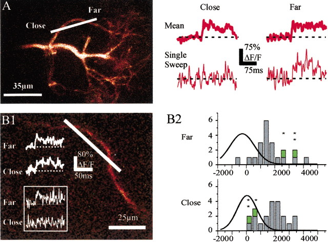
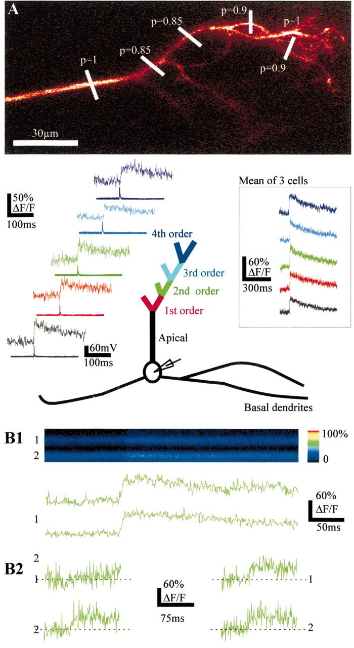
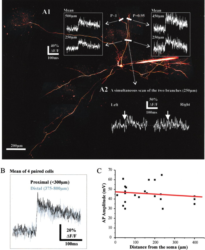
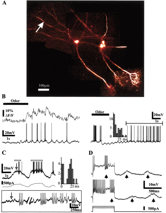
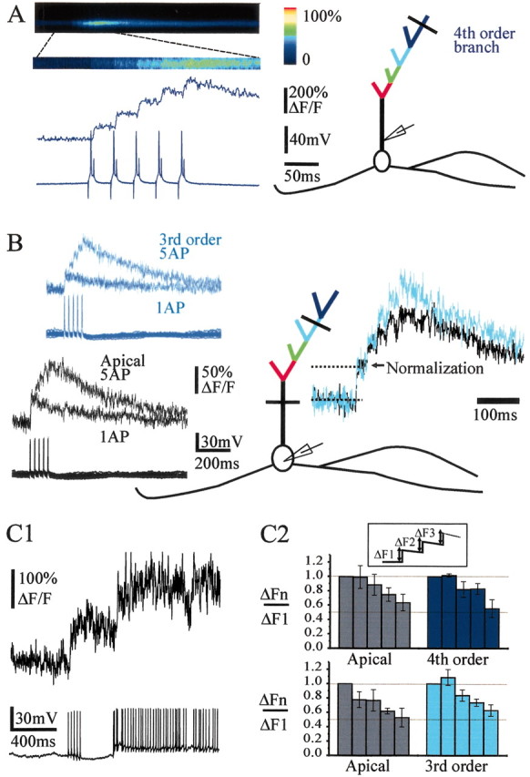
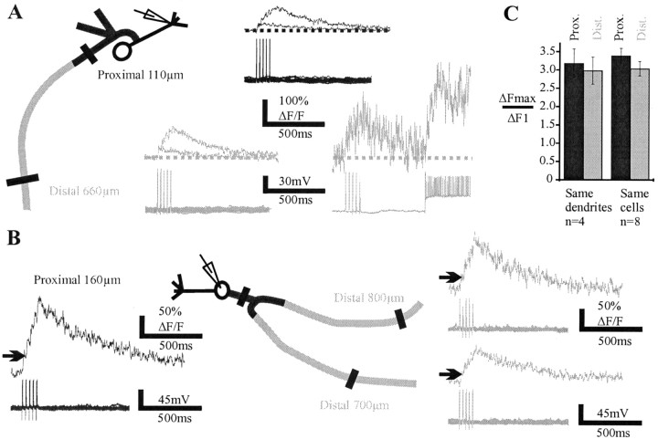
Odors evoke beta-gamma frequency field potential oscillations in the olfactory systems of awake and anesthetized vertebrates. In the rat olfactory bulb, these oscillations reflect the synchronous discharges of mitral cells that result from both their intrinsic membrane properties and their dendrodendritic interactions with local inhibitory interneurons. Activation of dendrodendritic synapses is purportedly involved in odor memory and odor contrast enhancement. Here we investigate in vivo to what extent action potentials propagate to remote dendrodendritic sites in the entire dendritic tree and if this propagation is changed during discharges at 40 Hz. By combining intracellular recording and two-photon microscopy imaging of intracellular calcium ([Ca2+]i), we show that in remote branches of the apical tuft and basal dendrites, transient Ca2+ changes are triggered by single sodium action potentials. Neither the amplitude of these Ca2+ transients nor that of action potentials obtained from intradendritic recordings showed a significant attenuation as a function of the distance from the soma. Calcium channel density seemed homogeneous; however, propagating action potentials occasionally failed to trigger a Ca2+ transient at a site closer to the soma whereas it did farther. This suggests that measurements of calcium transients underestimate the occurrence of sodium action potentials. During 40 Hz bursts of action potentials, [Ca2+]i increases with the number of action potentials in all dendritic compartments. These results suggest that the presence of release sites in dendrites is accompanied by an "axonal-like behavior" of the entire dendritic tree of mitral cells, including their most distal dendritic branches.


Figures

Figure 3.
Figure 3.
Measurements of fast Ca2+ transients underestimate action potential backpropagation. A, Ca2+ signals were recorded simultaneously at two points from the same first-order branch of a tuft. Average Ca2+ transients (including failures) were similar at both locations (top traces). In some cases, however, an action potential did not trigger any Ca2+ signal at the site close to the soma whereas it did farther (bottom traces). B1, Ca2+ signals were recorded simultaneously at two close points from the same branch of a basal dendrite. Average Ca2+ transients (including failures) were similar at both locations (top traces). Occasionally, an action potential did not trigger any Ca2+ signal at the site closer to the soma whereas it did farther (bottom traces, inset). B2, Integral histograms of ΔF/F measured over 50 msec after the action potential peak at the recording sites closer to (Close) and farther from (Far) the soma on the basal dendrite shown in B1. The green symbols (and the above stars) identify two cases of apparent failures at the proximal site and the corresponding calcium transients at the distal site. The superimposed curves correspond to the Gaussian fits of the noise integral distributions.
Figure 1.
Figure 1.
Single action potentials backpropagate reliably in remote branches of the apical dendrite. A, Top, morphology of a mitral cell tuft. The recording electrode was placed in the soma. The detection probabilities of fast Ca2+ transients evoked by single action potentials are indicated at the levels where the line scan recordings were done. Bottom, Averages (n = 30) of fast Ca2+ transients (top traces) triggered by single action potentials (bottom traces) in branches of successive order represented by different colors. Inset, Mean of three cells. B, Ca2+ transients can occur independently in two branches (second order) originating from the same branch and recorded simultaneously. B1, Single action potentials evoke transient changes in fluorescence (F, x = time, y = distance scanned) seen as color changes in the two branches (top panel) or as changes in ΔF/F (bottom two traces). B2, Traces on the left show an example of a Ca2+ failure occurring in branch 1 and not in branch 2. Traces on the right show single Ca2+ transients recorded simultaneously in branches 1 and 2.
Figure 2.
Figure 2.
Single action potentials backpropagate in remote sites of the basal dendrites. A1, Ca2+ transients were detected in two basal branches (top) in response to single action potentials. The probability of detection at the most distal sites is indicated at the levels where the line scans were done (450 and 500 μm from the soma). Note that the average Ca2+ transients (n = 30) recorded proximally (250μm) and distally (∼500 μm) in the two basal branches were very similar. A2 illustrates a case in which a failure occurred only in the right proximal branch. B, Mean values of the Ca2+ transients observed proximally and distally. In one case, the signal was measured at 950 μm from the soma. C, The amplitude of sodium action potentials did not significantly decrease with the distance from the soma at which the recording electrode was placed in the basal dendrite.
Figure 4.
Figure 4.
Mitral cells fire at β-γ frequencies. A, Three mitral cells were successively impaled in vivo, labeled with Oregon Green-1, and imaged with two-photon microscopy. B, The left cell was impaled in a basal dendrite and fired small high-frequency bursts of action potentials (left panel, bottom trace) during odor stimulation. Simultaneous recording of the Ca2+ signal (left panel, top trace, movie recording) from the basal dendrite site indicated by an arrow reveals that action potentials backpropagate during odor stimulation. The trace on the right shows the recording from another cell that exhibited bursts of action potentials after an initial odor-evoked inhibition. The distribution of the interspike intervals corresponded to firing frequencies in the β-γ range. C, Intrinsic membrane properties favor cell firing at 40 Hz. Intracellular depolarizing DC current injections induced bursts of action potentials triggered by subthreshold membrane potential oscillations (white arrow) with an interspike interval centered on 25 msec. Right, Distribution of the interspike intervals measured over the parts of the trace indicated by the white rectangles. Inset, Enlargement of these two parts of the above trace indicated with white rectangles. D, Spontaneous synaptic inputs entrain burst discharges. Two depolarizing current pulses were applied to induce spike accommodation. Additional bursts of action potentials (∼50 Hz) were triggered by the synaptic inputs locked to air inhalations (arrows).
Figure 5.
Figure 5.
Bursts of action potentials backpropagate in the entire apical tuft. A, Left, Illustration of the protocol type used to mimic bursts of action potentials in the β-γ range, which induces a summation of Ca2+ transients (average of 18 bursts). Right, Schematic representation of the recorded mitral cell with the location of the recording pipette in the proximal part of the apical trunk and the line scan in a fourth-order branch. B, Another mitral cell recorded with an intracellular pipette placed in the soma. Left, Superpositions of Ca2+ signals evoked by one and five action potentials (interspike interval = 25 msec) in the apical trunk and in a third-order branch. Right, Superposition of the traces obtained with five action potentials at both locations and normalized to the first Ca2+ transients. Note that summation of fluorescence was sublinear in the apical trunk and in the third-order branch. C1, The sublinearity was not caused by saturation of the dye because the Ca2+ signal could still increase during higher firing frequency. C2, Histograms of ΔFn/ΔF1 (inset) in the apical trunk and branches of the third (bottom histograms) and fourth (top histograms) order. In the two series, measurements were performed at two different sites of the same cell (apical trunk vs third-order branch, n = 3, or apical trunk vs fourth-order branch, n = 3).
Figure 6.
Figure 6.
Burst of action potentials backpropagate distally in basal dendrites. A, Left, Superposition of Ca2+ signals evoked by one and five action potentials (interspike interval = 25 msec) in the proximal (top traces) and distal (bottom traces) parts of abasal dendrite (average of 10–18 sweeps). The recording electrode was located near the first branching point of the apical dendrite. The summation of fluorescence was sublinear at each site. The sublinearity was not caused by saturation of the dye because the Ca2+ signal could still increase significantly during higher firing frequency, as shown for the most distal measurement. B, Another cell in which the amplitude of ΔF/F differed in two distal branches originating from the same proximal branch. In that case summation was nearly linear. C, Means of ΔFmax/ΔF1 recorded in the proximal and distal parts of basal dendrites. Histograms on the left are from four different cells in which measurements were obtained on the same dendritic branch (no branching points in between). Histograms on the right include additional recordings in which branching points were present between the proximal and distal recording sites.

References

    1. Aroniadou-Anderjaska V, Zhou FM, Priest CA, Ennis M, Shipley MT ( 2000) Tonic and synaptically evoked presynaptic inhibition of sensory input to the rat olfactory bulb via GABA(B) heteroreceptors. J Neurophysiol 84: 1194–1203. - PubMed
    1. Bischofberger J, Jonas P ( 1997) Action potential propagation into the presynaptic dendrites of rat mitral cells. J Physiol (Lond) 504: 359–365. - PMC - PubMed
    1. Charpak S, Mertz J, Beaurepaire E, Moreaux L, Delaney K ( 2001) Odor-evoked calcium signals in dendrites of rat mitral cells. Proc Natl Acad Sci USA 98: 1230–1234. - PMC - PubMed
    1. Chen WR, Shepherd GM ( 1997) Membrane and synaptic properties of mitral cells in slices of rat olfactory bulb. Brain Res 745: 189–196. - PubMed
    1. Chen WR, Midtgaard J, Shepherd GM ( 1997) Forward and backward propagation of dendritic impulses and their synaptic control in mitral cells. Science 278: 463–467. - PubMed

Publication types

LinkOut - more resources