Clinical Trial
. 2003 Jul 2;23(13):5627-33.
doi: 10.1523/JNEUROSCI.23-13-05627.2003.

Neural correlates of the automatic processing of threat facial signals

Affiliations
Clinical Trial

Neural correlates of the automatic processing of threat facial signals

Adam K Anderson et al. J Neurosci. .

Abstract

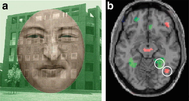
The present study examined whether automaticity, defined here as independence from attentional modulation, is a fundamental principle of the neural systems specialized for processing social signals of environmental threat. Attention was focused on either scenes or faces presented in a single overlapping display. Facial expressions were neutral, fearful, or disgusted. Amygdala responses to facial expressions of fear, a signifier of potential physical attack, were not reduced with reduced attention to faces. In contrast, anterior insular responses to facial expressions of disgust, a signifier of potential physical contamination, were reduced with reduced attention. However, reduced attention enhanced the amygdala response to disgust expressions; this enhanced amygdala response to disgust correlated with the magnitude of attentional reduction in the anterior insular response to disgust. These results suggest that automaticity is not fundamental to the processing of all facial signals of threat, but is unique to amygdala processing of fear. Furthermore, amygdala processing of fear was not entirely automatic, coming at the expense of specificity of response. Amygdala processing is thus specific to fear only during attended processing, when cortical processing is undiminished, and more broadly tuned to threat during unattended processing, when cortical processing is diminished.


Figures

Figure 1.
Figure 1.
Face–place object selection attention task. a, Example stimulus. Observers were presented with color-coded superimposed faces (disgusted, fearful, and neutral expressions in red) and places (inside and outside of buildings in green). Before each test stimulus, observers were presented with a color-coded prompt indicating which task they were to perform on that trial: indicate the gender of the face (attend trials) or indicate the location of the place (unattend trials). b, A representative subject demonstrated a greater response when attending to places (in green) in a bilateral region along the collateral sulcus, consistent with the PPA and a greater response when attending to faces (in red) in the right middle fusiform gyrus, consistent with the FFA.
Figure 2.
Figure 2.
Attentional dependence of amygdala and anterior insular responses to facial expressions. a, The amygdala was functionally defined by the group level contrast of fear relative to neutral trials when faces were attended. This resulted in a prominent activation in the right amygdala (at a peak height x,22; y, 1; z, –28; F(1, 11) =20.52; p < 0.0001). b, Effect of stimulus and attention on amygdala response. Peak amygdala response is displayed for each facial stimulus type during attended (red) and unattended (green) conditions. Attention did not significantly reduce the magnitude of amygdala response to fear, but the enhanced response to disgust during reduced attention suggests attention influenced the specificity of amygdala response. c, The insula was functionally defined by contrasting activation on disgust trials compared with neutral trials when faces were attended. This resulted in a prominent activation in the right anterior insula (at a peak height x, 44; y, 5; z, –14; F(1, 11) = 32.72, p < 0.0001). d, Effect of stimulus and attention on anterior insular response. Peak anterior insular response is displayed for each facial stimulus type during attended (red) and unattended (green) conditions. Reduced attention significantly reduced the magnitude of anterior insular response to disgust.
Figure 3.
Figure 3.
Response to disgust faces when unattended. a, Amygdala response to disgust relative to neutral faces when observers were attending to faces. No significant activation was found when faces were attended. b, Amygdala response to disgust relative to neutral faces when observers were attending to places. Activation was present when disgust faces were unattended. c, Time course of the disgust response difference score (unattended minus attended). A negative deflection of time course represents a decreased response when faces were attended. A positive deflection represents an increased response when faces were unattended. An inverse effect of attention on anterior insula and amygdala response to disgust faces peaked ∼ 6 sec after the stimulus onset.
Figure 4.
Figure 4.
Effect of inattention on FFA, LOCf, anterior insula (INS), and amygdala (AMYG) ROI responses to facial expressions. Bars represent the difference score between attended and unattended conditions (unattend minus attend) for each fear, disgust, and neutral face. The predominant effect of inattention was to reduce cortical responsiveness in the FFA, LOCf, and insula. In contrast, the amygdala demonstrated a marked increased response to disgust.

References

    1. Adolphs R, Tranel D, Damasio H, Damasio A ( 1994) Impaired recognition of emotion in facial expressions following bilateral damage to the human amygdala. Nature 372: 669–672. - PubMed
    1. Anderson AK, Phelps EA ( 2001) Lesions of the human amygdala impair enhanced perception of emotionally salient events. Nature 411: 305–309. - PubMed
    1. Birbaumer N, Grodd W, Diedrich O, Klose U, Erb M, Lotze M, Schneider F, Weiss U, Flor H ( 1998) fMRI reveals amygdala activation to human faces in social phobics. NeuroReport 9: 1223–1226. - PubMed
    1. Breiter HC, Etcoff NL, Whalen PJ, Kennedy WA, Rauch SL, Buckner RL, Strauss MM, Hyman SE, Rosen BR ( 1996) Response and habituation of the human amygdala during visual processing of facial expression. Neuron 17: 875–887. - PubMed
    1. Cahill L, Haier RJ, White NS, Fallon J, Kilpatrick L, Lawrence C, Potkin SG, Alkire MT ( 2001) Sex-related difference in amygdala activity during emotionally influenced memory storage. Neurobiol Learn Mem 75: 1–9. - PubMed

Publication types

LinkOut - more resources