. 2003 Jul 2;23(13):5723-31.
doi: 10.1523/JNEUROSCI.23-13-05723.2003.

Caspase-independent photoreceptor apoptosis in mouse models of retinal degeneration

Affiliations

Caspase-independent photoreceptor apoptosis in mouse models of retinal degeneration

Francesca Doonan et al. J Neurosci. .

Abstract

Apoptosis is the mode of cell death in retinitis pigmentosa, a group of retinal degenerative disorders primarily affecting rod photoreceptors. Although caspases have been demonstrated to play a central role in many incidences of apoptosis, accumulating evidence suggests that they may not be required for all forms of apoptotic cell death. The present study examined the mechanism of cell death in two in vivo models of photoreceptor apoptosis: the retinal degeneration (rd) mouse, a naturally occurring mutant model, and N-methyl-N-nitrosourea-induced retinal degeneration. Specifically, we examined the activation status of caspase-9, -8, -7, -3, and -2 and determined the caspase requirements for cytochrome c release, DNA fragmentation, and apoptosis-associated proteolysis of specific caspase substrates. We show that apoptosis in both in vivo models is independent of caspase-9, -8, -7, -3, and -2 activation. DNA fragmentation occurs in the absence of caspase-mediated ICAD (inhibitor of caspase-activated DNase) proteolysis, suggesting that an alternative endonuclease is responsible for DNA cleavage in these models. Importantly, we show that apoptosome activation is prevented because of an absence of mitochondrial cytochrome c release. Experiments performed using a cell-free system indicate that cytochrome c-dependent proteolysis and activation of caspase-9 can be restored in a neonatal cell-free system. However, we found that cytochrome c-dependent proteolysis and activation of caspase-9 could not be restored in an adult cell-free system because of an age-related decrease in the expression of Apaf-1 in the normal developing mouse retina. In the rd mouse, however, this age-related downregulation of apoptotic proteins was not observed, highlighting a critical feature of this model and the prevention of cytochrome c release as an apical event in caspase-independent apoptosis in this system.


Figures
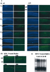
Figure 1.
Figure 1.
Detection of rod photoreceptor apoptosis in both the rd mouse and MNU-treated BALB/c mice. A, Detection of rod photoreceptor apoptosis in rd and MNU-treated BALB/c mice. Apoptotic cell death was assessed by detection of DNA strand breaks in photoreceptor nuclei by TUNEL. i, Retinas of rd mice at P9 show scattered labeling of the inner nuclear layer (INL) because of a developmentally associated reduction in cell number. This developmental apoptosis is essentially complete by P10. A similar pattern is seen in C57 mice at P9 and P10. Scattered photoreceptor apoptosis is visible at P11 in the ONL, with significant apoptosis observed at P12 and P13. By P14, the number of TUNEL-labeled cells has diminished, correlating with the loss of photoreceptor layers. ii, Retina of untreated control BALB/c mice did not exhibit any TUNEL-positive cells; however, 14 hr after MNU treatment, scattered apoptosis is observed in the ONL. Time points examined 24 and 48 hr after treatment reveal large numbers of apoptotic photoreceptor cells. B, Apoptosis was confirmed through detection of DNA fragmentation by agarose gel electrophoresis. The presence of a DNA ladder in MNU-treated mice 24 and 48 hr after exposure (lanes 4 and 5, respectively) confirms that apoptosis is the mode of cell death. Untd, Untreated.
Figure 2.
Figure 2.
Caspase-3 is not activated during photoreceptor apoptosis in the rd mouse or in MNU-treated BALB/c mice. A, Analysis of caspase-3 activity by immunoblot. Retinal-cell lysates were taken from rd mice between P9 and P15 (i) and MNU-treated BALB/c mice 14, 24, and 48 hr after exposure (ii). Untreated (–) and UV-treated (+) 32D cells (apoptotic index, <10%) served as negative and positive controls, respectively, to confirm the ability of the antibody to detect the large 17–20 kDa active fragment of caspase-3. Bi, Bii, These cells demonstrated the processing of procaspase-3 (32 kDa) to its active fragment of 17–20 kDa. i, Although procaspase-3 was detectable in retinal-cell lysates from rd mice at all of the time points examined, the 17–20 kDa fragment was absent up to P15. A similar pattern was observed in C57 retinal-cell lysates. ii, Equally, procaspase-3 was detectable in retinal-cell lysates from MNU-treated BALB/c mice at all of the time points examined; however, the 17–20 kDa fragment was absent up to 48 hr. Each blot was reprobed with an antibody to β-actin to demonstrate equal protein loading, followed by an antibody to GAPDH to validate the use of actin as a loading control. B, Analysis of caspase-3-like activity by detection of DEVD-ρNA cleavage. The measurement of DVED-ρNA cleavage was performed in a spectrophotometric assay by monitoring the liberation of ρNA caused by caspase activity in rd retinal-cell lysates from P9 to P15 (rd, filled bars; C57, open bars) (i) and retinal-cell lysates from MNU-treated BALB/c mice 14, 24, and 48 hr after exposure (open bars) (ii). Untreated (–) and anti-Fas IgM-treated (+) Jurkat cells (hatched bars) were used as negative and positive controls, respectively (i). Similarly, untreated (–) and UV-treated (+) 32D cells (hatched bars) were used as negative and positive controls, respectively (ii). An equal quantity of protein was loaded into each well. Data are expressed as the mean + SE of three independent experiments. Untd, Untreated; Abs., absorption.
Figure 6.
Figure 6.
Cytochrome c is not released from mitochondria during photoreceptor apoptosis in the rd mouse or MNU-treated BALB/c mice. A, Cells were fractionated at each time point from P9–P15 in the rd mouse (i) and 14, 24, and 48 hr after MNU treatment in BALB/c mice (ii). [Mitochondrial fractions were collected and analyzed for each time point (data not shown)]. The first lane was loaded with a representative mitochondrial fraction (M) as a positive control for cytochrome c. Cytochrome c was readily detectable in the mitochondrial fraction but was absent from the cytosol of rd mice at all of the time points examined. Cytochrome c was also absent from the cytosol of MNU-treated BALB/c mice up to 48 hr after treatment. Untreated and UV-treated 32D cells served as negative and positive controls, respectively, to demonstrate the release of cytochrome c from the mitochondria into the cytosol. B, Retinal-cell-free extracts were prepared from rd mice between P9 and P15 (i) and MNU-treated BALB/c mice 14, 24, and 48 hr after exposure (ii). Equivalent quantities of protein were incubated with cytochrome c and dATP for 2 hr and then resolved by SDS-PAGE, transferred onto nitrocellulose membrane, and probed with an antibody to caspase-9. Untreated (–) and 32D-cell-free extracts incubated with cytochrome c (+) were used as a positive control to demonstrate the processing of procaspase-9 to its large 37–39 kDa fragment. Caspase-9 was cleaved in the rd mouse at P10 and also at P15 after addition of cytochrome c. In C57 mice, however, caspase-9 could be cleaved at P10, but not at P15, after cytochrome c treatment. MNU-treated BALB/c mice retain caspase-9 in its inactive form; however, it was not cleaved on addition of cytochrome c. All of the blots were reprobed with an antibody to β-actin to demonstrate equal protein loading. A representative result of three experiments is shown. C, Apaf-1 and caspase-3 are downregulated at P15 in C57 mice, but not in the rd mouse. Retinal-cell lysates were taken from rd and C57 mice at P10 and P15. Analysis of Apaf-1 protein levels indicated expression dropped significantly from P10 to P15 in C57 mice, but not in the rd mouse (Fig. 5A). Similarly, the levels of procaspase-3 decreased in C57 mice from P10 to P15, but not in rd mice (Fig. 5B). All of the blots were reprobed with an antibody to β-actin to demonstrate equal protein loading. A representative result of three experiments is shown. Untd, Untreated; Td, treated.
Figure 3.
Figure 3.
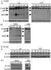
PARP and ICAD are not cleaved by caspase-3 in the rd mouse and in MNU-treated BALB/c mice. Western blot analysis was performed to detect cleavage of PARP and ICAD. Retinal-cell lysates were taken from rd mice between P9 and P15 (Ai, Bi) and MNU-treated BALB/c mice 14, 24, and 48 hr after exposure (Aii, Bii). Untreated (–) and UV-treated (+) 32D cells (apoptotic index, <10%) served as negative and positive controls, respectively, to demonstrate the processing of PARP (116 kDa) to its cleaved fragment (85 kDa) and to confirm the ability of the antibody to detect murine ICAD (45 kDa). Ai, PARP levels decreased in the rd mouse from P12 onward; however, the 85 kDa cleaved fragment was absent from P9 to P15. Levels of PARP remained constant in C57 mice from P9 to P15. Aii, Native PARP levels decreased in treated BALB/c mice, whereas the p85 fragment, present at basal levels in untreated BALB/c mice, did not increase. Bi, ICAD did not undergo cleavage in the rd mouse at any of the time points examined. Bii, Similarly, cleavage of ICAD was not observed in MNU-treated mice at any of the time points examined. All of the blots were reprobed with an antibody to β-actin to demonstrate equal protein loading. A representative result of three experiments is shown. Untd, Untreated.
Figure 4.
Figure 4.
Caspase-7 and caspase-2 are not activated during photoreceptor apoptosis in the rd mouse or in MNU-treated BALB/c mice. A, Analysis of caspase-7 activity by immunoblot. Retinal-cell lysates were taken from rd mice between P9 and P15 (i) and MNU-treated BALB/c mice 14, 24, and 48 hr after exposure (ii). Untreated (–) and UV-treated (+) 32D cells (apoptotic index, <10%) served as negative and positive controls, respectively, to demonstrate the processing of procaspase-7 (35 kDa) to its large 20 kDa and small 12 kDa fragments. The 35 kDa species was present at all of the time points analyzed in the rd mouse; however, both cleaved fragments were absent up to P15. Procaspase-7 was detectable in retinal-cell lysates from MNU-treated BALB/c mice at all of the time points examined; however, both large and small cleaved fragments were absent even up to 48hr. A positive 32D control has been included in this blot to rule out any ambiguity regarding the nonspecific bands observed in the MNU-treated samples. B, Analysis of caspase-2 activity by immunoblot. Retinal-cell lysates were taken from rd mice between P9 and P15 (i) and MNU-treated BALB/c mice 14, 24, and 48 hr after exposure (ii). Untreated (–) and UV-treated (+) 32D cells (apoptotic index, <10%) served as negative and positive controls, respectively, to demonstrate the processing of procaspase-2 (48 kDa) to its 12 kDa product. The 48 kDa species was present at all of the time points analyzed in the rd mouse; however, both cleaved fragments were absent up to P15. Procaspase-2 was detectable in retinal-cell lysates from MNU-treated BALB/c mice at all of the time points examined; however, the large cleaved fragment was absent even up to 48 hr. All of the blots were reprobed with an antibody to β-actin to demonstrate equal protein loading. A representative result of three experiments is shown. Untd, Untreated.
Figure 5.
Figure 5.
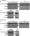
Neither caspase-8 nor caspase-9 is activated during photoreceptor apoptosis in the rd mouse or MNU-treated BALB/c mice. A, Analysis of caspase-8 activity by immunoblot. Retinal-cell lysates were taken from rd mice between P9 and P15 (i) and MNU-treated BALB/c mice 14, 24, and 48 hr after exposure (ii). Untreated (–) and anti-Fas IgM-treated (+) Jurkat cells were used as negative and positive controls, respectively, to demonstrate the processing of procaspase-8 (53 kDa) to its 42 kDa cleaved intermediate. The 53 kDa species was present at all of the time points analyzed in the rd mouse; however, the 42 kDa fragment was absent up to P15. Equally, procaspase-8 was detectable in retinal-cell lysates from MNU-treated BALB/c mice at all of the time points examined; however, the 42 kDa active fragment was absent up to 48 hr after treatment. *Nonspecific bands recognized by the antibody. B, Retinal-cell lysates were taken from rd mice between P9 and P15 (i) and MNU-treated BALB/c mice 14, 24, and 48 hr after exposure (ii). Untreated (–) and UV-treated (+) 32D cells (apoptotic index, <10%) served as negative and positive controls, respectively, to demonstrate the processing of procaspase-9 (46 kDa) to its large 37–39 kDa active fragment. Although procaspase-9 was detectable in retinal-cell lysates from rd mice at all of the time points examined, the 37–39 kDa large fragment was absent up to P15. A similar pattern was observed in C57 retinal-cell lysates. Equally, procaspase-9 was detectable in retinal-cell lysates from MNU-treated BALB/c mice at all of the time points examined; however, the 37–39 kDa fragment was absent even at 48 hr. All of the blots were reprobed with an antibody to β-actin to demonstrate equal protein loading. A representative result of three experiments is shown. *This band represents a caspase-9 splice variant present only in adult mice. Untd, Untreated.

Similar articles

Cited by

References

    1. Azarian SM, Williams DS ( 1995) Calpain activity in the retinas of normal and RCS rats. Curr Eye Res 14: 731–735. - PubMed
    1. Bascom RA, Manara S, Collins L, Molday RS, Kalnins VI, McInnes RR ( 1992) Cloning of the cDNA for a novel photoreceptor membrane protein (rom-1) identifies a disk rim protein family implicated in human retinopathies. Neuron 8: 1171–1184. - PubMed
    1. Borner C, Monney L ( 1999) Apoptosis without caspases: an inefficient molecular guillotine? Cell Death Differ 6: 497–507. - PubMed
    1. Boulares AH, Zoltoski AJ, Contreras FJ, Yakovlev AG, Yoshihara K, Smulson ME ( 2002) Regulation of DNAS1L3 endonuclease activity by poly(ADP-ribosyl)ation during etoposide-induced apoptosis. Role of poly(ADP-ribose) polymerase-1 cleavage in endonuclease activation. J Biol Chem 277: 372–378. - PubMed
    1. Bowes C, Li T, Danciger M, Baxter LC, Applebury ML, Farber DB ( 1990) Retinal degeneration in the rd mouse is caused by a defect in the beta subunit of rod cGMP-phosphodiesterase. Nature 347: 677–680. - PubMed

Publication types

MeSH terms

LinkOut - more resources