. 2003 Jul 2;23(13):5789-98.
doi: 10.1523/JNEUROSCI.23-13-05789.2003.

High threshold for induction of the stress response in motor neurons is associated with failure to activate HSF1

Affiliations

High threshold for induction of the stress response in motor neurons is associated with failure to activate HSF1

Zarah Batulan et al. J Neurosci. .

Abstract

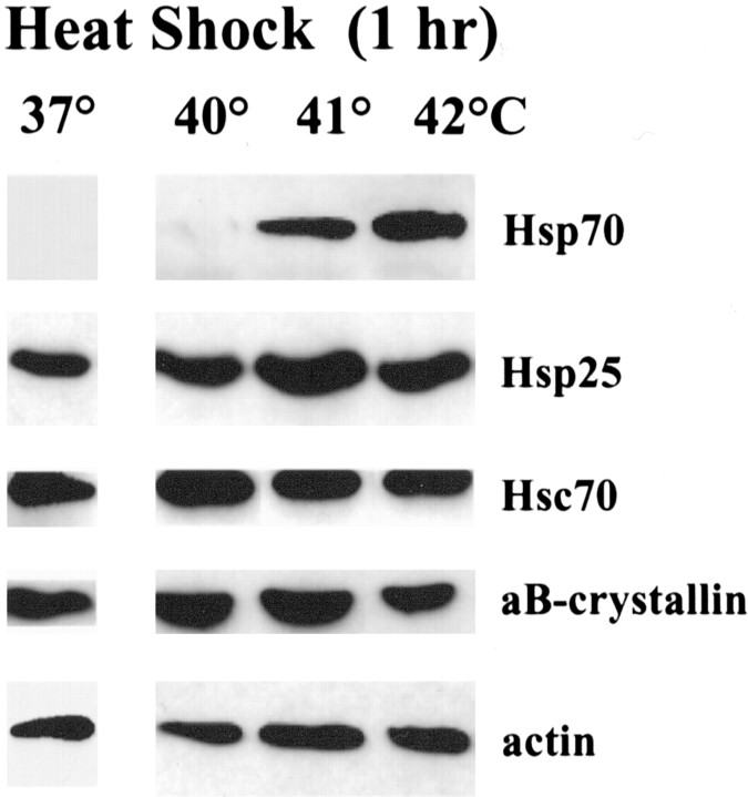
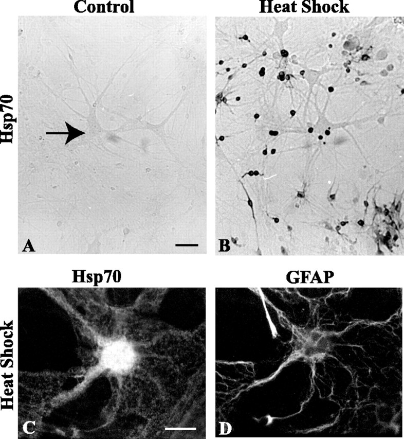
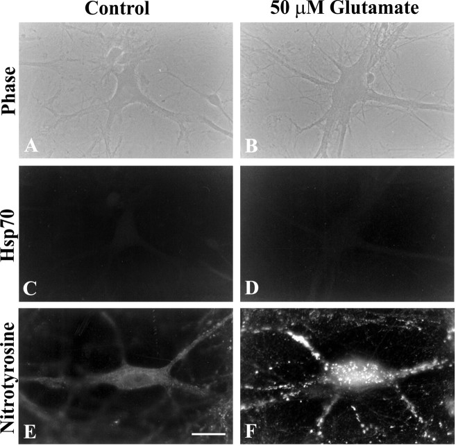
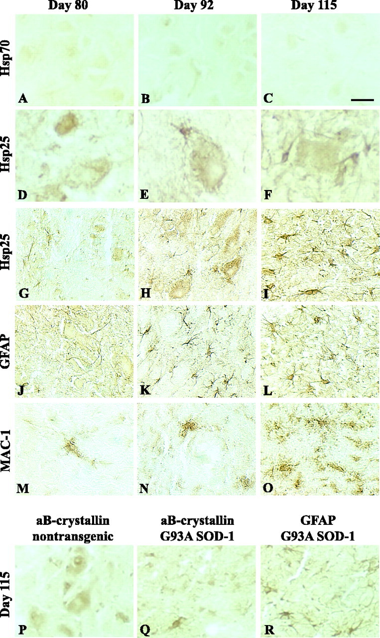
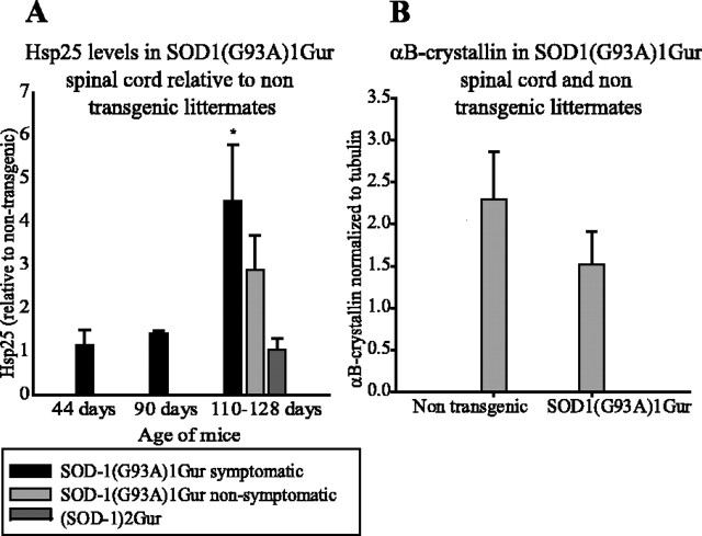
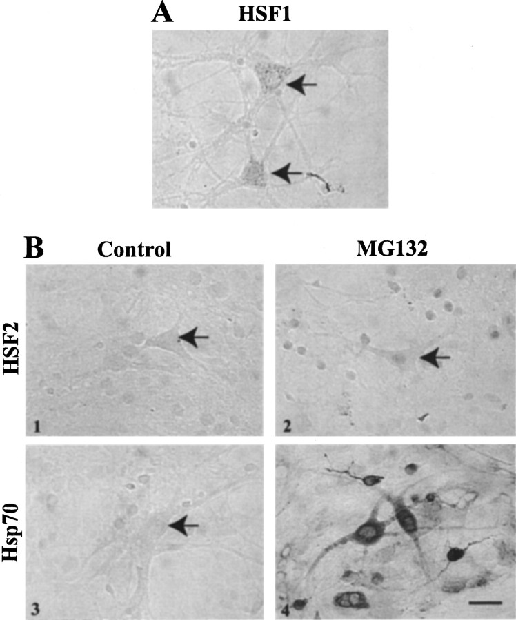
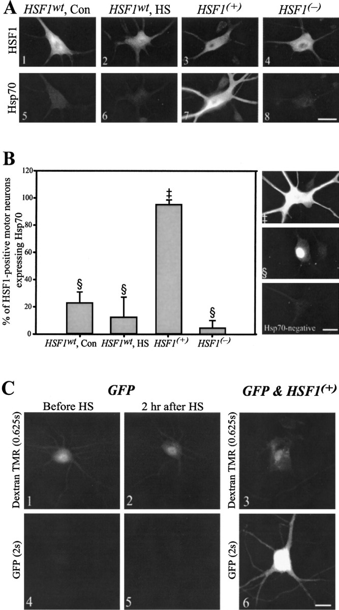
Heat shock protein 70 (Hsp70) protects cultured motor neurons from the toxic effects of mutations in Cu/Zn-superoxide dismutase (SOD-1), which is responsible for a familial form of the disease, amyotrophic lateral sclerosis (ALS). Here, the endogenous heat shock response of motor neurons was investigated to determine whether a high threshold for activating this protective mechanism contributes to their vulnerability to stresses associated with ALS. When heat shocked, cultured motor neurons failed to express Hsp70 or transactivate a green fluorescent protein reporter gene driven by the Hsp70 promoter, although Hsp70 was induced in glial cells. No increase in Hsp70 occurred in motor neurons after exposure to excitotoxic glutamate or expression of mutant SOD-1 with a glycine--> alanine substitution at residue 93 (G93A), nor was Hsp70 increased in spinal cords of G93A SOD-1 transgenic mice or sporadic or familial ALS patients. In contrast, strong Hsp70 induction occurred in motor neurons with expression of a constitutively active form of heat shock transcription factor (HSF)-1 or when proteasome activity was sufficiently inhibited to induce accumulation of an alternative transcription factor HSF2. These results indicate that the high threshold for induction of the stress response in motor neurons stems from an impaired ability to activate the main heat shock-stress sensor, HSF1.


Figures

Figure 1.
Figure 1.
Heat shock induces Hsp70 in spinal cord cultures. Spinal cord cultures were subjected to increasing heat shock temperatures (40–42°C) for 1 hr, followed by 2 hr recovery at 37°C. Western blots were probed with antibodies to HSPs [Hsp70 (SPA-810), Hsp25 (SPA-801), Hsc70 (K19), andαB-crystallin (SPA-222)] and actin as loading control. Blots shown are representative of three different experiments. Only Hsp70 was upregulated.
Figure 2.
Figure 2.
Glial cells, but not motor neurons, upregulate Hsp70 after heat shock. AD, Spinal cord cultures were subjected to 37°C control temperature (A) or 42°C heat shock (BD) for 1 hr, followed by 2 hr recovery at 37°C. Hsp70 induction was assessed by immunocytochemistry using biotin-conjugated secondary antibodies and DAB as substrate (A, B) or by fluorescence-conjugated secondary antibodies (C). Heat-shocked spinal cord cultures were also double labeled with antibodies against GFAP (D) to verify that glial cells induced Hsp70. The arrow in A points to a motor neuron. Scale bars: A, B, 50 μm; C, D, 20 μm.
Figure 3.
Figure 3.
Motor neurons failed to express Hsp70 in response to glutamate. AF, Spinal cord cultures were treated with 50μm glutamic acid (B, D,F) or vehicle (A, C,E) for 30 min and then assessed for Hsp70 induction by immunocytochemistry (SPA-810). Nitrotyrosine labeling demonstrated that glutamate treatment had an effect on motor neurons (E, F). Scale bar, 20 μm
Figure 4.
Figure 4.
Expression of HSPs in lumbar spinal cord of G93A mutant SOD-1 transgenic mice. The 20 μm sections of lumbar spinal cord from 80, 92, and 115-d-old mice were labeled with antibodies to Hsp70 (SPA-810), Hsp25 (M20; recognizes primate Hsp27 and rodent Hsp25), GFAP, MAC-1, or αB-crystallin. No induction of Hsp70 was observed (AC). Motor neurons constitutively expressed Hsp25 (DF), but no qualitative changes were noted with disease progression. There was a progressive increase in Hsp25 expressing astrocytes with age (GI). Motor neurons of 115-d-old nontransgenic mice constitutively expressed αB-crystallin (P). In lumbar cord from 115-d-old symptomatic G93A SOD-1 transgenics,αB-crystallin was detected in some but not all reactive astrocytes (Q,R). Reactive astrocytes and microglia were identified by expression of GFAP (J, K, L, R) and MAC-1 (MO) labeling of adjacent sections, respectively. Scale bars: AC, G–R, 40 μm; D–F, 15.4 μm.
Figure 5.
Figure 5.
Increase in Hsp25 with age and disease progression in G93A SOD-1 transgenic mice. A, Quantitation of Hsp25 on Western blots of spinal cord homogenates from wild-type and G93A SOD-1 mice. Ratios of Hsp25 to tubulin in samples from transgenic mice were normalized to ratios in matched samples of nontransgenic littermates. Note the threefold increase in Hsp25 in symptomatic G93A SOD-1 mice relative to those at 44 d of age. The mean Hsp25 level in the spinal cord of symptomatic animals was also higher than in samples from nonsymptomatic littermates. No inducible Hsp70 was detected on immunoblots (our unpublished data). Shown are means ± SD for three to nine animals per group. The asterisk indicates a significant difference from all other values; p ≤ 0.05 (t test; one-tailed; unequal variance). B, Quantitation of αB-crystallin immunolabeling relative to tubulin on Western blots of lumbar spinal cord homogenates from 115-d-old G93A SOD-1 mice and age-matched nontransgenic littermates.
Figure 6.
Figure 6.
HSP expression in cervical spinal cord of control, sporadic, and FALS (A4V SOD-1 mutation). Hsp27 was detected in spinal motor neurons and neuritic processes in the anterior horn of control (A), sporadic (B), and FALS (C) patients; the arrow in C points to a spinal motor neuron containing a Bunina body. Hsp70 immunolabeling was not observed in the cytoplasm of motor neurons of FALS (D) or sporadic ALS (our unpublished data). Hsp27 labeling in the neuropil (E) does not correspond to the pattern of GFAP immunoreactivity (F). Scale bars: A, 100 μm; B, 40 μm; CF, 20 μm.
Figure 7.
Figure 7.
Motor neurons express the heat shock transcription factors HSF1 and HSF2. A, Motor neurons and other cells in primary spinal cord cultures were labeled by a monoclonal antibody to HSF1 (SPA-950). B, Overnight treatment with the proteasomal inhibitor MG132 (0.1 μm) caused HSF2 to accumulate and translocate into the nuclei of spinal cord cells, including motor neurons (1,2).In motor neurons and other cells treated with MG132 (1μm), strong Hsp70 (SPA-810) expression was also detected by immunocytochemistry using DAB as substrate (3,4). Arrows indicate motor neurons. Scale bar, 30 μm.
Figure 8.
Figure 8.
Expression of activated HSF1, but not HSF1 wt, by gene transfer induces HSP70 expression in motor neurons. A, B, Motor neurons were microinjected with plasmid expression vectors encoding HSF1 wt, constitutively activated HSF1 (HSF1 (+)), or nonactivatable HSF1 (HSF1 (-)). After 48 hr, motor neurons injected with HSF1 wt were heat shocked (42°C; 1 hr) or subjected to control conditions (37°C; 1 hr) and recovered at 37°C for 2 hr. All cultures were double labeled with antibodies against HSF1 and Hsp70. A, Although motor neurons expressed HSF1 wt (1,2) at levels comparable with HSF1 (+) (3), only HSF1 (+) induced Hsp70 (5–7). B, Percentage of motor neurons coexpressing HSF1 and Hsp70 (n ≥ 4 cultures per group). Intense Hsp70 immunolabeling (‡) occurred only in motor neurons injected with HSF1(+). In the other three conditions, lower intensity labeling was observed frequently in the nucleus (§). Only HSF1 (+) significantly increased the percentage of motor neurons expressing Hsp70 (t test; one-tailed; unequal variance; p < 0.0001). C, HSF1 (+) also caused strong expression of a GFP reporter gene under the control of the Hsp70 promoter in motor neurons. Left, Motor neurons injected with the dextran TMR marker and GFP vector were imaged before and 2 hr after heat shock. Right, Heat shock failed to induce GFP (5), in contrast to coexpression of HSF1 (+), which resulted in strong GFP expression (6). Scale bar, 30 μm.

References

    1. Bechtold DA, Rush SJ, Brown IR ( 2000) Localization of the heat-shock protein Hsp70 to the synapse following hyperthermic stress in the brain. J Neurochem 74: 641-646. - PubMed
    1. Bence NF, Sampat RM, Kopito RR ( 2001) Impairment of the ubiquitin-proteasome system by protein aggregation. Science 292: 1552-1555. - PubMed
    1. Benn SC, Perrelet D, Kato AC, Scholz J, Decosterd I, Mannion RJ, Bakowska JC, Woolf CJ ( 2002) Hsp27 upregulation and phosphorylation is required for injured sensory and motor neuron survival. Neuron 36: 45-56. - PubMed
    1. Brown IR, Rush SJ ( 1999) Cellular localization of the heat shock transcription factors HSF1 and HSF2 in the rat brain during postnatal development and following hyperthermia. Brain Res 821: 333-340. - PubMed
    1. Bruening W, Roy J, Giasson B, Figlewicz DA, Mushynski WE, Durham HD ( 1999) Up-regulation of protein chaperones preserves viability of cells expressing toxic Cu/Zn-superoxide dismutase mutants associated with amyotrophic lateral sclerosis. J Neurochem 72: 693-699. - PubMed

Publication types

MeSH terms