. 2003 Jul 2;23(13):5816-26.
doi: 10.1523/JNEUROSCI.23-13-05816.2003.

Novel role of vitamin k in preventing oxidative injury to developing oligodendrocytes and neurons

Affiliations

Novel role of vitamin k in preventing oxidative injury to developing oligodendrocytes and neurons

Jianrong Li et al. J Neurosci. .

Abstract

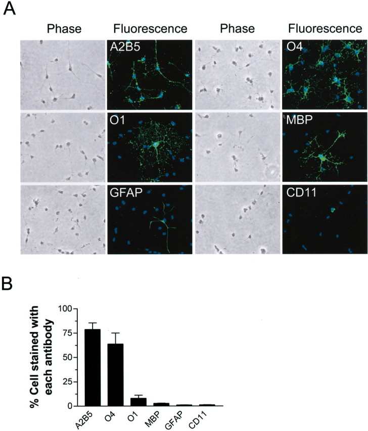
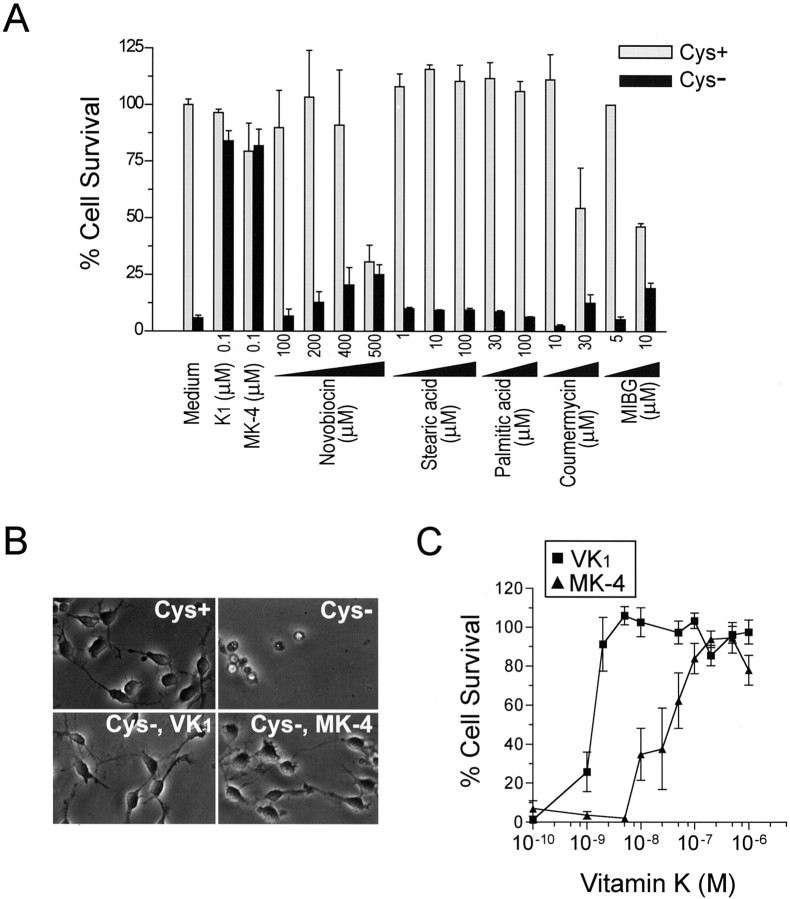
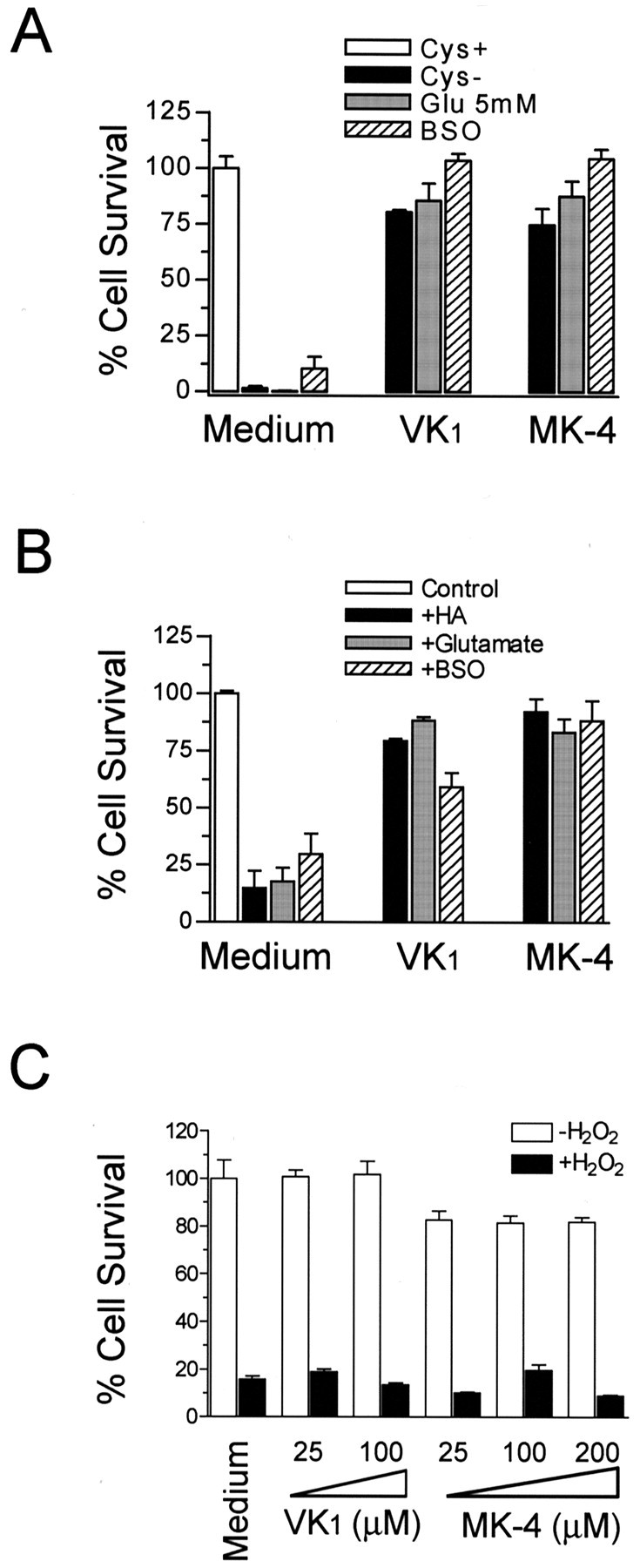
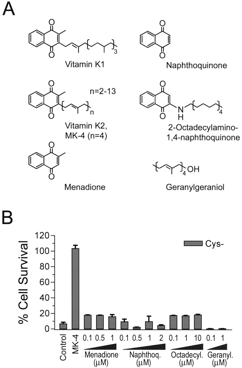
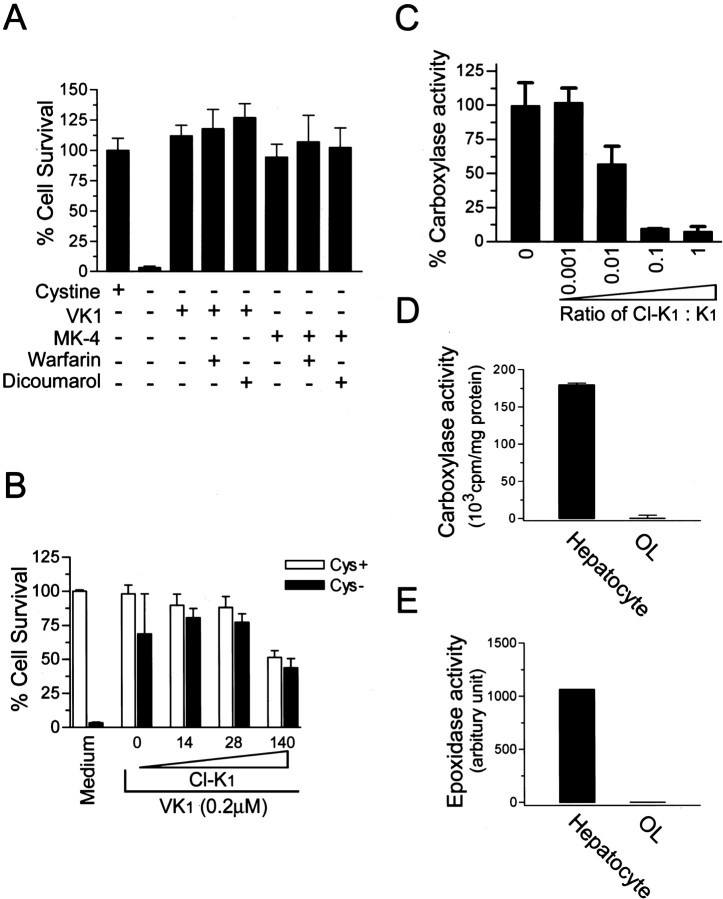
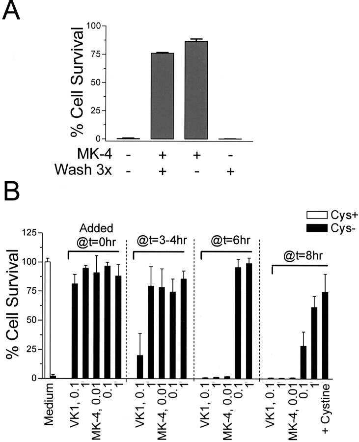
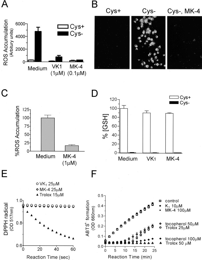
Oxidative stress is believed to be the cause of cell death in multiple disorders of the brain, including perinatal hypoxia/ischemia. Glutamate, cystine deprivation, homocysteic acid, and the glutathione synthesis inhibitor buthionine sulfoximine all cause oxidative injury to immature neurons and oligodendrocytes by depleting intracellular glutathione. Although vitamin K is not a classical antioxidant, we report here the novel finding that vitamin K1 and K2 (menaquinone-4) potently inhibit glutathione depletion-mediated oxidative cell death in primary cultures of oligodendrocyte precursors and immature fetal cortical neurons with EC50 values of 30 nm and 2 nm, respectively. The mechanism by which vitamin K blocks oxidative injury is independent of its only known biological function as a cofactor for gamma-glutamylcarboxylase, an enzyme responsible for posttranslational modification of specific proteins. Neither oligodendrocytes nor neurons possess significant vitamin K-dependent carboxylase or epoxidase activity. Furthermore, the vitamin K antagonists warfarin and dicoumarol and the direct carboxylase inhibitor 2-chloro-vitamin K1 have no effect on the protective function of vitamin K against oxidative injury. Vitamin K does not prevent the depletion of intracellular glutathione caused by cystine deprivation but completely blocks free radical accumulation and cell death. The protective and potent efficacy of this naturally occurring vitamin, with no established clinical side effects, suggests a potential therapeutic application in preventing oxidative damage to undifferentiated oligodendrocytes in perinatal hypoxic/ischemic brain injury.


Figures

Figure 1.
Figure 1.
Characterization of primary OL cultures.A,Representative microphotographs of morphology and immunocytochemistry of cells (7–9 d in vitro) labeled with indicated antibodies. B, Composition of developing OL cultures as determined by immunostaining for indicated markers. Total cell number was determined by counting all cells labeled with the nuclei dye Hoechst 33258. Values represent mean ± SEM from three separate experiments.
Figure 2.
Figure 2.
Vitamin K protected against cystine deprivation-induced OL death independent of MART. A, Effects of various MART inhibitors on cystine depletion-induced OL death. OLs were subjected to either normal medium (Cys +) or cystine-free medium (Cys -) in the presence of indicated concentrations of various MART inhibitors. Cell viability was evaluated after 24 hr. Results are representative of four independent experiments. B, Phase contrast photomicrographs of OLs exposed to normal or cystine-depleted medium with or without 0.1μm vitamin K1 or MK-4 for 18 hr. Magnification, 400×. Data are representative of at least 10 separate experiments. C, Vitamin K prevented OL death in a concentration-dependent manner. Cell viability was assayed 24 hr after cystine deprivation in the presence of increasing concentrations of K1 and MK-4. Values are mean ± SEM of six separate experiments.
Figure 3.
Figure 3.
Vitamin K1 and MK-4 protected both OL precursors and neurons from cell death induced by various GSH depletion methods. A, Vitamin K prevented OL cell death induced by cystine depletion, by glutamate (5 mm), and by BSO (1 mm). B, Vitamin K1 and MK-4 prevented immaturecorticalneuronaldeathinducedbyhomocysteicacid(HA,2.5mm),glutamate(5mm), or BSO (1 mm). Primary cells were incubated with indicated agents for 24 hr in the presence or absence of K1 (0.1μm) or MK-4 (0.1μm), and cell viability was analyzed after 24 hr. Results are mean ± SEM of three separate experiments. C, Effect of vitamin K1 and MK-4 on H2O2-induced toxicity. OL precursors were incubated with or without 800μm H2O2 in the presence of indicated concentrations of vitamin K for 15 hr, and cell viability was evaluated. Data are representative of at least four independent experiments with similar results.
Figure 4.
Figure 4.
Structural requirement for vitamin K-dependent protection against cystine depletion-induced cell death. A, Structures of vitamin K1, K2, and compounds tested in B for their effects on cystine depletion-induced OL death. B, Effect of various compounds on OL cell death induced by cystine depletion. Representative results of two to three independent experiments are shown. No cytotoxicity to OLs in normal culture medium was seen with the test compounds at the concentration used.
Figure 5.
Figure 5.
Vitamin K protected OLs via a mechanism independent of vitamin K-dependentγ-glutamylcarboxylase.A,Inhibitors of vitamin K cycling, warfarin and dicoumarol, had no effect on vitamin K protection. OL precursors were induced to undergo cystine depletion-induced cell death in the presence or absence of indicated treatment for 24 hr, and cell viability was assayed. Concentrations used were: vitamin K1, 0.1μm; MK-4, 0.1μm; warfarin, 500μm; dicoumarol, 200μm. Results are representative of at least two separate experiments. B, Carboxylase inhibitor Cl-K1 did not reverse vitamin K protection. Cells were treated as indicated with or without Cl-K1 (0–140 μm) and vitamin K1 (0.2 μm) for 24 hr, and cell viability was analyzed. C, Cl-K1 directly inhibited enzymatic activity of purified γ-glutamylcarboxylase in vitro. Purified γ-glutamylcarboxylase was incubated in the presence of increasing concentrations of Cl-K1 versus K1 and assayed in duplicates for its enzyme activity. Results are mean ± SEM of three independent experiments. D, E, OL precursors express little, if any, γ-glutamylcarboxylase and vitamin K epoxidase activity. Microsomal proteins were extracted from cultured primary OLs, and vitamin K-dependent carboxylase and epoxidase activity of the microsomal preparation were analyzed. Primary rat hepatocyte microsomal preparation was performed in parallel as positive control for the enzyme assays. Results are representative of two independent experiments.
Figure 6.
Figure 6.
Persistent presence of MK-4 was not required for protection. A, Effect of pretreatment with MK-4 on subsequent cystine depletion-induced cell death. OL precursors were preincubated with or without MK-4 (0.1 μm) for 1 hr in cystine-free medium, followed by wash three times with cystine-free medium, when indicated. Cell viability was determined 24 hr later, and the percentage of cell survival was based on the control in which cells were maintained in normal medium. Results are one representative of three independent experiments with similar results. B, Effect of vitamin K treatment at various time intervals after initiation of cystine depletion. Cells were treated with increasing amounts of K1 or MK-4 (μm) at the time of cystine depletion or the indicated time after cystine depletion. The total cystine deprivation time is 24 hr, and the cell viability was evaluated. l-Cystine (200 μm) was added back to the cystine-free medium at 8 hr after cystine depletion and was used as a control. Results are representative of three experiments.
Figure 7.
Figure 7.
Vitamin K blocked cystine deprivation-induced free radical accumulation. OLs were subjected to Cys + or Cys - medium as indicated, with or without vitamin K1 or MK-4 for 10–15 hr, and cells were then loaded with DCFH-DA (A) or Rho123 (B), and the free radical production was evaluated as described. Both DCFH-DA and Rho123 are nonfluorescent but become fluorescent on oxidation. DCF fluorescence was measured using a microplate reader, and oxidized Rho123 was visualized under a fluorescence microscope with fixed exposure settings to compare the relative production of ROS among cells with different treatments. C, Effect of vitamin K on ROS accumulation when added 8 hr after initiation of cystine depletion. Cells were first subjected to cystine deprivation for 8 hr, followed by the addition of MK-4. ROS accumulation in cells was evaluated 3 hr later, as inA.D,Effect of vitam in K on the loss of intracellular GSH induced by cystine depletion. Cells were treated as indicated for 15 hr, and the GSH level was evaluated as described. E, F, Vitamin K did not react with free radicals. Free radicals are generated by DDPH in solution and have absorption at 517 nm. The scavenging of the radicals was monitored spectrophotometrically after the addition of vitamin K or Trolox. Trolox was used as a positive control. Trolox rapidly scavenged free radicals, whereas vitamin K was unable to react directly with the radicals, and the absorption of free radical remained constant over time (E). Trolox equivalent antioxidant activity was determined by monitoring inhibition by specified reagents of ABTS cation formation with H2O2 as oxidant trigger as described (F). Data are representative of at least three independent experiments.

Similar articles

Cited by

References

    1. Back SA, Gan X, Li Y, Rosenberg PA, Volpe JJ ( 1998) Maturation-dependent vulnerability of oligodendrocytes to oxidative stress-induced death caused by glutathione depletion. J Neurosci 18: 6241-6253. - PMC - PubMed
    1. Back SA, Khan R, Gan X, Rosenberg PA, Volpe JJ ( 1999) A new Alamar Blue viability assay to rapidly quantify oligodendrocyte death. J Neurosci Methods 91: 47-54. - PubMed
    1. Back SA, Luo NL, Borenstein NS, Levine JM, Volpe JJ, Kinney HC ( 2001) Late oligodendrocyte progenitors coincide with the developmental window of vulnerability for human perinatal white matter injury. J Neurosci 21: 1302-1312. - PMC - PubMed
    1. Back SA, Han BH, Luo NL, Chricton CA, Xanthoudakis S, Tam J, Arvin KL, Holtzman DM ( 2002) Selective vulnerability of late oligodendrocyte progenitors to hypoxia-ischemia. J Neurosci 22: 455-463. - PMC - PubMed
    1. Banasik M, Komura H, Shimoyama M, Ueda K ( 1992) Specific inhibitors of poly(ADP-ribose) synthetase and mono(ADP-ribosyl)transferase. J Biol Chem 267: 1569-1575. - PubMed

Publication types

MeSH terms

LinkOut - more resources