. 2003 Jul 2;23(13):5846-53.
doi: 10.1523/JNEUROSCI.23-13-05846.2003.

The trophic role of oligodendrocytes in the basal forebrain

Affiliations

The trophic role of oligodendrocytes in the basal forebrain

Xudong Dai et al. J Neurosci. .

Abstract

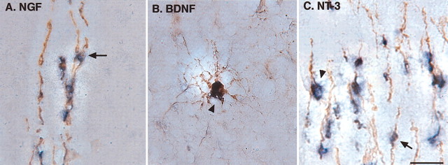
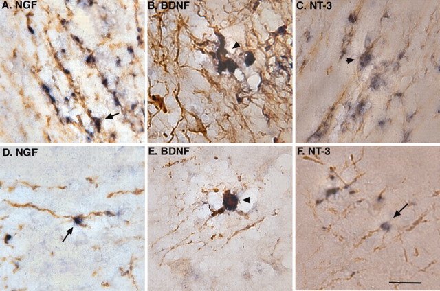
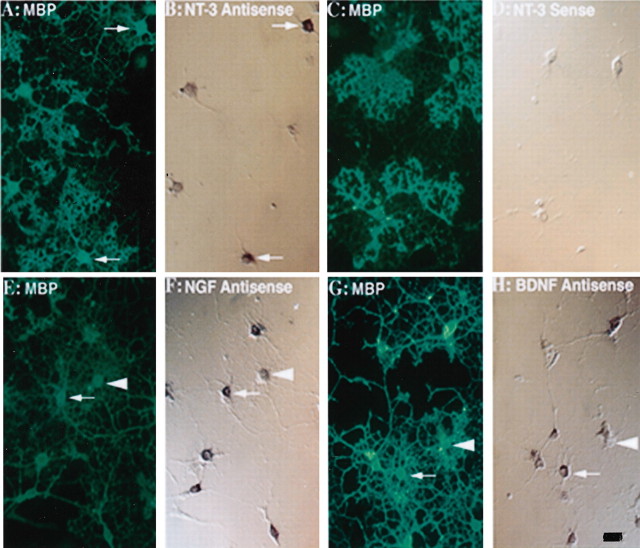
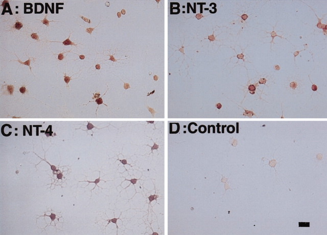
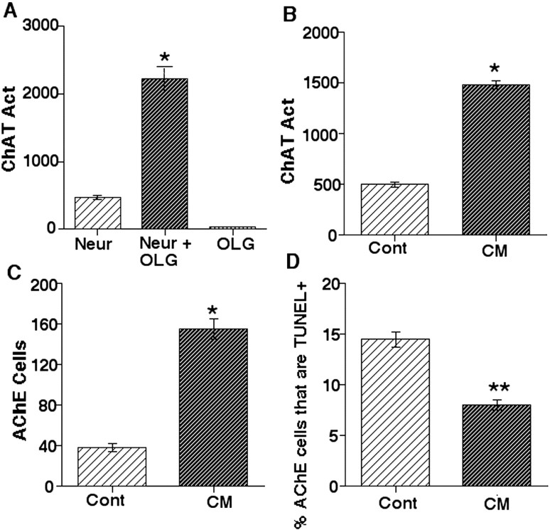
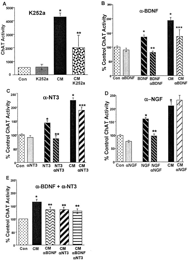
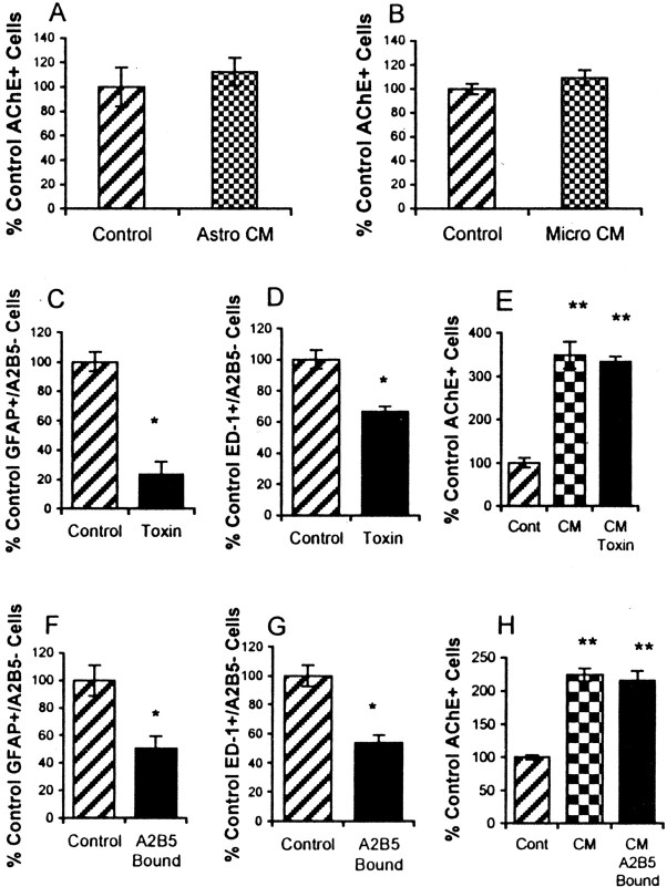
Traditionally, the primary function of oligodendrocytes (OLGs) in the CNS has been considered to be myelination. Here, we investigated whether OLGs may play a trophic role, particularly during development. Neurotrophin expression was assessed in postnatal day 7 basal forebrain (BF) OLGs, using in situ hybridization and detection of myelin basic protein. Nerve growth factor, brain-derived neurotrophic factor (BDNF) and neurotrophin-3 (NT-3) mRNAs were revealed in OLGs in vivo and in culture. To determine whether OLGs support nearby neurons, we examined the influence of OLGs on BF cholinergic neurons. Neuronal function was enhanced by cocultured OLGs and OLG conditioned medium. Moreover, trophic effects of OLG conditioned medium were partially blocked by K252a, a trk tyrosine kinase inhibitor, and by neutralizing anti-BDNF or anti-NT-3 antisera, indicating that neurotrophins may mediate these effects, perhaps in concert with other signals. Our studies support a novel role for OLGs in providing local trophic support for neurons in the CNS.


Figures

Figure 1.
Figure 1.
Expression of neurotrophins by MBP-positive OLGs in the P7 BF. Nonradioactive in situ hybridization reveals that NGF, BDNF, and NT-3 mRNA are coexpressed with MBP in vivo. Coronal sections were prepared from P7 rats and examined by in situ hybridization in combination with an immunocytochemical staining technique. A subset of MBP-positive cells, visualized by brown DAB precipitation, is colocalized with NGF (A), BDNF (B), or NT-3 (C) mRNA, visualized by purple precipitation. Scale bar, 50 μm. Arrows indicate small cells. Arrowheads indicate large cells.
Figure 2.
Figure 2.
Expression of neurotrophins by MBP-positive OLGs in other forebrain structures. In the corpus callosum, MBP positivity, visualized by brown DAB precipitation, is colocalized with NGF (A), BDNF (B), or NT-3 (C) mRNA, visualized by purple precipitation. In the frontal cortex, some MBP-positive cells also express NGF (D), BDNF (E), and NT-3 (F) mRNA. Such labeling was also observed in parietal and cingulate cortex. Scale bar, 50 μm. Arrows indicate small cells. Arrowheads indicate large cells.
Figure 3.
Figure 3.
Expression of neurotrophin mRNAs by MBP-positive cells in culture. MBP-positive cells, visualized by FITC-conjugated horse anti-rabbit antibody (A, E, G) express mRNAs for NGF (F), BDNF (H), or NT-3 (B), revealed by digoxigenin-11-UTP-labeled riboprobes and visualized as purple precipitation (arrows). Not all MBP-positive cells are positive for neurotrophin (arrowheads). The sense probes for the neurotrophins served as negative controls (D). Similar patterns were observed in other controls (data not shown). Scale bar, 50 μm.
Figure 4.
Figure 4.
Expression of neurotrophin protein in OLG-like cells. P1 OLG-enriched cultures were immunocytochemically stained by primary antisera against BDNF (A), NT-3 (B), or NT-4 (C). BDNF, NT-3, and NT-4 immunoreactivity is detected on most of the oligodendrocyte-like cells. In negative controls, preimmune serum (D) or antibodies preadsorbed with appropriate neurotrophins reveal no staining. Scale bar, 50 μm.
Figure 5.
Figure 5.
Trophic effects of OLGs on BF cholinergic neurons. A, Coculturing OLGs with E17 BF neurons for 5 d increases the activity (Act) of ChAT in culture. B, C, BF OLG CM exposure for 5 d increases both ChAT activity (B) and the number of AChE-positive cells (C). CM exposure for 7 d decreases the percentage of AChE-positive cells that are TUNEL positive (D). One of three (A–C) or two (D) independent experiments that yielded similar results are shown. Sample sizes for experiments: n = 15 (A); n = 10 (B–D) (5 dishes per group). Neur, Neuron; Cont, control. *Significantly different from control at p < 0.05. Data were analyzed by Student's t test (B–D) or ANOVA and the Scheffe's test (A). **Significantly different from control at p < 0.01.
Figure 6.
Figure 6.
Contribution of neurotrophins to the effects of CM on ChAT activity. A, K252a partially blocks the effects of CM on ChAT activity. One of two independent experiments that yielded similar results are shown. Sample size was ≥20 (5 dishes per group). *Significantly different from control plus DMSO at p < 0.05. **Significantly different from CM plus DMSO at p < 0.05. Data were analyzed by ANOVA and the Scheffe's test. B, C, Concentrations of anti-BDNF (α-BDNF; B) or anti-NT-3 (α-NT-3; C) that completely block the effects of the appropriate neurotrophin partially block the effects of CM on ChAT activity. Combined data from four independent experiments are shown. D, Concentrations of anti-NGF (α-NGF) that completely block the actions of NGF fail to block the effects of CM. Combined data from three independent experiments are shown. E, When added together, concentrations of anti-BDNF and anti-NT-3 that block the effects of CM individually have no additional effect on blockade of CM activity. Combined data from three independent experiments are shown. For each neurotrophin tested, sample sizes were as follows: 89 (B), 93 (C), 72 (D), and 59 (E); three or four dishes per group per experiment. *Significantly different from control at p < 0.05. **Significantly different from neurotrophin-treated cultures. ***Significantly different from CM (B, C)at p < 0.05. Data were analyzed by ANOVA and Scheffe's test. Con, Control.
Figure 7.
Figure 7.
Lack of trophic support by astrocytes and microglia. A, B, Numbers of AChE-positive cells are unaffected by astrocyte (Astro) CM (A) or microglia (Micro) CM (B) compared with control serum-free medium. Data represent one of two independent experiments that yielded similar results. Sample size is four (A) or three (B) dishes per group per experiment. C–E, Combined treatment with two toxins, α-amino adipic acid and leucine methyl ester, significantly reduces numbers of GFAP-positive/A2B5-negative astrocytes (C) and ED-1 microglia (D). However, the effects of CM from toxin-treated cultures (CM Toxin) are not different from effects of CM derived from untreated oligodendrocytes (CM) (E). Data represent one of two experiments that yielded similar results. F–H, Immunopanning with A2B5 antibody (A2B5 Bound) significantly reduces GFAP-positive/A2B5-negative astrocytes (F) and ED-1-positive A2B5-negative microglia (G) compared with untreated cells (Control). However, CM effects from the depleted cultures (CM A2B5 Bound) are not different from CM derived from untreated oligodendrocytes (CM). Data represent one of three experiments that yielded similar results. Sample size was four (C–E) or three (F–H) dishes per group per experiment. *Significantly different from control at p < 0.05; data were analyzed by Student's t test. **Significantly different from control at p < 0.05; data were analyzed by ANOVA and Fisher's test. Under control conditions, astrocytes made up 2% of total cells, and microglia made up 7% (C–H).

References

    1. Abiru Y, Katoh-Semba R, Nishio C, Hatanaka H ( 1998) High potassium enhances secretion of neurotrophic factors from cultured astrocytes. Brain Res 809: 115-126. - PubMed
    1. Alderson RF, Alterman AL, Barde YA, Lindsay RM ( 1990) Brain-derived neurotrophic factor increases survival and differentiated functions of rat septal cholinergic neurons in culture. Neuron 5: 297-306. - PubMed
    1. Arendt T, Bruckner MK, Krell T, Pagliusi S, Kruska L, Heumann R ( 1995) Degeneration of rat cholinergic basal forebrain neurons and reactive changes in nerve growth factor expression after chronic neurotoxic injury. II. Reactive expression of the nerve growth factor gene in astrocytes. Neuroscience 65: 647-659. - PubMed
    1. Byravan S, Foster LM, Phan T, Verity AN, Campagnoni AT ( 1994) Murine oligodendroglial cells express nerve growth factor. Proc Natl Acad Sci USA 91: 8812-8816. - PMC - PubMed
    1. Cannella B, Pitt D, Marchionni M, Raine CS ( 1999) Neuregulin and erbB receptor expression in normal and diseased human white matter. J Neuroimmunol 100: 233-242. - PubMed

Publication types

MeSH terms