. 2003 Jul 2;23(13):5887-96.
doi: 10.1523/JNEUROSCI.23-13-05887.2003.

Alivin 1, a novel neuronal activity-dependent gene, inhibits apoptosis and promotes survival of cerebellar granule neurons

Affiliations

Alivin 1, a novel neuronal activity-dependent gene, inhibits apoptosis and promotes survival of cerebellar granule neurons

Tomio Ono et al. J Neurosci. .

Abstract

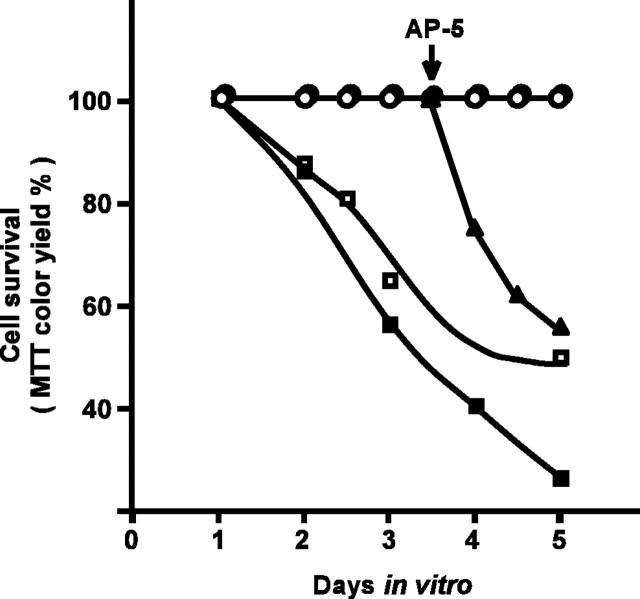
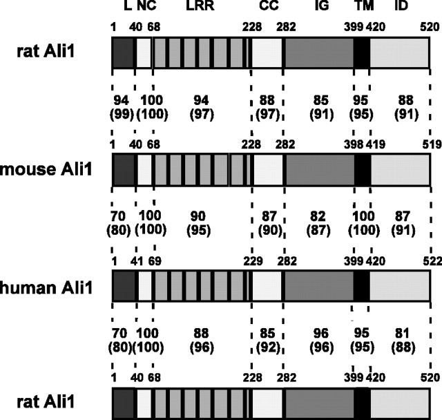
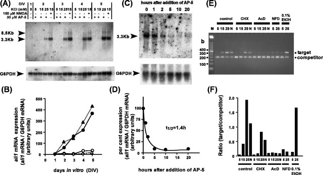
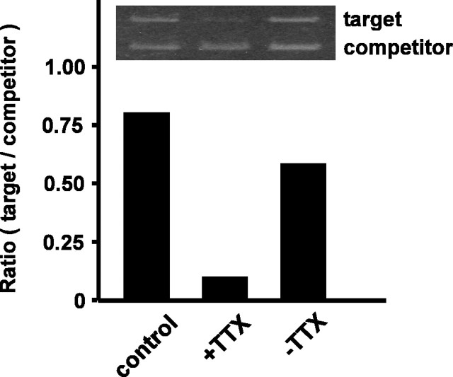
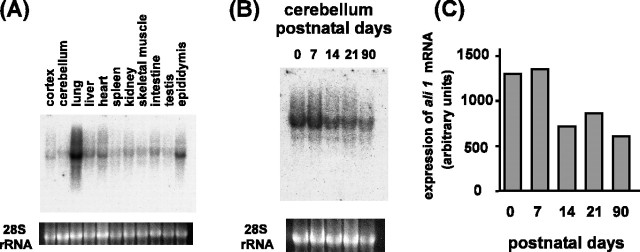
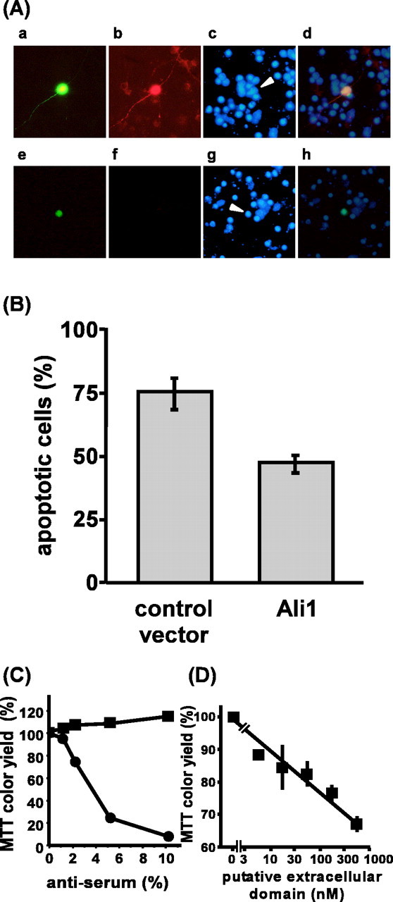
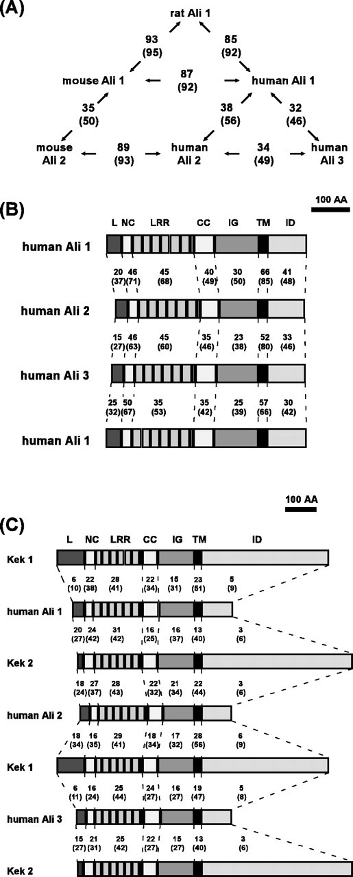
Neurons require Ca2+-dependent gene transcription for their activity-dependent survival, the mechanisms of which have not been fully elucidated yet. Here, we demonstrate that a novel primary response gene, alivin 1 (ali1), is an activity-dependent gene and promotes survival of neurons. Sequence analyses reveal that rat, mouse, and human Ali1 proteins contain seven leucine-rich repeats, one IgC2-like loop and a transmembrane domain, and display homology to Kek and Trk families. Expression of ali1 mRNA in cultured cerebellar granule neurons is rigidly regulated by KCl and/or NMDA concentrations in the culture medium and tightly correlated to depolarization-dependent survival and/or NMDA-dependent survival of the granule neuron. ali1 mRNA expression was regulated at the transcriptional step by the Ca2+ influx through voltage-dependent L-type Ca2+ channels when the cells were stimulated by 25 mm KCl. Expression of ali1 mRNA in cultured cortical neurons was inhibited when their spontaneous electrical activity was blocked by tetrodotoxin. Thus, the expression is neuronal activity dependent. Overexpression of Ali1 in cerebellar granule neurons inhibited apoptosis that was induced by the medium containing 5 mm KCl. The addition of anti-Ali1 antiserum or the soluble putative extracellular Ali1 domain to the 25 mm KCl-supported culture inhibited the survival of the granule neuron. These results suggest that expression of ali1 promotes depolarization-dependent survival of the granule neuron. Mouse ali1 was mapped to a locus approximately 55.3 cM from the centromere on chromosome 15 that is syntenic to positional candidate loci for familial Alzheimer's disease type 5 and Parkinson's disease 8 on human chromosome 12.


Figures

Figure 1.
Figure 1.
Time course of depolarization-dependent survival and/or NMDA-dependent survival of cerebellar granule neurons. Cerebellar granule neurons were cultured in medium containing 5 mm KCl and 30 μm AP-5 (▪), 15 mm KCl and 30 μm AP-5 (□), 25 mm KCl and 30 μm AP-5 (○), or 150μm NMDA and 15 mm KCl(•). At 3.5 DIV, 300μm AP-5 were added to some of the NMDA-supported cultures and apoptosis was induced (▴). Relative cell survival rate (MTT color yield) of each culture was plotted against the survival rate in the culture medium containing 25 mm KCl and 30 μm AP-5.
Figure2.
Figure2.
Structure of Alivin 1 proteins. The domain organizations of the deduced amino acid sequence of Ali1 proteins are shown schematically. The numbers on the Ali1 structures indicate positions of amino acid residues. The numbers between Ali1 structures indicate homology: numbers outside the parentheses indicate percentage identity, whereas numbers inside represent percentage similarity. L, Leader sequence including the signal peptide sequence; NC, NH2-terminal flanking cysteine-rich domain; CC, COOH-terminal flanking cysteine-rich domain; IG, IgC2-like loop; TM, transmembrane domain; ID, intracellular domain.
Figure3.
Figure3.
Expression of ali1 mRNA is tightly associated with depolarization-dependent survival and/or NMDA-dependent survival of cerebellar granule neurons.A,Northern blotting of ali1 mRNA expression in granule neurons. Total RNA samples (10μg) extracted from granule neurons cultured in the presence of 5 mm KCl, 30μm AP-5; 15 mm KCl, 30μm AP-5; 25 mm KCl, 30μm AP-5; or 150 μm NMDA, 15 mm KCl were subjected to Northern blotting. The cDNA obtained by differential display analysis (which corresponds to the 3′-untranslated region of rat ali1 cDNA) was used as a probe. The bottom panel depicts expression levels of G6PDH mRNA. B, Quantification of data shown in A. Granule neurons were cultured in the presence of 5 mm KCl,30μm AP-5(▵); 15 mm KCl,30μm AP-5 (○); 25 mm KCl, 30μm AP-5 (▴); or 15 mm KCl, 150μm NMDA (•). C, Rapid downregulation of ali1 mRNA expression by blocking the NMDA receptor with AP-5. Granule neurons were cultured for 4.5 d in medium containing 15 mm KCland150μm NMDA, and the NMDA receptor was blocked by the addition of AP-5 to a final concentration of 300μm. Total RNA (10μg) extracted at the indicated times was subjected to Northern blotting analyses. The bottom panel displays G6PDH mRNA expression levels. D, The data shown in C were quantified. E, Effects of various inhibitors on ali1 mRNA expression. Cerebellar granule neurons were cultured for 4 d in medium containing 25 mm KCl, as described in Materials and Methods. The medium was replaced with one containing 5 mm KCl, and the culture was continued for another 24 hr, after which cells were stimulated with 5 mm KCl, 30μm AP-5 (5); 15 mm KCl, 30μm AP-5 (15); 25 mm KCl, 30μm AP-5 (25); or 15 mm KCl, 150μm NMDA (N). Inhibitors were added to the culture medium 30 min before stimulation with KCl and/or NMDA. After stimulation (24 hr), total RNA was extracted and subjected to competitive PCR analysis. The inhibitors used included 35 μm cycloheximide (CHX), 4.0 μm actinomycin D (AcD), and 0.2 μm nifedipine (NFD). Ethanol was used as vehicle for nifedipine. F, The data shown in E were quantified.
Figure 4.
Figure 4.
Expression of ali1 is activity dependent. Rat cortical neurons were cultured for 14 d (control). TTX (1μm) was added to the culture medium, and the cells were continued to culture for another 30 hr (+TTX). Then, the medium was replaced with fresh one and the cells were continued to culture for another 30 hr(-TTX). Total RNA was extracted from each experimental group and subjected to competitive PCR analysis, as described in Materials and Methods. The data shown are representative results of two experiments.
Figure 5.
Figure 5.
Expression of ali1 mRNA in rat tissue. A, Tissue distribution of ali1 mRNA. Total RNA (10 μg) extracted from various tissues of adult rat was subjected to Northern blotting. The bottom panel shows ethidium bromide staining of 28S rRNA. B, Developmental expression of ali1 mRNA in the cerebellum. Total RNA was extracted from the cerebellum on specified postnatal days, and 10μg of RNA were subjected to Northern blotting analysis. The bottom panel shows ethidium bromide staining of 28S rRNA. C, The data shown in B were quantified.
Figure 6.
Figure 6.
Immunohistochemical localization of Ali1 in brain. A, Total brain extract (100μg) was subjected to 10% SDS-PAGE, and Western blotting was performed with anti-rat Ali1 antiserum. B, Immunohistochemical localization of Ali1 in the rat cerebellum. ML, Molecular layer; PL, Purkinje cell layer; GL, granule cell layer. C, Immunohistochemical localization of Ali1 in the rat hippocampus. D, Negative control staining of the rat cerebellum. E, Negative control staining of the rat hippocampus.
Figure 7.
Figure 7.
Subcellular distribution of Ali1 in brain. Whole brains of rats, 10 weeks of age, were separated into various subcellular fractions. The protein (100 μg) was subjected to 10% SDS-PAGE. Western blotting was performed with anti-rat Ali1 antiserum.
Figure 8.
Figure 8.
Ali1 is involved in depolarization-dependent survival of cerebellar granule neurons. A, Ali1 promotes survival of neurons. Expression vectors were transfected to cells at 0 DIV, and the cells were cultured in medium containing 5 mm KCl and 30μm AP-5. At 3.0 DIV, the cells were fixed with 4% paraformaldehyde. Transfected cells were monitored by staining with anti-Flag antibody and EGFP fluorescence. Apoptotic cells were monitored by condensed and/or fragmented nuclei by staining with Hoechst dye. ad, Ali1-Flag expression vector; eh, control vector; a, e, EGFP fluorescence; b, f, staining with anti-Flag antibody and Alexa Flour 594-coupled secondary antibody; c, g, staining with Hoechst dye; d, h, overlay of EGFP fluorescence, staining with anti-Flag antibody, and Hoechst staining. Arrowheads indicate transfected cells. B, Quantification of the effect of ali1 transfection on apoptosis in cerebellar granule neurons. Results are presented as mean ± SD. The cultures transfected with ali1 expression vector contained a significantly reduced rate of apoptotic cells (Student's t test; p < 0.001; n = 4). C, Effect of anti-Ali1 antiserum on cerebellar granule neuron survival. Anti-Ali1 antiserum was dialyzed extensively against BME and sterilized by filtering through a 0.22 μm membrane (Millipore, Billerica, MA). Granule neurons were cultured in medium containing 25 mm KCl and 30 μm AP-5, as described in Materials and Methods. Antiserum or nonimmune serum was added to the culture at 1.0 DIV, and survival was assayed on 5.0 DIV. The data shown are representative of two independent experiments. Each value is the mean ± SD of duplicate analyses. The horizontal axis (anti-serum %) indicates that percentage of the volume of the antiserum or control serum in the total volume of the culture medium. •, Anti-Ali1 antiserum; ▪, nonimmune serum. D, Effect of Ali1 (57–402) on the survival of cerebellar granule neurons. Rat Ali1 (52–402) was dialyzed extensively against BME and sterilized by filtering through a 0.22 μm membrane (Millipore). Granule neurons were cultured in medium containing 25 mm KCl and 30 μm AP-5, as described in Materials and Methods. Ali1 (57–402) was added to the culture at 2.0 DIV, and neuronal survival was assayed at 6.0 DIV. The data shown are representative of two independent experiments. Each value is the mean ± SD of duplicate analyses.
Figure 9.
Figure 9.
Homology comparisons.A, The homology between each Ali family member is shown in the schematic diagram. The numbers outside the parentheses indicate percentage identity, whereas the numbers inside the parentheses represent percentage similarity. B, The domain organization of members of the human Ali family are shown schematically, and the homology between each homologous domain is compared. The numbers outside the parentheses indicate percentage identity, whereas the numbers inside represent percentage similarity. L, Leader sequence including the signal peptide sequence; NC, NH2-terminal flanking cysteine-rich domain; CC, COOH-terminal flanking cysteine-rich domain; IG, IgC2-like loop; TM, transmembrane domain; ID, intracellular domain. C, Domain organization of human Ali1 and the Drosophila Kek family is shown schematically, and the homology between the each domain is compared as in B.

References

    1. Bonni A, Brunet A, West AE, Datta SR, Takasu MA, Greenberg ME ( 1999) Cell survival promoted by the Ras-MAPK signaling pathway by transcription-dependent and -independent mechanisms. Science 286: 1358-1362. - PubMed
    1. Brosenitsch TA, Katz DM ( 2001) Physiological patterns of electrical stimulation can induce neuronal gene expression by activating N-type calcium channels. J Neurosci 21: 2571-2579. - PMC - PubMed
    1. Chomczynski P, Sacchi N ( 1987) Single-step method of RNA isolation by acid guanidinium thiocyanate-phenol-chloroform extraction. Anal Biochem 162: 156-159. - PubMed
    1. Franke B, Bayatti N, Engele J ( 2000) Neurotrophins require distinct extracellular signals to promote the survival of CNS neurons in vitro Exp Neurol 165: 125-135. - PubMed
    1. Freter RR, Irminger JC, Porter JA, Jones SD, Stiles CD ( 1992) A novel 7-nucleotide motif located in 3′ untranslated sequences of the immediate-early gene set mediates platelet-derived growth factor induction of the JE gene. Mol Cell Biol 12: 5288-5300. - PMC - PubMed

MeSH terms

Associated data