. 2003 Jul 2;23(13):5928-35.
doi: 10.1523/JNEUROSCI.23-13-05928.2003.

Energy contribution of octanoate to intact rat brain metabolism measured by 13C nuclear magnetic resonance spectroscopy

Affiliations

Energy contribution of octanoate to intact rat brain metabolism measured by 13C nuclear magnetic resonance spectroscopy

Douglas Ebert et al. J Neurosci. .

Abstract

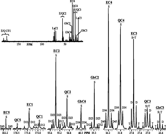
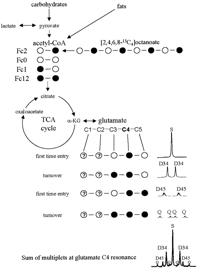
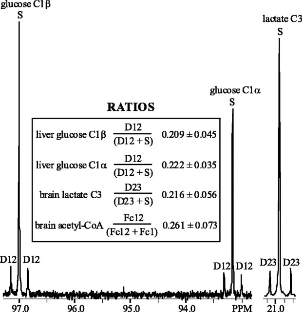
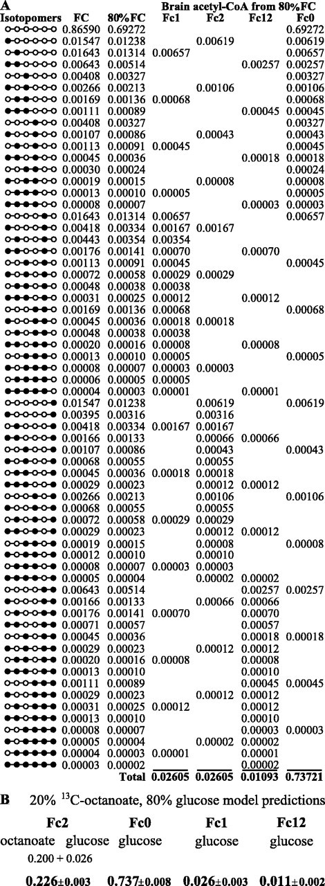
Glucose is the dominant oxidative fuel for brain, but studies have indicated that fatty acids are used by brain as well. We postulated that fatty acid oxidation in brain could contribute significantly to overall energy usage and account for non-glucose-derived energy production. [2,4,6,8-13C4]octanoate oxidation in intact rats was determined by nuclear magnetic resonance spectroscopy. We found that oxidation of 13C-octanoate in brain is avid and contributes approximately 20% to total brain oxidative energy production. Labeling patterns of glutamate and glutamine were distinct, and analysis of these metabolites indicated compartmentalized oxidation of octanoate in brain. Examination of liver and blood spectra revealed that label from 13C-octanoate was incorporated into glucose and ketones, which enabled calculation of its overall energy contribution to brain metabolism: glucose (predominantly unlabeled) and 13C-labeled octanoate can account for the entire oxidative metabolism of brain. Additionally, flux through anaplerotic pathways relative to tricarboxylic acid cycle flux (Y) was calculated to be 0.08 +/- 0.039 in brain, indicating that anaplerotic flux is significant and should be considered when assessing brain metabolism. Y was associated with the glutamine synthesis compartment, consistent with the view that anaplerotic flux occurs primarily in astrocytes.


Figures

Figure 1.
Figure 1.
13C spectrum of representative extract of intact rat brain infused with [2,4,6,8-13C4]octanoate for 105 min. Glutamate (E) and glutamine (Q) regions of carbons 1–5 (C1–C5) and GABA (Gb) C2–C4 are shown. Multiplets are depicted as follows: singlet (S), doublet (D), and triplet (T). Doublets resulting from carbon–carbon coupling of adjacent carbons are noted [e.g., C3 and C4 (D34) in C4 region].Inset,Full13C spectrum; regions of E, Q, Gb, and lactate (La) carbon resonances are indicated. PPM, Parts per million.
Figure 2.
Figure 2.
13C isotope isomer (isotopomer) analysis is based on the appearance of 13C label (•) in the carbons of glutamate originating from labeled acetyl-CoA. Glutamate is in rapid exchange with the TCA cycle intermediate, α-ketoglutarate (α-KG), and is present in concentrations high enough to be readily detected using magnetic resonance spectroscopy. Label from β-oxidation of exogenously administered [2,4,6,8-13C4]octanoate gives rise to label in the carbon 2 of acetyl-CoA (Fc2). When this label enters the TCA cycle the first time, it gives rise to a single peak (singlet) in the glutamate carbon 4 (C4) region. As the TCA cycle turns over, 13C label is mixed at symmetrical intermediates giving rise to label in adjacent carbons, which splits the signal of those carbons into multiple peaks (multiplets). For example,if C2-labeled oxaloacetate condenses with methyl-labeled acetyl-CoA (from exogenous octanoate), a doublet (D34) will be seen (attributable to J34) in the area where glutamate C4 resonates in the 13C spectrum as shown. Different starting populations of labeled acetyl-CoA yield distinct glutamate labeling patterns. If the glutamate C4 resonance is considered, doubly labeled acetyl-CoA first gives rise to D45 in this region. As the TCA cycle turns over and this 13C label is mixed, it is possible to generate label in carbons 3, 4, and 5 of glutamate, leading to a doublet of doublets or quartet (Q) in the glutamate C4 resonance. No label is generated in the C4 area from the unlabeled acetyl-CoA populations (Fc0) or those labeled only in carbon 1 (Fc1). Thus, the combination of the S, D34, D45, and Q in the glutamate C4 region will be nine peaks. This analysis can be done with other carbons of glutamate (e.g., C3) or other metabolites (e.g., glucose).
Figure 3.
Figure 3.
13C spectral regions of glucose carbon 1 (C1) α and β anomers from representative liver extract and lactate C3 from representative brain extract of intact rat infused with [2,4,6,8-13C4]octanoate for 105 min. Fractional contribution of glucose doublet arising from carbon–carbon coupling of C1 and C2 (D12) is shown in the inset for each anomer. The fractional contribution of the corresponding lactate doublet (D23) and the ratio of the analogous contribution of doubly labeled acetyl-CoA population (Fc12) to acetyl-CoA pool labeled in C1 (Fc1) plus Fc12 in brain is depicted. None of these ratios were different from each other (p ≤ 0.01).
Figure 4.
Figure 4.
A, Glucose isotopomers and resultant fractional isotopomer contribution to the glucose pool (FC) as generated from TCAsim software using liver data from rats infused with [2,4,6,8-13C4]octanoate for 105 min. Brain energy metabolism was modeled using an 80% glucose contribution (80% of FC) giving rise to acetyl-CoA populations as shown (Fc1, Fc2, Fc12, and Fc0). B, By combining 80% glucose contribution with 20% from exogenous [2,4,6,8-13C4]octanoate, 100% of brain energy metabolism is accounted for (not different from steady-state analysis values in Table 1; p ≤ 0.01).

References

    1. Auestad N, Korsak RA, Morrow JW, Edmond J ( 1991) Fatty acid oxidation and ketogenesis by astrocytes in primary culture. J Neurochem 56: 1376-1386. - PubMed
    1. Aureli T, di Cocco ME, Calvani M, Conti F ( 1997) The entry of [1- 13C]glucose into biochemical pathways reveals a complex compartmentation and metabolite trafficking between glia and neurons: a study by 13C-NMR spectroscopy. Brain Res 765: 218-227. - PubMed
    1. Badar-Goffer RS, Bachelard HS, Morris PG ( 1990) Cerebral metabolism of acetate and glucose studied by 13C-n.m.r. spectroscopy. A technique for investigating metabolic compartmentation in the brain. Biochemical J 266: 133-139. - PMC - PubMed
    1. Brand A, Richter-Landsberg C, Leibfritz D ( 1997) Metabolism of acetate in rat brain neurons, astrocytes and cocultures: metabolic interactions between neurons and glia cells, monitored by NMR spectroscopy. Cell Mol Biol 43: 645-657. - PubMed
    1. Cerdan S, Künnecke B, Seelig J ( 1990) Cerebral metabolism of [1,2- 13C2]acetate as detected by in vivo and in vitro13C NMR. J Biol Chem 265: 12916-12926. - PubMed

Publication types

LinkOut - more resources