. 2003 Jul 2;23(13):5963-73.
doi: 10.1523/JNEUROSCI.23-13-05963.2003.

Connexins are critical for normal myelination in the CNS

Affiliations

Connexins are critical for normal myelination in the CNS

Daniela M Menichella et al. J Neurosci. .

Abstract

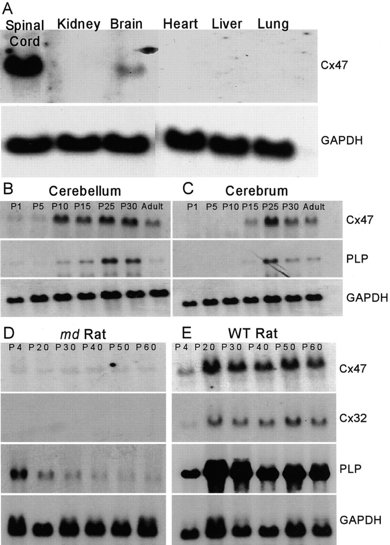
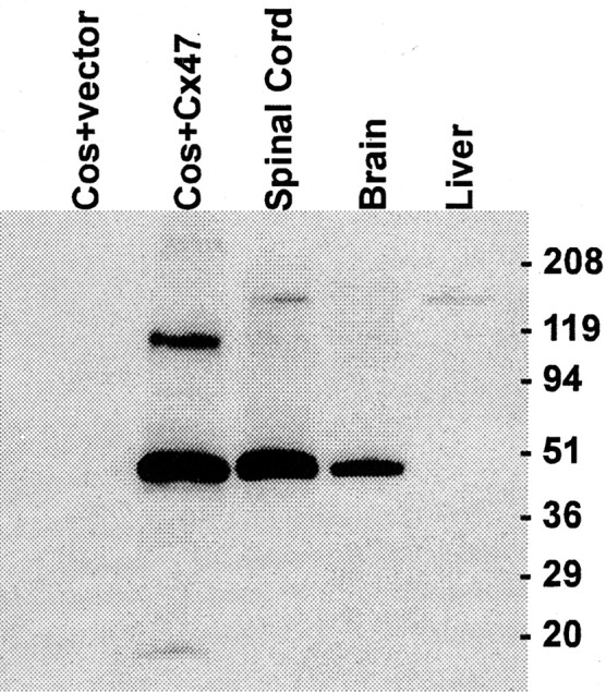
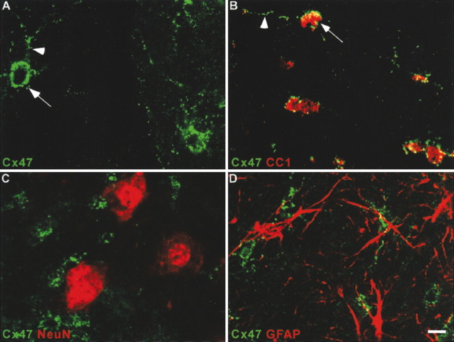
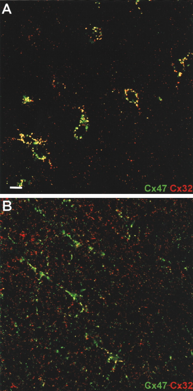
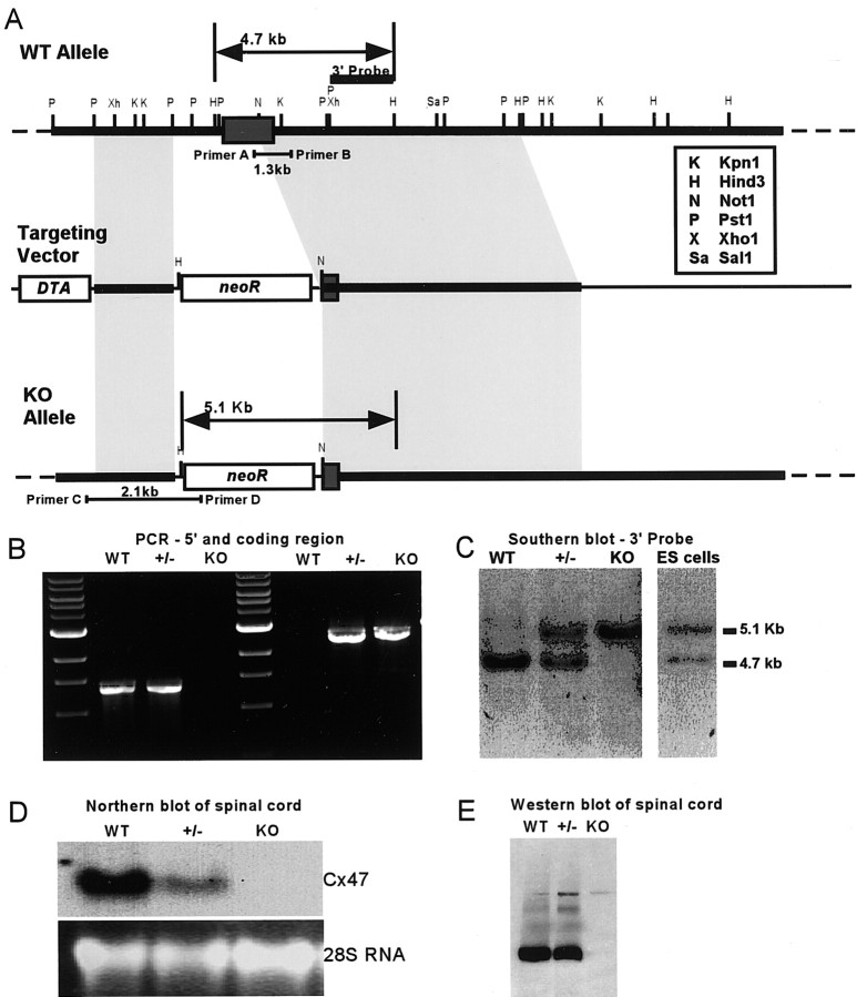
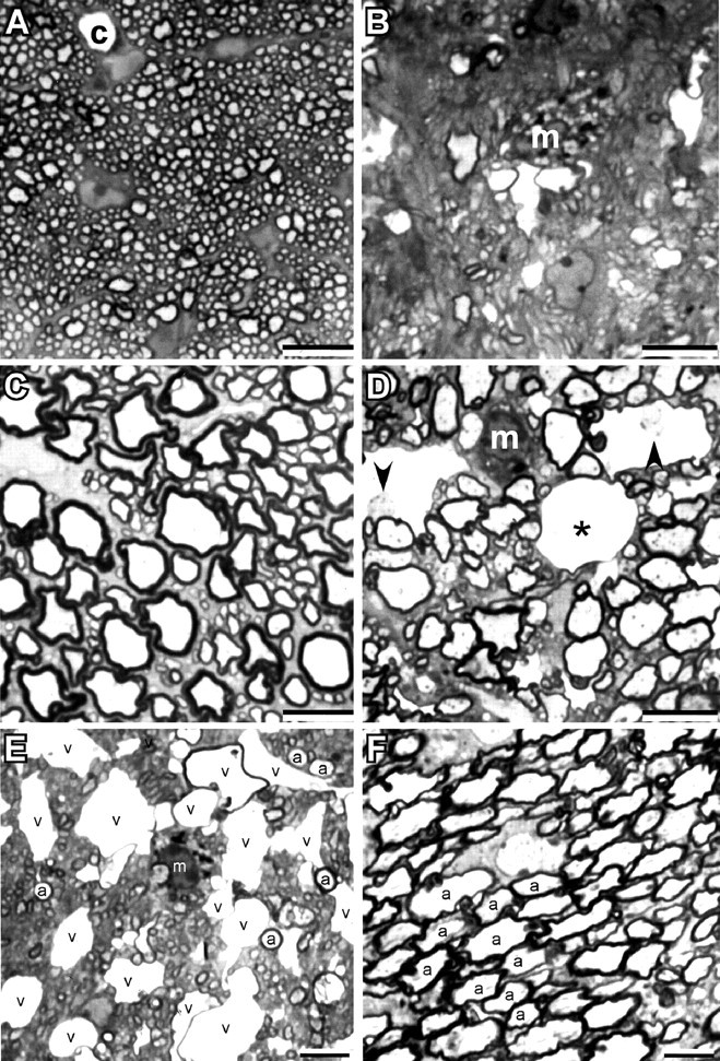
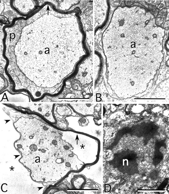
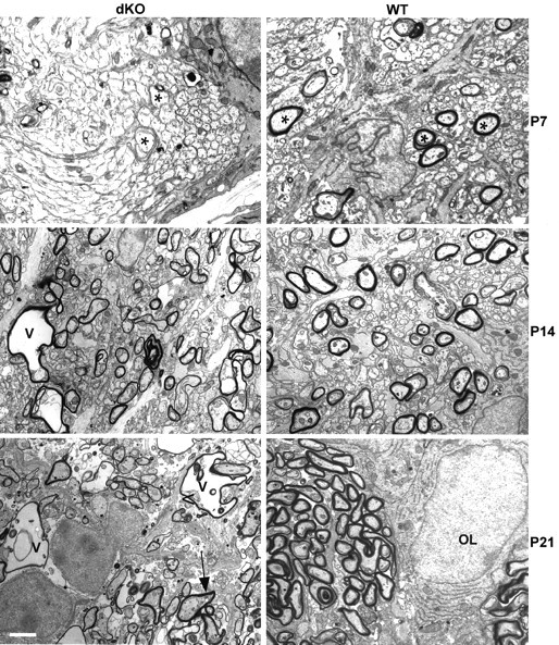
Mutations in Cx32, a gap-junction channel-forming protein, result in X-linked Charcot-Marie-Tooth disease, a demyelinating disease of the peripheral nervous system. However, although oligodendrocytes express Cx32, central myelination is unaffected. To explore this discrepancy, we searched for additional oligodendrocyte connexins. We found Cx47, which is expressed specifically in oligodendrocytes, regulated in parallel with myelin genes and partially colocalized with Cx32 in oligodendrocytes. Mice lacking either Cx47 or Cx32 are viable. However, animals lacking both connexins die by postnatal week 6 from profound abnormalities in central myelin, characterized by thin or absent myelin sheaths, vacuolation, enlarged periaxonal collars, oligodendrocyte cell death, and axonal loss. These data provide the first evidence that gap-junction communication is crucial for normal central myelination.


Figures

Figure 1.
Figure 1.
Cx47 mRNA is predominantly expressed in the CNS and regulated like a myelin-related gene. A, Northern blot of total RNA from various adult mouse tissues. Cx47 mRNA is detected only in spinal cord and brain. B, C, Northern blot of total RNA from developing mouse cerebellum and cerebrum. Temporal regulation of Cx47 transcript parallels that of PLP. D, E, Northern blot of total RNA from md and WT rat brain during postnatal development. md and WT RNA samples were obtained from littermates and processed for blotting under identical conditions. The levels of Cx47, Cx32, and PLP mRNA are lower in md rats, which carry a point mutation in PLP, resulting in profound CNS dysmyelination. GAPDH, Glyceraldehyde-3-phosphate dehydrogenase.
Figure 2.
Figure 2.
ISH for Cx47 mRNA in brain and spinal cord. A–C, Coronal sections through adult mouse brain hybridized with a Cx47 antisense riboprobe (A), a control Cx47 sense riboprobe (B), or a PLP antisense riboprobe (C). Cx47 is most abundant in white-matter tracts, such as the corpus callosum, a pattern similar to that of PLP. D, E, Longitudinal sections of adult mouse spinal cord hybridized for Cx47 (D) or PLP (E). Labeled cells are arranged in chains, characteristic of intrafascicular oligodendrocytes. Scale bars: A–C, 500 μm; D, E, 150 μm.
Figure 3.
Figure 3.
Western blot characterization of an anti-Cx47 serum. Bands at ∼47 and at ∼100 kDa are evident in COS cells transiently transfected with an expression vector containing the mouse Cx47 open reading frame, but not with an empty vector. The higher-molecular-mass band is most likely a dimerization artifact (see text). A specific signal was observed in adult mouse brain and spinal cord but not in liver.
Figure 4.
Figure 4.
Cx47 is expressed specifically in oligodendrocytes. A–D, Confocal micrographs of adult mouse spinal cord immunostained for Cx47 (green) (A) or double-stained for Cx47 (green) and CC1 (B), NeuN (C) or GFAP (D) (red). Cx47 immunoreactivity, composed of numerous intensely labeled puncta around the cell body (arrows) and processes (arrowheads), is associated with oligodendrocytes (B), but not neurons (C) or astrocytes (D). Scale bar, 6 μm.
Figure 5.
Figure 5.
Cx47 and Cx32 colocalize in oligodendrocyte cell bodies and processes. Confocal micrographs of adult mouse spinal cord gray matter (A) or white matter (B) double-stained for Cx47 (green) and Cx32 (red). In gray matter, numerous puncta around oligodendrocyte cell bodies and processes contain both Cx47 and Cx32 (yellow). However, away from the cell body in the neuropil, Cx47 and Cx32 do not colocalize. Scale bar, 6 μm.
Figure 6.
Figure 6.
Targeting strategy for Cx47. A, Top, WT Cx47 locus showing the single exon of Cx47 (dark gray box). Middle, A neo cassette replaced most of the Cx47 coding region in the targeting vector. A, Bottom, The Cx47 knock-out allele after homologous recombination. B, 5′ recombination was verified by PCR of tail DNA. Primer pair A/B (A, top) produced a 1.3 kb amplicon only in WT and +/- samples, whereas primer pair C/D produced a 2.1 kb amplicon only in +/- and KO samples. C, 3′ recombination was verified by Southern blotting HindIII digests of WT, +/-, and KO tail DNA, and recombinant ES-cell DNA using the 3′ probe designated in A. A 4.7 kb WT allele and a 5.1 kb recombined allele were detected. D, Loss of Cx47 expression in vivo.A single band was detected in WT RNA. Cx47 RNA levels were reduced in the +/- and were undetectable in the KO. Similarly, a Western blot confirmed the absence of Cx47 protein in the KO (E).
Figure 7.
Figure 7.
Variable involvement of myelinated axons in Cx32/Cx47 dKO CNS. Photomicrographs of semithin sections from optic nerve (A, B) and spinal cord ventral funiculus (C, D) of a P31 WT (A, C) and Cx32/47 double-null (B, D) mouse. Note the marked loss of myelinated axons in the optic nerve, the thinner myelin sheaths in the ventral funiculus, and the vacuoles in the myelin sheaths of the mutant. Two of these contain axons (arrowheads) and one is empty (asterisk); a capillary (c) is shown for comparison. m, Macrophage. E, F, Photomicrographs of the same semithin section from the pons of a P31 dKO. E, A portion of the corticobulbar tract, which contains numerous small myelinated axons; some of the largest ones are labeled (a). Note the large number of vacuolated myelin sheaths, and macrophage containing myelin debris. F, A portion of intramedullary trigeminal nerve, which contains many large myelinated axons, some of which are labeled (a). No vacuolated myelin sheaths are present. Scale bars, 10μm.
Figure 8.
Figure 8.
Myelin ultrastructure in the Cx32/Cx47 dKO. These are electron micrographs of the ventral funiculus of a P31 dKO. A, An axon (a) surrounded by a periaxonal collar of cytoplasm and a thin myelin sheath (arrow). B, A demyelinated axon, with neurofilaments that are more tightly packed than in A. C, An axon still partially surrounded by the adaxonal membrane (arrowheads), but separated from its myelin sheath (arrow) by extracellular space (asterisks). D, An apoptotic oligodendrocyte nucleus (n). Scale bar, 1 μm.
Figure 9.
Figure 9.
Myelin development in WT and dKO optic nerve. At P7, a transverse section through optic nerve shows myelin sheaths surrounding large-diameter axons (asterisk) in WT but not in dKO, indicating a delay in myelination. At P14, more axons are myelinated in WT optic nerves; myelin sheaths can also be seen in dKO optic nerves, but even at this early stage myelin abnormalities such as vacuolation (v) are present. At P21, most axons are surrounded by well formed myelin sheaths in the WT; in contrast dKO optic nerves at this stage show a rapid progression of the pathology, with thin myelin sheaths, pronounced vacuolation, and oligodendrocyte cytoplasm inclusions between the axons and the myelin sheath (arrow). OL, Oligodendrocyte nucleus. Scale bar, 2 μm.

Similar articles

Cited by

References

    1. Altevogt BM, Kleopa KA, Postma FR, Scherer SS, Paul DL ( 2002) Connexin29 is uniquely distributed within myelinating glial cells of the central and peripheral nervous systems. J Neurosci 22: 6458-6470. - PMC - PubMed
    1. Balice-Gordon RJ, Bone LJ, Scherer SS ( 1998) Functional gap junctions in the Schwann cell myelin sheath. J Cell Biol 142: 1095-1104. - PMC - PubMed
    1. Bao ZZ, Cepko CL ( 1997) The expression and function of Notch pathway genes in the developing rat eye. J Neurosci 17: 1425-1434. - PMC - PubMed
    1. Bergoffen J, Scherer SS, Wang S, Scott MO, Bone LJ, Paul DL, Chen K, Lensch MW, Chance PF, Fischbeck KH ( 1993) Connexin mutations in X-linked Charcot–Marie–Tooth disease. Science 262: 2039-2042. - PubMed
    1. Bhat RV, Axt KJ, Fosnaugh JS, Smith KJ, Johnson KA, Hill DE, Kinzler KW, Baraban JM ( 1996) Expression of the APC tumor suppressor protein in oligodendroglia. Glia 17: 169-174. - PubMed

Publication types

MeSH terms