Review
. 2003;4(7):223.
doi: 10.1186/gb-2003-4-7-223. Epub 2003 Jul 1.

Poly(A)-binding proteins: multifunctional scaffolds for the post-transcriptional control of gene expression

Affiliations
Review

Poly(A)-binding proteins: multifunctional scaffolds for the post-transcriptional control of gene expression

David A Mangus et al. Genome Biol. 2003.

Abstract

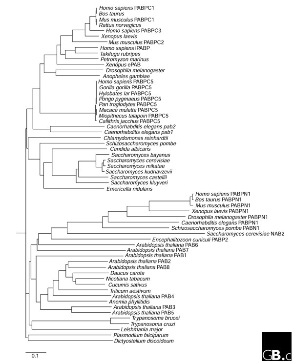
Most eukaryotic mRNAs are subject to considerable post-transcriptional modification, including capping, splicing, and polyadenylation. The process of polyadenylation adds a 3' poly(A) tail and provides the mRNA with a binding site for a major class of regulatory factors, the poly(A)-binding proteins (PABPs). These highly conserved polypeptides are found only in eukaryotes; single-celled eukaryotes each have a single PABP, whereas humans have five and Arabidopis has eight. They typically bind poly(A) using one or more RNA-recognition motifs, globular domains common to numerous other eukaryotic RNA-binding proteins. Although they lack catalytic activity, PABPs have several roles in mediating gene expression. Nuclear PABPs are necessary for the synthesis of the poly(A) tail, regulating its ultimate length and stimulating maturation of the mRNA. Association with PABP is also a requirement for some mRNAs to be exported from the nucleus. In the cytoplasm, PABPs facilitate the formation of the 'closed loop' structure of the messenger ribonucleoprotein particle that is crucial for additional PABP activities that promote translation initiation and termination, recycling of ribosomes, and stability of the mRNA. Collectively, these sequential nuclear and cytoplasmic contributions comprise a cycle in which PABPs and the poly(A) tail first create and then eliminate a network of cis- acting interactions that control mRNA function.


Figures

Figure 1
Figure 1
Predicted evolutionary relationships of PABPs. Full-length PABP sequences were compiled from various databases (see Additional data files) and aligned using the CLUSTALW program at the European Bioinformatics Institute [122]. The tree was constructed using the neighbor-joining method [123] and drawn using Phylodendron [124]. The scale bar represents 0.1 substitutions. In the instances where no PABP name is given, only a single PABP protein has been identified in that organism.
Figure 2
Figure 2
The domains of human PABPs. PABPC1, PABPC3, iPABP, PABPC5 and PABPN1 are shown, aligned on their first RNA-recognition motifs (RRMs). White capsules represent individual RRMs; black hexagons (5H) represent the five conserved helices at the carboxyl terminus. Inverted brackets indicate the site of expansion of a run of alanines in PABN1 that leads to the synthesis of PABPN1 with 12-17 alanines and results in the autosomal recessive disease oculopharyngeal muscular dystrophy (OMPD) [24,25]. PABPN1 accumulates in OMPD patients and forms intranuclear inclusions that appear to sequester mRNAs and associated factors and promote cell death [25].
Figure 3
Figure 3
Structures of the domains of human PABPC1. (a) Crystal structure of RRMs 1 and 2 in association with poly(A) [17]. The central two 3 strands of each RRM include two highly conserved sequence motifs, octomeric RNP1 ((K/R)-G-(F/Y)-(G/A)-F-V-X-(F/Y), where X is any amino acid) and hexameric RNP2 ((L/I)-(F/Y)-(V/I)-(G/K)-(N/G)-(L/M)), which is repeated six times. (b) The RNA-binding trough that is present when RRM1 and RRM2 of human associate with poly(A). (c) NMR structure of the five carboxy-terminal helices [21]. Figures were generated by MOLSCRIPT 2.0 using data from Protein data bank (PDB) files (a,b) 1CVJ and (c) 1G9L [125,126].
Figure 4
Figure 4
Roles of PABP in mRNA translation and stability. This model depicts different stages of a cytoplasmic mRNA 'life cycle', in which distinct roles can be ascribed to PABP. (a) Association of PABP with the mRNA poly(A) tail. (b) Interaction of PABP with elongation initiation factor eIF4G to promote formation of the 'closed loop', thus (c) initiating translation and antagonizing decapping. (d) Interaction of PABP with the termination factor eRF3 and recycling of the ribosome from the 5' to the 3' end of the same mRNA. (e) Poly(A) shortening by the Ccr4p-Pop2p-Notp deadenylase complex. (f) Loss of the poly(A) tail and PABP, facilitating (g) dissociation of the proteins of the mRNP, binding of the Lsm1-7p-Pat1p complex, and decapping by the decapping proteins Dcp1p and Dcp2p, and subsequent (h) 5'-to-3' degradation of the mRNA by the exonuclease Xrn1p or (i) 3'-to-5' degradation by the exosome.

References

    1. Burd CG, Matunis EL, Dreyfuss G. The multiple RNA-binding domains of the mRNA poly(A)-binding protein have different RNA-binding activities. Mol Cell Biol. 1991;11:3419–3424. Characterization of the RNA-binding domains of the yeast poly(A)-binding protein. - PMC - PubMed
    1. The Jacobson lab - additional data http://jacobsonlab.umassmed.edu/cgi-bin/dbcontents.cgi?gencon-tents=PABPs Fasta data and links to poly(A)-binding protein sequences are available on this website.
    1. Kleene KC, Mulligan E, Steiger D, Donohue K, Mastrangelo MA. The mouse gene encoding the testis-specific isoform of poly(A) binding protein (Pabp2) is an expressed retroposon: intimations that gene expression in spermatogenic cells facilitates the creation of new genes. J Mol Evol. 1998;47:275–281. This article reports the characterization of mouse testis-specific PABP2 isoform. - PubMed
    1. Feral C, Guellaen G, Pawlak A. Human testis expresses a specific poly(A)-binding protein. Nucleic Acids Res. 2001;29:1872–1883. This article describes the characterization of human testis-specific PABP3 isoform. - PMC - PubMed
    1. Blanco P, Sargent CA, Boucher CA, Howell G, Ross M, Affara NA. A novel poly(A)-binding protein gene (PABPC5) maps to an X-specific subinterval in the Xq21.3/Yp11.2 homology block of the human sex chromosomes. Genomics. 2001;74:1–11. The human PABPC5 was localized to the X chromosome. - PubMed

Publication types

LinkOut - more resources