

doi: 10.1046/j.1469-7580.2003.00183.x.
Morphogenesis and growth of the soft tissue and cartilage of the vomeronasal organ in pigs
Affiliations
- PMID: 12846472
- PMCID: PMC1571109
- DOI: 10.1046/j.1469-7580.2003.00183.x
Item in Clipboard
Morphogenesis and growth of the soft tissue and cartilage of the vomeronasal organ in pigs
J Anat.
2003 Jun.
Abstract
The morphology of the soft tissue and supporting cartilage of the vomeronasal organ of the fetal pig was studied from early stages to term. Specimens obtained from an abattoir were aged by crown-to-rump distance. Series of transverse sections show that some time before birth all structures--cartilage, connective tissue, blood vessels, nerves, glands and epithelia--are well developed and very similar in appearance to those of the adult. Furthermore, in transmission electron microscopy photomicrographs obtained at this stage the vomeronasal glands exhibit secretory activity.
Figures

Transverse sections through the VNO of the prenatal pig. Centre, camara lucida drawing of a section through the whole VNO of a 12-cm specimen. Clockwise from top left: HE-stained sections showing the development of the vomeronasal epithelia; SE, sensory epithelium; VNCg, vomeronasal cartilage; VNDc, vomeronasal duct; d, dorsal; l, lateral. Scale bar = 100 µm.

HE-stained transverse sections through the VNO of 3.1-cm and 22-cm pig fetuses, showing communication between the VNDc and the nasal (NC) and oral (OC) cavities via the incisive duct (asterisk). PI, papilla incisiva; VNDc, vomeronasal duct; d, dorsal; l, lateral. Scale bar = 100 µm.

Transverse sections through the VNO of prenatal pig at two different levels and magnifications (A and B), showing the typical appearance and arrangement of connective tissue (arrows). VNCg, vomeronasal cartilage. Stain: Weigert's resorcin-fuchsin. Scale bar = 100 µm.
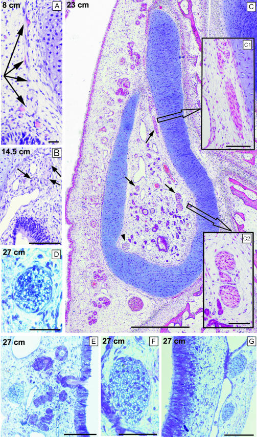
Transverse sections through the VNO of several prenatal pig specimens stained with haematoxylin-eosin (A–C) or toluidine blue (D–G). The general topography of the soft tissue is illustrated in the main picture (C), which shows a section taken caudally. The other pictures show the myelinization status of the nasocaudal nerves (D, E, and arrowhead in C) and the vomeronasal nerves (F,G, and arrows in A–C). Scale bars = 20 µm (A), 50 µm (C1,D,F), 100 µm (B,C2,E,G) and 500 µm (C).

Transverse sections through the VNO of several prenatal pig specimens stained with Weigert's resorcin-fuchsin (A,B) or haematoxylin-eosin (the others), showing arteries (A,B, arrows) with a clear tunica intima, subepithelial capillaries (C, arrows), veins (V) and venous sinuses (asterisks). LE, lateral epithelium; SE, sensory epithelium. Scale bar = 20 µm (C,D), 50 µm (A,B) and 100 µm(E–G).

Transverse sections through the VNO of several prenatal pig specimens stained with haematoxylin-eosin (A–C), PAS (D,F,H) or PAS and Alcian blue (E,G,I), showing vomeronasal glands opening into the lumen of the VNDc (A–C) and reactivity of vomeronasal glands (D–I, arrows). Scale bar = 20 µm (A) and 100 µm (others).

TEM micrographs of the apical cytoplasm of VNGl cells showing the release of the contents of secretory granules by fusion of granule and cell membrane (arrows). L, lumen. Scale bar = 1 µm.

Selected members of series of HE-stained transverse sections through the VNO of several prenatal pig specimens, showing the vomeronasal cartilage (in black) at various levels (I–VI) as described in the text.

Cellular components of the vomeronasal cartilage at various stages of prenatal development, showing the fibrous layer of the perichondrium (A,B, double arrow), mitotic figures (C,D, arrows), cell nests (E, arrows) and round and oval chondrocytes (F, arrows). Scale bar = 50 µm.
Similar articles
-
The prenatal maturity of the accessory olfactory bulb in pigs.Chem Senses. 2004 Jan;29(1):3-11. doi: 10.1093/chemse/bjh001. Chem Senses. 2004. PMID: 14752035
-
The soft-tissue components of the vomeronasal organ in pigs, cows and horses.Anat Histol Embryol. 1997 Sep;26(3):179-86. doi: 10.1111/j.1439-0264.1997.tb00122.x. Anat Histol Embryol. 1997. PMID: 9334496
-
The human vomeronasal organ. Part II: prenatal development.J Anat. 2000 Oct;197 Pt 3(Pt 3):421-36. doi: 10.1046/j.1469-7580.2000.19730421.x. J Anat. 2000. PMID: 11117628 Free PMC article.
-
Prenatal development of the mammalian vomeronasal organ.Microsc Res Tech. 1998 Jun 15;41(6):456-70. doi: 10.1002/(SICI)1097-0029(19980615)41:6<456::AID-JEMT2>3.0.CO;2-L. Microsc Res Tech. 1998. PMID: 9712194 Review.
-
Mechanisms underlying pre- and postnatal development of the vomeronasal organ.Cell Mol Life Sci. 2021 Jun;78(12):5069-5082. doi: 10.1007/s00018-021-03829-3. Epub 2021 Apr 19. Cell Mol Life Sci. 2021. PMID: 33871676 Free PMC article. Review.
Cited by
-
Odor exploration behavior of the domestic pig (Sus scrofa) as indicator of enriching properties of odors.Front Behav Neurosci. 2023 May 5;17:1173298. doi: 10.3389/fnbeh.2023.1173298. eCollection 2023. Front Behav Neurosci. 2023. PMID: 37214639 Free PMC article.
-
The vomeronasal system of the wolf (Canis lupus signatus): The singularities of a wild canid.J Anat. 2024 Jul;245(1):109-136. doi: 10.1111/joa.14024. Epub 2024 Feb 16. J Anat. 2024. PMID: 38366249 Free PMC article.
-
Development of a New Swine Model Resembling Human Empty Nose Syndrome.Medicina (Kaunas). 2024 Sep 24;60(10):1559. doi: 10.3390/medicina60101559. Medicina (Kaunas). 2024. PMID: 39459347 Free PMC article.
-
The Pig Olfactory Brain: A Primer.Chem Senses. 2016 Jun;41(5):415-25. doi: 10.1093/chemse/bjw016. Epub 2016 Mar 2. Chem Senses. 2016. PMID: 26936231 Free PMC article.
-
The olfactory limbus of the red fox (Vulpes vulpes). New insights regarding a noncanonical olfactory bulb pathway.Front Neuroanat. 2023 Jan 10;16:1097467. doi: 10.3389/fnana.2022.1097467. eCollection 2022. Front Neuroanat. 2023. PMID: 36704406 Free PMC article.
References
-
- Adams DR. Fine structure of the vomeronasal and septal olfactory epithelia and of glandular structures. Microscopy Res. Technique. 1992;23:86–97. - PubMed
-
- Bertmar G. Evolution of vomeronasal organs in vertebrates. Evolution. 1981;35:359–366. - PubMed
-
- Bonenfant C, Vallee I, Sun J, Brossay A, Thibault G, Guillaumin JM, et al. Analysis of human CD4 T lymphocyte proliferation induced by porcine lymphoblastoid B cell lines. Xenotransplantation. 2003;10:107–119. - PubMed
-
- Buck LB. The molecular architecture of odor and pheromone sensing in mammals. Cell. 2000;100:611–618. - PubMed
Publication types
MeSH terms
LinkOut - more resources
Full Text Sources
