. 2003 Jun;202(6):563-71.
doi: 10.1046/j.1469-7580.2003.00189.x.

Polyploid cells in the mouse ovary

Affiliations

Polyploid cells in the mouse ovary

Margaret A Keighren et al. J Anat. 2003 Jun.

Abstract

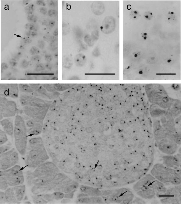
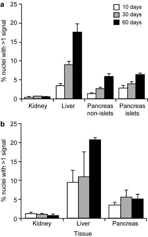
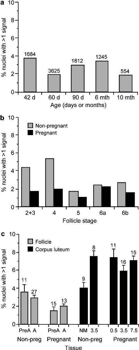
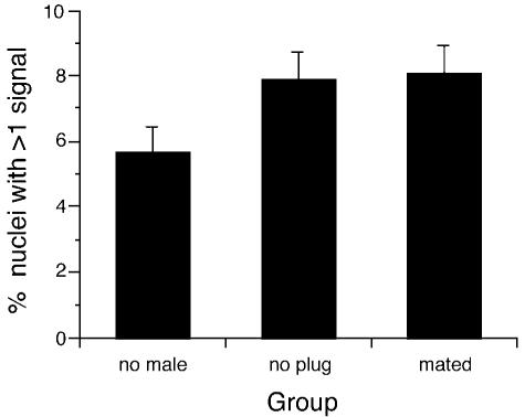
Cell ploidy in the ovarian follicle and corpus luteum was investigated by DNA in situ hybridization to a reiterated, chromosome 3 transgene in mice that were hemizygous for the transgene. This approach was first validated by analysis of mouse kidney, pancreas and liver control tissues, which contain different frequencies of polyploid nuclei. Polyploid nuclei (with multiple hybridization signals) were seen in histological sections of both ovarian follicles and corpora lutea. The frequency of polyploid nuclei in follicles showed no consistent relationship with age (between 6 weeks and 10 months) but polyploid nuclei were significantly more abundant in corpora lutea than follicles (6.3% vs. 2.5%). This implies that production of polyploid cells is more closely associated with differentiation of ovarian follicles into corpora lutea than with the age of the female. Polyploidy tended to be more frequent in corpora lutea of mice that had mated even if they did not become pregnant. This study has highlighted the presence of polyploid cells in the mouse ovarian follicle and corpus luteum and has identified mating as a possible trigger for polyploidy in the corpus luteum. Further work is required to determine the physiological role of polyploid ovarian cells in reproduction.


Figures

Fig. 1
Fig. 1
Nuclei with two hybridization signals (some are arrowed) in (a) a section of ovarian follicle, (b) a section of ovarian corpus luteum, (c) a cell spread from pancreas of 60-day-old mouse and (d) a section of pancreas of 60-day-old mouse showing an islet of Langerhans (small, paler stained cells). Some positive nuclei appear negative in the photographs because the hybridization signals are out of the plane of focus. Scale bar = 20 µm.
Fig. 2
Fig. 2
Histograms showing the percentages of putatively polyploid nuclei (with multiple hybridization signals) in kidney, pancreas and liver from mice (males and females pooled) at different ages in (a) histological sections and (b) cell spreads. Pancreases were analysed from six mice at each age and kidneys were analysed from three mice at each age. Livers were analysed from three mice at each age, except that only two samples were analysed at 10 days by histological sections and at 30 days by cell spreads.
Fig. 3
Fig. 3
Histogram showing the mean percentages of putatively polyploid nuclei (with multiple hybridization signals) in mouse ovaries. (a) Ovarian follicles from non-pregnant females of different ages (the number of nuclei with hybridization signals analysed are shown above the bars). (b) Ovarian follicles at different stages (stages 6a and 6b are antral follicles; others are pre-antral follicles). A total of 8920 nuclei with hybridization signals were analysed from non-pregnant mice (ranging from 125 for stage 5 follicles to 5917 for stage 6a); 6236 positive nuclei were scored from non-pregnant mice (ranging from 299 for stages 2 and 3 to 2896 for stage 6a). (c) Ovarian follicles and corpora lutea from pregnant and non-pregnant mice. (The mean and standard error are shown for percentages calculated separately for each follicle or corpus luteum and the numbers of follicles and corpora lutea analysed are shown above the bars.) Abbreviations: Non-preg, non-pregnant; PreA, pre-antral follicles; A, antral follicles; NM, female not housed with male; 0.5, 3.5 and 7.5 indicate number of days post-coitum. No embryos were found in three mice, 3 days after finding vaginal plugs (3.5 non-pregnant group).
Fig. 4
Fig. 4
Histogram showing the mean percentages of putatively polyploid nuclei (with multiple hybridization signals) in mouse corpora lutea in three groups of superovulated females. The mean and standard error are shown for percentages calculated separately for each corpus luteum. Females in the ‘no male’ group were not housed with males. Females in the ‘no plug’ group were housed overnight with vasectomized males but did not have vaginal plugs the following morning so there was no evidence of mating. Females in the ‘mated’ group were housed overnight with vasectomized males and had vaginal plugs the next morning (evidence of mating).

Similar articles

Cited by

References

    1. Alfert M, Geschwind I. The development of polysomaty in the rat liver. Exp. Cell Res. 1958;15:230–232. - PubMed
    1. Brodsky VY, Uryvaeva IV. Cell polyploidy: its relation to tissue growth and function. Int. Rev. Cytol. 1977;50:275–332. - PubMed
    1. Brodsky VY, Uryvaeva IV. Genome Multiplication in Growth and Development. Biology of Polyploid and Polytene Cells. Cambridge: Cambridge University Press; 1985.
    1. Coulson PB. Characterization of polyploidy in ovarian granulosa cells. In: Midgley AR, Sadler WA, editors. Ovarian Follicular Development and Function. New York: Raven Press; 1979. pp. 385–394.
    1. Epstein CJ. Cell size, nuclear content and the development of polyploidy in the mammalian liver. Proc. Natl. Acad. Sci. USA. 1967;57:327–334. - PMC - PubMed

Publication types

LinkOut - more resources