. 2003 Jul 7;162(1):15-22.
doi: 10.1083/jcb.200212136.

The Caenorhabditis elegans p120 catenin homologue, JAC-1, modulates cadherin-catenin function during epidermal morphogenesis

Affiliations

The Caenorhabditis elegans p120 catenin homologue, JAC-1, modulates cadherin-catenin function during epidermal morphogenesis

Jonathan Pettitt et al. J Cell Biol. .

Abstract

The cadherin-catenin complex is essential for tissue morphogenesis during animal development. In cultured mammalian cells, p120 catenin (p120ctn) is an important regulator of cadherin-catenin complex function. However, information on the role of p120ctn family members in cadherin-dependent events in vivo is limited. We have examined the role of the single Caenorhabditis elegans p120ctn homologue JAC-1 (juxtamembrane domain [JMD]-associated catenin) during epidermal morphogenesis. Similar to other p120ctn family members, JAC-1 binds the JMD of the classical cadherin HMR-1, and GFP-tagged JAC-1 localizes to adherens junctions in an HMR-1-dependent manner. Surprisingly, depleting JAC-1 expression using RNA interference (RNAi) does not result in any obvious defects in embryonic or postembryonic development. However, jac-1(RNAi) does increase the severity and penetrance of morphogenetic defects caused by a hypomorphic mutation in the hmp-1/alpha-catenin gene. In these hmp-1 mutants, jac-1 depletion causes failure of the embryo to elongate into a worm-like shape, a process that involves contraction of the epidermis. Associated with failed elongation is the detachment of actin bundles from epidermal adherens junctions and failure to maintain cadherin in adherens junctions. These results suggest that JAC-1 acts as a positive modulator of cadherin function in C. elegans.


Figures
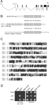
Figure 1.
Figure 1.
jac-1 encodes a C. elegans p120ctn homologue that interacts specifically with the JMD of HMR-1. (A) Structure of the jac-1 gene. Exons are represented by shaded boxes; two putative alternative exons are lightly shaded. The spliced-leader sequences found at the start of each alternative jac-1 transcript are indicated. (B) Schematic structure of JAC-1 compared with Dp120ctn and human δ-catenin (GenBank/EMBL/DDBJ accession nos. AAF33245 and Q9UQB3, respectively). Fn3 domains and Arm repeats are represented as open and shaded boxes, respectively. Interruptions in Arm repeats 4, 6, and 9 are indicated by unshaded regions. The numbers indicate the percent amino acid identity shared between corresponding Arm repeats. Predicted alternative start codons for JAC-1 are indicated by vertical lines. (C) ClustalW alignment of the Arm repeat regions (as defined by Anastasiadis and Reynolds, 2000) of JAC-1, Dp120, and δ-catenin (beginning at amino acids 594, 201, and 527, respectively). Black countershading indicates identical residues; gray, similarity. The borders of each Arm repeat are indicated by vertical lines. (D) JAC-1 interacts specifically with the JMD of HMR-1 in the yeast two-hybrid system. Growth on minus Ura, Leu, His media containing 1 mM 3-AT indicates interaction between the activation domain fusion proteins (JAC-1 and HMP-2) and the binding domain fusion proteins (HMR-1).
Figure 2.
Figure 2.
jac-1 is expressed in the early epidermis and JAC-1–GFP localizes to epidermal cell borders during morphogenesis. (A) Representative images from in situ hybridization with a control jac-1 sense probe (left) and jac-1 antisense probe (right). (B) Confocal images of JAC-1–GFP expression. (a) Dorsal view showing JAC-1–GFP localization during dorsal intercalation (two interdigitating cells are marked with asterisks). (b and c) Lateral views showing JAC-1–GFP localization during elongation. (d–f) Time course of an embryo undergoing ventral enclosure. Bar, 10 μm.
Figure 3.
Figure 3.
JAC-1–GFP localizes to adherens junctions in an HMR-1–dependent manner. (A–C) Confocal images showing a lateral view of an embryo expressing JAC-1–GFP (green) stained for HMR-1 (red). The merged image is shown in C, and the boxed region is enlarged 2.5× in C'. (D–F) Confocal images showing a lateral view of an embryo expressing JAC-1–GFP (green) stained for AJM-1 (red). The merged image is shown in F, and the boxed region is enlarged 2.5× in F'. (G and H) JAC-1–GFP expression in a wild-type (G) and hmr-1(zu389) embryo (H) of approximately the same age. Bar, 10 μm.
Figure 4.
Figure 4.
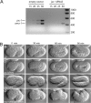
jac-1(RNAi) enhances the elongation defects of hmp-1(fe4) mutants. (A) Agarose gel electrophoresis of semiquantitative RT-PCR reactions derived from worms grown on HT115(DE3) bacteria carrying either the empty RNAi feeding vector or the jac-1(RNAi) feeding vector. Numbers on top indicate the number of amplification cycles. Numbers on the side indicate the base pair size of the marker DNAs. (B) Nomarski images at 30-min intervals of representative embryos undergoing elongation. Corresponding videos (Videos 1–4) are available at http://www.jcb.org/cgi/content/full/jcb.200212136/DC1. (a) Wild-type embryo. (b) jac-1(RNAi) embryo. (c) hmp-1(fe4) embryo. (d) hmp-1(fe4); jac-1(RNAi) embryo. Bar, 10 μm.
Figure 5.
Figure 5.
JAC-1 cooperates with HMP-1 to promote the anchorage of CFBs and the maintenance of HMR-1 distribution during elongation of the embryonic epidermis. (A) Images showing representative embryos stained for actin (a–d) or HMR-1 (e–h). (a and e) Wild-type embryos. (b and f) jac-1(RNAi) embryos. (c and g) hmp-1(fe4) embryos. (d and h) hmp-1(fe4); jac-1(RNAi) embryos. In c, the arrow points to an abnormally thick actin filament bundle. In g, the arrow indicates an area with no HMR-1 staining, and the arrowhead indicates an area of punctate HMR-1 staining. All images are lateral views with the dorsal side up. Bar, 10 μm. (B) An epidermal seam cell from an hmp-1(fe4); jac-1(RNAi) embryo double labeled for HMR-1 and actin. Bar, 5 μm.

References

    1. Anastasiadis, P.Z., and A.B. Reynolds. 2000. The p120 catenin family: complex roles in adhesion, signaling and cancer. J. Cell Sci. 113:1319–1334. - PubMed
    1. Anastasiadis, P.Z., and A.B. Reynolds. 2001. Regulation of Rho GTPases by p120-catenin. Curr. Opin. Cell Biol. 13:604–610. - PubMed
    1. Brenner, S. 1974. The genetics of Caenorhabditis elegans. Genetics. 77:71–94. - PMC - PubMed
    1. Chin-Sang, I.D., and A.D. Chisholm. 2000. Form of the worm: genetics of epidermal morphogenesis in C. elegans. Trends Genet. 16:544–551. - PubMed
    1. Costa, M., B.W. Draper, and J.R. Priess. 1997. The role of actin filaments in patterning the Caenorhabditis elegans cuticle. Dev. Biol. 184:373–384. - PubMed

Publication types

MeSH terms