. 2003 Jul 7;162(1):85-97.
doi: 10.1083/jcb.200301022.

Lack of GTP-bound Rho1p in secretory vesicles of Saccharomyces cerevisiae

Affiliations

Lack of GTP-bound Rho1p in secretory vesicles of Saccharomyces cerevisiae

Mitsuhiro Abe et al. J Cell Biol. .

Abstract

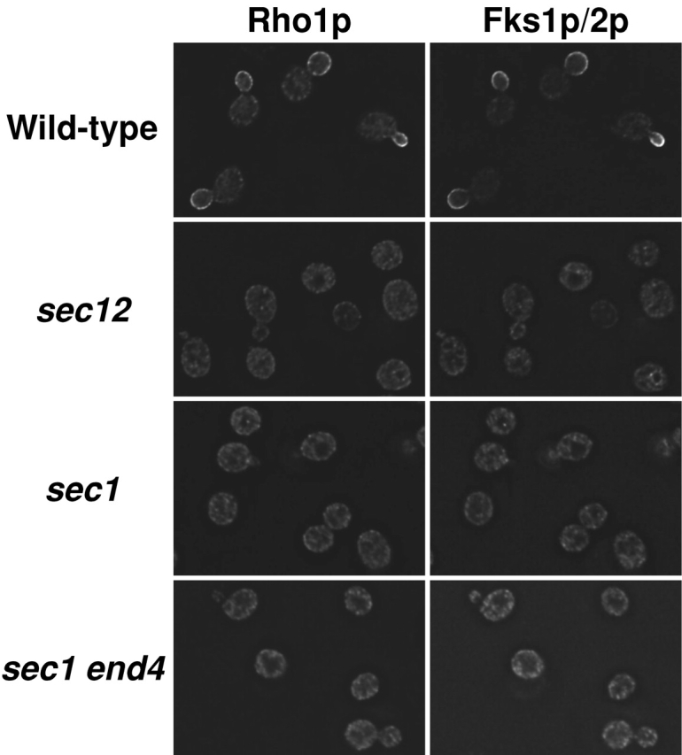
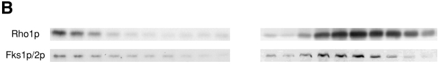
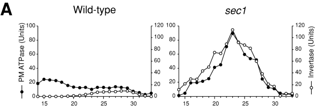
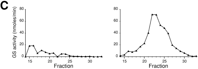
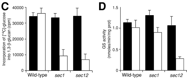
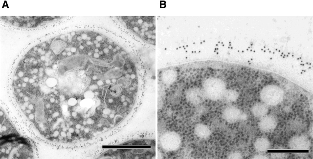
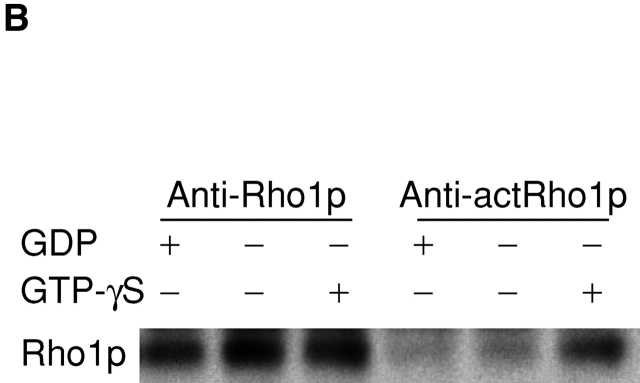
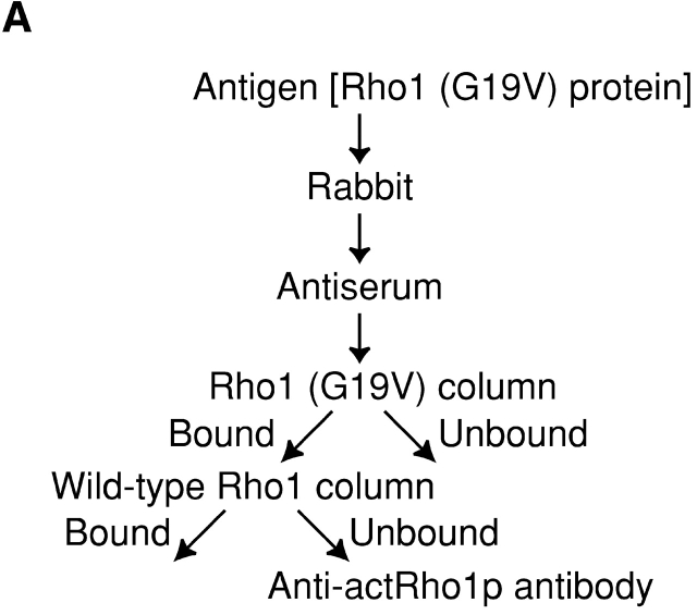
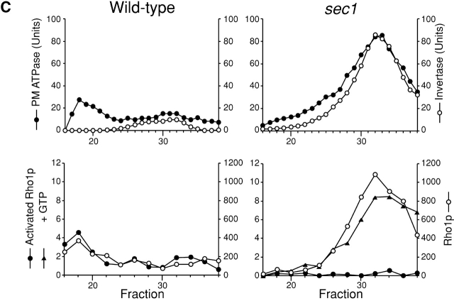
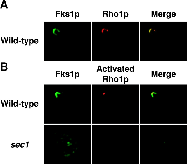
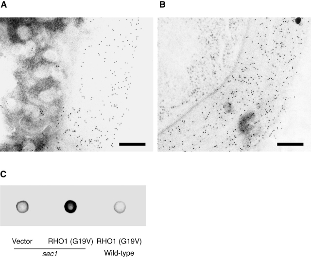
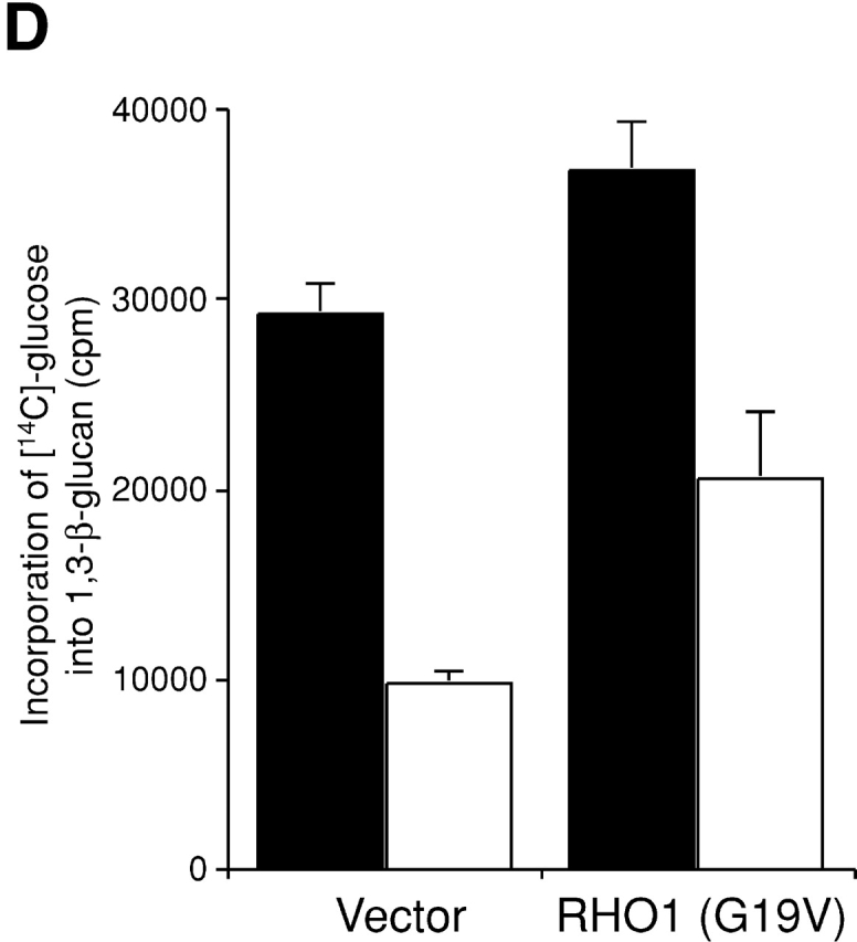
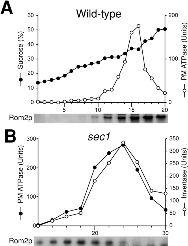
Rho1p, an essential Rho-type GTPase in Saccharomyces cerevisiae, activates its effectors in the GTP-bound form. Here, we show that Rho1p in secretory vesicles cannot activate 1,3-beta-glucan synthase, a cell wall synthesizing enzyme, during vesicular transport to the plasma membrane. Analyses with an antibody preferentially reacting with the GTP-bound form of Rho1p revealed that Rho1p remains in the inactive form in secretory vesicles. Rom2p, the GDP/GTP exchange factor of Rho1p, is preferentially localized on the plasma membrane even when vesicular transport is blocked. Overexpression of Rom2p results in delocalization of Rom2p and accumulation of 1,3-beta-glucan in secretory vesicles. Based on these results, we propose that Rho1p is kept inactive in intracellular secretory organelles, resulting in repression of the activity of the cell wall-synthesizing enzyme within cells.


Figures

Figure 1.
Figure 1.
Localization of Rho1p and Fks1p/2p in cells shifted to 37°C. Cells were cultured in YPD at 25°C, shifted to 37°C and cultured for 2 h. Cultured cells were fixed with formaldehyde and then stained for immunofluorescence microscopy with the anti-Rho1p antibody (left) or the anti-Fks1p/2p antibody (right). Strains used were as follows: wild-type (YPH500), sec12, sec1, and sec1 end4.
Figure 2.
Figure 2.
Secretory vesicle fractions of sec1 mutant and wild-type cells. Wild-type (left) or sec1 (right) cells were incubated at 37°C for 2 h, lysed, and subjected to differential centrifugations. The high-spin pellet was applied to a Sephacryl™ S-1000 column, and 4-ml fractions were collected. (A) Distributions of plasma membrane ATPase (closed circles) and invertase activity (open circles). (B) Immunoblotting analysis of GS-containing fractions. The amounts of Rho1p and Fks1p/2p were estimated with the guinea pig antiserum against Rho1p and the mouse mAb against Fks1p/2p, respectively. (C) Distributions of GS activity in the presence of GTP-γS.
Figure 2.
Figure 2.
Secretory vesicle fractions of sec1 mutant and wild-type cells. Wild-type (left) or sec1 (right) cells were incubated at 37°C for 2 h, lysed, and subjected to differential centrifugations. The high-spin pellet was applied to a Sephacryl™ S-1000 column, and 4-ml fractions were collected. (A) Distributions of plasma membrane ATPase (closed circles) and invertase activity (open circles). (B) Immunoblotting analysis of GS-containing fractions. The amounts of Rho1p and Fks1p/2p were estimated with the guinea pig antiserum against Rho1p and the mouse mAb against Fks1p/2p, respectively. (C) Distributions of GS activity in the presence of GTP-γS.
Figure 3.
Figure 3.
1,3-β-Glucan is not synthesized in intracellular organelles. (A) Immunogold labeling with the anti-1,3-β-glucan antibody in sec1 cells shifted to 37°C and cultured for 2 h. Bar, 1 μm. (B) The magnification image. Bar, 200 nm. (C) Reduced incorporation of glucose into 1,3-β-glucan in sec cells. Cells were cultured in YPD in the presence of [14C]glucose either at 25°C (black bars) or at 34°C for 2 h (white bars). The data represent the means and SDs of at least three experiments. (D) GS activity of the membrane fractions isolated from sec mutant cells. Cells were cultured either at 25°C (black bars) or at 37°C for 2 h (white bars), from which membrane fractions were isolated and assayed for GS activity in the presence of UDP-[14C]glucose and GTP-γS. The data represent the means and SDs of at least four experiments.
Figure 3.
Figure 3.
1,3-β-Glucan is not synthesized in intracellular organelles. (A) Immunogold labeling with the anti-1,3-β-glucan antibody in sec1 cells shifted to 37°C and cultured for 2 h. Bar, 1 μm. (B) The magnification image. Bar, 200 nm. (C) Reduced incorporation of glucose into 1,3-β-glucan in sec cells. Cells were cultured in YPD in the presence of [14C]glucose either at 25°C (black bars) or at 34°C for 2 h (white bars). The data represent the means and SDs of at least three experiments. (D) GS activity of the membrane fractions isolated from sec mutant cells. Cells were cultured either at 25°C (black bars) or at 37°C for 2 h (white bars), from which membrane fractions were isolated and assayed for GS activity in the presence of UDP-[14C]glucose and GTP-γS. The data represent the means and SDs of at least four experiments.
Figure 4.
Figure 4.
Localization of activated Rho1p detected with the purified anti-actRho1p antibody. (A) Affinity purification of the anti-actRho1p antibody. Antibody was raised against purified recombinant Rho1 (G19V) protein. Antiserum was loaded on an affinity column to which Rho1 (G19V) protein had been bound. The bound antibody was eluted, applied to another column charged with wild-type Rho1 protein, and the flow-through fractions were collected. (B) Immunoprecipitation of Rho1p with the anti-Rho1p (left), or the anti-actRho1p (right) antibody in the presence (+) or absence (−) of GDP and GTP-γS. Rho1p was detected by immunoblotting analysis with the guinea pig antiserum against Rho1p. (C) Immunoprecipitation of activated Rho1p. Wild-type (left) or sec1 mutant (right) cells were incubated at 37°C for 2 h. The cells were lysed in the presence or absence of 4 μM GTP, and fractionated by differential centrifugations. The high-spin pellet was applied to a Sephacryl™ S-1000 column without GTP, and 3-ml fractions were collected. Top panels, distribution of plasma membrane ATPase (closed circles) and invertase (open circles). Bottom panels, the distributions of activated Rho1p (closed circles and triangles) and total Rho1p (open circles). Aliquots of each fraction were assayed by immunoprecipitation with the anti-actRho1p, and detected by immunoblotting analysis with the guinea pig antiserum against Rho1p. The relative amount was quantified with a cooled CCD camera (LAS-1000plus; Fuji Photo Film).
Figure 4.
Figure 4.
Localization of activated Rho1p detected with the purified anti-actRho1p antibody. (A) Affinity purification of the anti-actRho1p antibody. Antibody was raised against purified recombinant Rho1 (G19V) protein. Antiserum was loaded on an affinity column to which Rho1 (G19V) protein had been bound. The bound antibody was eluted, applied to another column charged with wild-type Rho1 protein, and the flow-through fractions were collected. (B) Immunoprecipitation of Rho1p with the anti-Rho1p (left), or the anti-actRho1p (right) antibody in the presence (+) or absence (−) of GDP and GTP-γS. Rho1p was detected by immunoblotting analysis with the guinea pig antiserum against Rho1p. (C) Immunoprecipitation of activated Rho1p. Wild-type (left) or sec1 mutant (right) cells were incubated at 37°C for 2 h. The cells were lysed in the presence or absence of 4 μM GTP, and fractionated by differential centrifugations. The high-spin pellet was applied to a Sephacryl™ S-1000 column without GTP, and 3-ml fractions were collected. Top panels, distribution of plasma membrane ATPase (closed circles) and invertase (open circles). Bottom panels, the distributions of activated Rho1p (closed circles and triangles) and total Rho1p (open circles). Aliquots of each fraction were assayed by immunoprecipitation with the anti-actRho1p, and detected by immunoblotting analysis with the guinea pig antiserum against Rho1p. The relative amount was quantified with a cooled CCD camera (LAS-1000plus; Fuji Photo Film).
Figure 5.
Figure 5.
Distribution of wild-type Rho1p and activated Rho1p. (A) Colocalization of wild-type Rho1p and Fks1p/2p. Wild-type cells were cultured at 25°C, fixed, and stained for immunofluorescence microscopy with the anti-Fks1p/2p antibody (green) or the anti-Rho1p antibody (red). (B) Localization of activated Rho1p to a restricted region on the plasma membrane in wild-type cells. Wild-type cells were cultured at 25°C (top panels), whereas sec1 mutant cells were incubated at 37°C for 2 h (bottom panels), and stained for immunofluorescence microscopy with the anti-Fks1p/2p antibody (green) or the anti-actRho1p antibody (red).
Figure 6.
Figure 6.
Artificial synthesis of 1,3-β-glucan by expression of activated RHO1 in secretory vesicles. (A) sec1 cells expressing the active form of RHO1. sec1 cells transformed with YCpU-RHO1 (G19V) were incubated at 37°C for 2 h, fixed by the freeze-substituted fixation method, and 1,3-β-glucan was detected by the mouse mAb to 1,3-β-glucan. Bar, 200 nm. (B) Wild-type cells expressing the active form of RHO1. Bar, 200 nm. (C) 1,3-β-Glucan in high-speed pellets. Cells were incubated at 37°C for 2 h. The high-spin pellet was resuspended and 10 μg protein was blotted. 1,3-β-Glucan was detected by the mouse mAb against 1,3-β-glucan. (D) Incorporation of [14C]glucose into 1,3-β-glucan in sec1 cells. Cells were cultured in the presence of [14C]glucose at 25°C (black bars) or at 34°C for 2 h (white bars).
Figure 6.
Figure 6.
Artificial synthesis of 1,3-β-glucan by expression of activated RHO1 in secretory vesicles. (A) sec1 cells expressing the active form of RHO1. sec1 cells transformed with YCpU-RHO1 (G19V) were incubated at 37°C for 2 h, fixed by the freeze-substituted fixation method, and 1,3-β-glucan was detected by the mouse mAb to 1,3-β-glucan. Bar, 200 nm. (B) Wild-type cells expressing the active form of RHO1. Bar, 200 nm. (C) 1,3-β-Glucan in high-speed pellets. Cells were incubated at 37°C for 2 h. The high-spin pellet was resuspended and 10 μg protein was blotted. 1,3-β-Glucan was detected by the mouse mAb against 1,3-β-glucan. (D) Incorporation of [14C]glucose into 1,3-β-glucan in sec1 cells. Cells were cultured in the presence of [14C]glucose at 25°C (black bars) or at 34°C for 2 h (white bars).
Figure 7.
Figure 7.
Rom2p is localized on the plasma membrane even when vesicular transport is blocked. (A–D) Localization of Rom2-GFP fusion protein. rom2Δ cells transformed with YCpU-ROM2-GFP were incubated at 37°C for 2 h (A). sec1 rom2Δ cells transformed with YCpU-ROM2-GFP were incubated at 37°C for 2 h in the absence (B) or presence (C) of cycloheximide. sec1 rom2Δ cells transformed with YEpU-ROM2-GFP were incubated at 37°C for 2 h (D). Cultured cells were all fixed in methanol, and were subjected to observation. (E) Immunogold labeling with the anti-1,3-β-glucan antibody in sec1 cells overexpressing ROM2. sec1 cells were transformed with YEpU-ROM2. Transformants were incubated at 37°C for 2 h, and 1,3-β-glucan was detected. Bar, 200 nm.
Figure 8.
Figure 8.
Subcellular fractionation analysis of Rom2p when vesicular transport is blocked. (A) Distribution of Rom2p on the plasma membrane in wild-type cells. Wild-type cells were incubated in YPD at 37°C for 2 h. Cell lysate was fractionated on a 20–60% sucrose density gradient. Top, distribution of plasma membrane ATPase (open circles) and sucrose density (closed circles). Bottom, immunoblotting analysis of Rom2-HA with an anti-HA antibody. (B) Distribution of Rom2p in Sephacryl™ S-1000 column fractions of sec1 cells. Top, distribution of plasma membrane ATPase (closed circles) and invertase (open circles). Bottom, immunoblotting analysis of Rom2-HA.
Figure 9.
Figure 9.
Effect of activated RHO1 overexpression on the growth of wild-type and sec mutant cells. (A) Inhibition of cell proliferation in sec1 and sec6 mutant cells. Wild-type (YPH500), sec1, and sec6 cells were transformed with a control vector, or YEpU-RHO1 (G19V), and incubated for 3 d at 31°C. [RHO1 (G19V)] indicates the transformants with YEpU-RHO1 (G19V). (B) Suppression of inhibition by disturbing 1,3-β-glucan synthesis. sec1 fks1–1154 fks2Δ cells were transformed with a control vector, YEpT-RHO1 (G19V), or YCpU-FKS1, and incubated for 3 d at 31°C. [RHO1 (G19V)] and [FKS1] indicate the transformants with YEpU-RHO1 (G19V) and YCpU-FKS1, respectively.

Similar articles

Cited by

References

    1. Abe, M., I. Nishida, M. Minemura, H. Qadota, Y. Seyama, T. Watanabe, and Y. Ohya. 2001. Yeast 1,3-beta-glucan synthase activity is inhibited by phytosphingosine localized to the endoplasmic reticulum. J. Biol. Chem. 276:26923–26930. - PubMed
    1. Audhya, A., and S.D. Emr. 2002. Stt4 PI 4-kinase localizes to the plasma membrane and functions in the Pkc1-mediated MAP kinase cascade. Dev. Cell. 2:593–605. - PubMed
    1. Ayscough, K.R., J.J. Eby, T. Lila, H. Dewar, K.G. Kozminski, and D.G. Drubin. 1999. Sla1p is a functionally modular component of the yeast cortical actin cytoskeleton required for correct localization of both Rho1p-GTPase and Sla2p, a protein with talin homology. Mol. Biol. Cell. 10:1061–1075. - PMC - PubMed
    1. Cabib, E., D.H. Roh, M. Schmidt, L.B. Crotti, and A. Varma. 2001. The yeast cell wall and septum as paradigms of cell growth and morphogenesis. J. Biol. Chem. 276:19679–19682. - PubMed
    1. Del Pozo, M.A., W.B. Kiosses, N.B. Alderson, N. Meller, K.M. Hahn, and M.A. Schwartz. 2002. Integrins regulate GTP-Rac localized effector interactions through dissociation of Rho-GDI. Nat. Cell Biol. 4:232–239. - PubMed

Publication types

MeSH terms