. 2003 Jul 22;100(15):8839-43.
doi: 10.1073/pnas.1533136100. Epub 2003 Jul 7.

Activation of yeast Snf1 and mammalian AMP-activated protein kinase by upstream kinases

Affiliations

Activation of yeast Snf1 and mammalian AMP-activated protein kinase by upstream kinases

Seung-Pyo Hong et al. Proc Natl Acad Sci U S A. .

Abstract

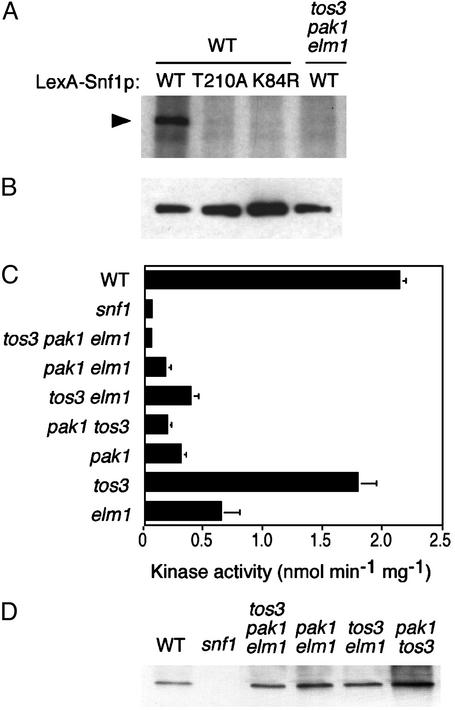
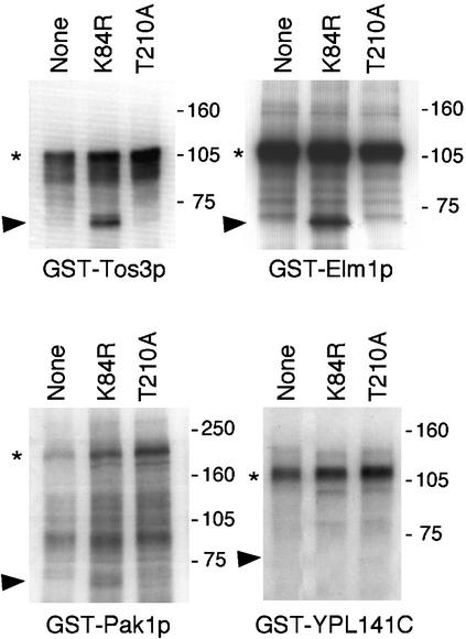
The Snf1/AMP-activated protein kinase (AMPK) family plays fundamental roles in cellular responses to metabolic stress in eukaryotes. In humans, AMPK regulates lipid and glucose metabolism and has been implicated in such metabolic disorders as diabetes and obesity and in cardiac abnormalities. Snf1 and AMPK are the downstream components of kinase cascades, but the upstream kinase(s) have remained elusive. We have here identified three yeast kinases, Pak1p, Tos3p, and Elm1p, that activate Snf1 kinase in vivo. Triple deletion of the cognate genes causes a Snf- mutant phenotype and abolishes Snf1 catalytic activity. All three kinases phosphorylate recombinant Snf1p on the activation-loop threonine. Moreover, Tos3p phosphorylates mammalian AMPK on the equivalent residue and activates the enzyme, suggesting functional conservation of the upstream kinases between yeast and mammals. We further show that the closely related mammalian LKB1 kinase, which is associated with Peutz-Jeghers cancer-susceptibility syndrome, phosphorylates and activates AMPK in vitro. Thus, the identification of the yeast upstream kinases should facilitate identification of the corresponding, physiologically important mammalian upstream kinases.


Figures

Fig. 1.
Fig. 1.
Overexpression and mutant phenotypes. (A) Overexpression of Tos3p, Pak1p, and Elm1p stimulates Snf1 function in a reporter assay. Transformants of a yeast strain (TAT7) carrying a lexAop-lacZ reporter expressed GST or GST-Tos3p, -Pak1p, or -Elm1p from a copper-inducible promoter (pOV85, pRH95, pRH98, and pRH94, respectively) and LexA-Snf1p (pOV8; ref. 37). Synthesis of β-galactosidase depended on catalytic activity of LexA-Snf1p (36). Transformants (n = 3) were grown to mid-log phase in selective SC plus 2% glucose, shifted to medium containing 0.5 mM CuSO4 in the presence of 2% glucose (open bars) or 0.05% glucose (filled bars) for 3 h, and assayed for β-galactosidase activity (36). Control transformants expressing LexA with each GST-kinase gave values <0.3 units. (B) Triple tos3Δ pak1Δ elm1Δ mutants exhibit growth defects. The tos3Δ pak1Δ and tos3Δ elm1Δ::URA3 double mutants (both carrying ura3) were crossed and subjected to tetrad analysis. Segregants were replicated from rich medium to SC-Ura plus 2% glucose and SC plus 2% glycerol/3% ethanol. Five tetrads are shown. The same pattern of growth was observed on SC plus 2% raffinose containing antimycin A (1 μg/ml). Asterisks indicate triple mutant segregants. Control strains: WT, snf1Δ mutant, and parental double mutants.
Fig. 2.
Fig. 2.
Assays of Snf1 kinase activity. (A and B) WT and triple mutant cells expressing LexA-Snf1p or its T210A and K84R mutant derivatives [pRJ55, pRJ217, and pRJ215 (16)] were grown in selective SC plus 2% glucose. Proteins were immunoprecipitated from extracts (200 μg) with anti-LexA. (A) Immunoprecipitates were incubated in kinase buffer containing [γ-32P]ATP and analyzed by SDS/PAGE and autoradiography. (B) Immunoprecipitates were analyzed by immunoblotting with anti-LexA. (C) Snf1 kinase activity was assayed by determining phosphorylation of the SAMS peptide (8, 30). A snf1-K84R mutant extract also showed no activity. (D) The assayed fractions were immunoblotted with anti-Snf1.
Fig. 3.
Fig. 3.
Tos3p, Pak1p, and Elm1p phosphorylate Snf1p on T210 in vitro. GST-Tos3p (31), -Pak1p (31), and -Elm1p (32) were purified from yeast cells. The GST-kinase, immobilized on beads, was incubated with bacterially expressed Snf1KD-K84R or -T210A (0.2 μg), or no substrate, in the presence of [γ-32P]ATP for 20 min at 25°C. Proteins were analyzed by SDS/PAGE and autoradiography. An autoradiogram is shown. An arbitrary GST-kinase, YPL141C, served as a control. Arrowheads, mutant Snf1KD; asterisks, autophosphorylated full-length GST-kinase. Molecular mass markers (kDa) are indicated.
Fig. 4.
Fig. 4.
Tos3p and LKB1 phosphorylate and activate AMPK in vitro.(A) Bacterially expressed AMPK (34) was incubated with MgATP and immobilized GST-kinase (expressed from pRH95, pRH98, and pRH94), and AMPK activity was measured by using the SAMS peptide assay (30). (B) A catalytically inactive mutant AMPK (D157A substitution) was incubated with GST-Tos3p or GST bound to glutathione-Sepharose beads in the presence of [γ-32P]ATP, and proteins were analyzed by SDS/PAGE and autoradiography. Molecular mass markers (kDa) are indicated. (C) Bacterially expressed AMPK (0.15 μg) was incubated with GST-Tos3p, GST, or partially purified rat liver AMPKK (21) and analyzed by immunoblotting with antibody specific to phosphothreonine 172. (D and E) Purified, immobilized FLAG-tagged LKB1, either WT or a catalytically inactive mutant (D194A), was incubated with bacterially expressed AMPK. The empty vector is pCDNA3 (Invitrogen) lacking LKB1 insert. (D) AMPK activity was measured (30). (E) Phosphorylation of T172 was determined by immunoblotting as in C.

References

    1. Hardie, D. G., Carling, D. & Carlson, M. (1998) Annu. Rev. Biochem. 67, 821–855. - PubMed
    1. Kemp, B. E., Stapleton, D., Campbell, D. J., Chen, Z. P., Murthy, S., Walter, M., Gupta, A., Adams, J. J., Katsis, F., Van Denderen, B., et al. (2003) Biochem. Soc. Trans. 31, 162–168. - PubMed
    1. Minokoshi, Y., Kim, Y. B., Peroni, O. D., Fryer, L. G., Muller, C., Carling, D. & Kahn, B. B. (2002) Nature 415, 339–343. - PubMed
    1. Zhou, G., Myers, R., Li, Y., Chen, Y., Shen, X., Fenyk-Melody, J., Wu, M., Ventre, J., Doebber, T., Fujii, N., et al. (2001) J. Clin. Invest. 108, 1167–1174. - PMC - PubMed
    1. Mu, J., Brozinick, J. T. J., Valladares, O., Bucan, M. & Birnbaum, M. J. (2001) Mol. Cell 7, 1085–1094. - PubMed

Publication types

MeSH terms