

Comparison of prefrontal cell pathology between depression and alcohol dependence
- PMID: 12849933
- PMCID: PMC3118500
- DOI: 10.1016/s0022-3956(03)00049-9
Comparison of prefrontal cell pathology between depression and alcohol dependence
Abstract
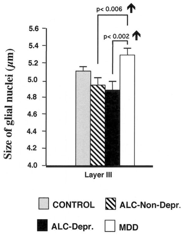
Chronic alcohol abuse is often co-morbid with depression symptoms and in many cases it appears to induce major depressive disorder. Structural and functional neuroimaging has provided evidence supporting some degree of neuropathological convergence of alcoholism and mood disorders. In order to understand the cellular neuropathology of alcohol dependence and mood disorders, postmortem morphometric studies have tested the possibility of alterations in the number and size of cells in the prefrontal cortex and other brain regions. The present review compares the cell pathology in the prefrontal cortex between alcohol dependence and depression, and reveals both similarities and differences. One of the most striking similarities is that, although pathology affects both neuronal and glial cells, effects on glia are more dramatic than on neurons in both alcohol dependence comorbid with depression and idiopathic depression. Moreover, prefrontal cortical regions are commonly affected in both depression and alcoholism. However, the cellular changes are more prominent and spread across cortical layers in alcohol dependent subjects than in subjects with mood disorders, and changes in glial nucleus size are opposite in alcoholism and depression. It could be argued that one defining factor in the manifestation of the depressive pathology is a reduction in the glial distribution in the dlPFC that is reflected in a reduced glial density. In alcoholism reduced glial nuclear size might be related to the cytotoxic effects of prolonged alcohol exposure, while in MDD, in the absence of alcohol abuse, other processes might be responsible for the increase in average size of glial nuclei. In either case abnormal function related to glial reduction would be associated with depression due to insufficient glial support to the surrounding neurons.
Figures

References
-
- Adams KM, Gilman S, Koeppe B, Kluin K, Junck L, Lohman M, et al. Correlation of neuropsychological function with cerebral metabolic rate in subdivisions of the frontal cortex of older alcoholics patients measured with [18F]fluorodeoxyglucose and positron emision tomography. Neuropsychology. 1995;9:275–80.
-
- Adams KM, Gilman S, Koeppe RA, Kluin KJ, Brunberg JA, Dede D, et al. Neuropsychological deficits are correlated with frontal hypometabolism in positron emission tomography studies of older alcoholic patients. Alcohol Clinical and Experimental Research. 1993;17:205–10. - PubMed
-
- Agartz I, Momenan R, Rawlings RR, Kerich MJ, Hommer DW. Hippocampal volume in patients with alcohol dependence. Archives of General Psychiatry. 1999;56:356–63. - PubMed
-
- Altshuler LL, Conrad A, Hauser P, Li XM, Guze BH, et al. Reduction of temporal lobe volume in bipolar disorder: a preliminary report of magnetic resonance imaging. Archives of General Psychiatry. 1991;48:482–3. - PubMed
-
- Arikuni T, Watanabe K, Kubota K. Connections of area 8 with area 6 in the brain of the macaque monkey. The Journal of Comparative Neurology. 1988;277:21–40. - PubMed
Publication types
MeSH terms
Grants and funding
LinkOut - more resources
Full Text Sources
Medical
