. 2003 Jul;5(1):45-57.
doi: 10.1016/s1534-5807(03)00169-2.

Neuropilin-1 conveys semaphorin and VEGF signaling during neural and cardiovascular development

Affiliations

Neuropilin-1 conveys semaphorin and VEGF signaling during neural and cardiovascular development

Chenghua Gu et al. Dev Cell. 2003 Jul.

Abstract

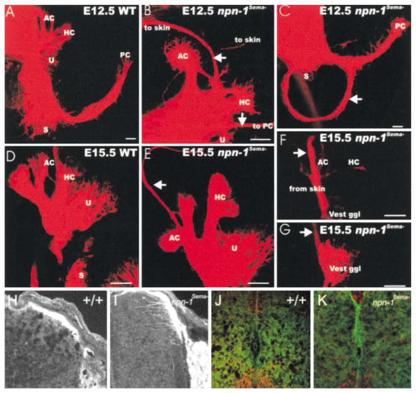
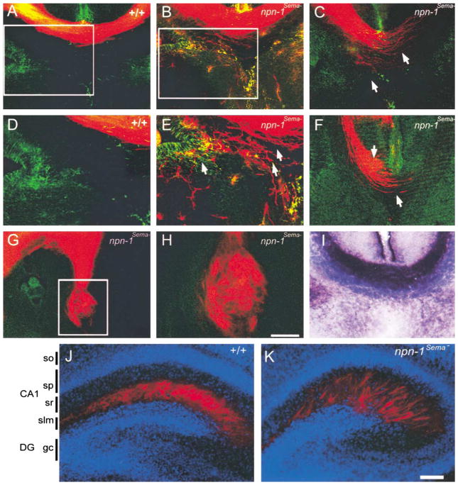
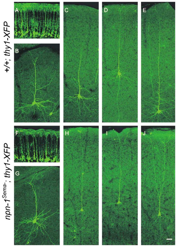
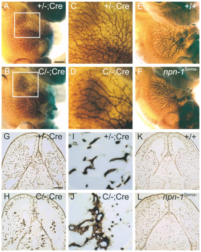
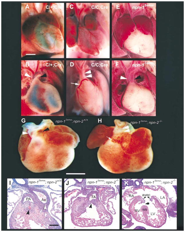
Neuropilin-1 (Npn-1) is a receptor that binds multiple ligands from structurally distinct families, including secreted semaphorins (Sema) and vascular endothelial growth factors (VEGF). We generated npn-1 knockin mice, which express an altered ligand binding site variant of Npn-1, and npn-1 conditional null mice to establish the cell-type- and ligand specificity of Npn-1 function in the developing cardiovascular and nervous systems. Our results show that VEGF-Npn-1 signaling in endothelial cells is required for angiogenesis. In striking contrast, Sema-Npn-1 signaling is not essential for general vascular development but is required for axonal pathfinding by several populations of neurons in the CNS and PNS. Remarkably, both Sema-Npn-1 signaling and VEGF-Npn-1 signaling are critical for heart development. Therefore, Npn-1 is a multifunctional receptor that mediates the activities of structurally distinct ligands during development of the heart, vasculature, and nervous system.


Figures

Figure 1
Figure 1. Generation and Characterization of npn-1Sema Mice
(A) Extracellular domain structure of the Npn-1 protein and a 7 amino acid substitution in the N terminus of the Npn-1 a1 CUB domain (Gu et al., 2002). (B) Gene targeting strategy used to generate npn-1Sema mice. The boxes represent npn-1 exons 1–3. The targeting vector (TV) contains a mutated exon 2 encoding a 7 amino acid substitution (three stars) and a pgk-neo cassette flanked by loxP sites (triangles) placed within the intron upstream of exon 2. The pgk-neo cassette was excised upon crossing mice with one TA with mice expressing germline Cre-recombinase. The final targeted allele (FTA) contains the mutated npn-2 exon 2 and one loxP site within the upstream intron. (C) Gene targeting strategy for generation of npn-1 null and conditional mutant mice. The targeted allele (TA) contains an FRT (squares) flanked neo cassette and loxP sites (triangles) flanking exon 2. After crossing mice harboring the TA with germline FlpE-recombinase mice, the pgk-neo cassette was removed and the conditional targeted allele (CA) contained two loxP sites flanking exon 2. After crossing with mice carrying Cre-recombinase under the control of the Tie-2 promoter, exon 2 was excised to generate a tissue-specific npn-1 null allele (TSNA). Npn-1 null mice were obtained by crossing mice harboring the CA with mice expressing Cre recombinase in the germline. (D–I) Alkaline phosphatase (AP)-tagged ligand binding to sections of the DREZ (arrowhead in [D] and [E]) from wild-type littermate controls (D, F, and H) and npn-1Sema mice (E, G, and I) with AP-Sema3A (D and E), AP-Sema3C (F and G), or AP-VEGF (H and I). (J–K) Expression of Npn-1 protein in npn-1Sema mice (K) and wild-type littermate control mice (J). (L–M) DRG sensory neuron axons from wild-type embryos are repelled by Sema3A-expressing COS cells (L) while DRG axons from npn-1Sema embryos are not repelled (M). (N) Quantitation of repulsion experiments. The repulsive activity is measured by the axon outgrowth ratio P/D, where P is the extent of axon outgrowth on the side proximal to the cell aggregate, and D is the extent of axon outgrowth distal to the cell aggregate. Shown are means ± SEM of 22 (wild-type) and 39 (npn-1Sema) explants from three independent experiments (p < 0.001, two-way ANOVA). Vec, vector-transfected COS cells; Sema3A, Sema3A-expressing COS cells. Scale bars: (D–I), 50 μm; (J–K), 40 μm; (L–M), 45 μm.
Figure 2
Figure 2. Peripheral Projections of Cranial and Spinal Nerves Are Severely Disorganized in Both npn-1 Null and npn-1Sema Mice
(A–D) Whole-mount antineurofilament staining of E11.5 npn-1 null (B), wild-type littermate (A), homozygous npn-1Sema (D), and wild-type littermate (C) embryos. The ophthalmic branch of the trigeminal nerve (upper arrowhead in [D]) and spinal nerves (lower arrowhead in [D]) are disorganized in both npn-1 null (B) and npn-1Sema mice (D). (E–H) Whole-mount antineurofilament staining of the ophthalmic nerve in E12.5 homozygous npn-1 null (F), wild-type littermate (E), homozygous npn-1Sema (H), and wild-type littermate (G) embryos. Note the exuberant extension and more regular distribution of ophthalmic nerve branches in npn-1Sema as compared to npn-1 null ophthalmic projections. Scale bar: (A–D), 0.5 mm; (E–H), 0.2 mm.
Figure 3
Figure 3. Sema–Npn-1 Signaling Is Required for Sensory Afferent Innervation of the Inner Ear and Spinal Cord
(A–C) In E12.5 npn-1Sema mice, vestibular nerve fibers extend beyond the anterior and horizontal cristae (AC, HC) and reach the skin above the ear ([B], top arrow). In addition, fiber bundles extend from the utricle (U) to the posterior crista (PC) where they may leave the ear through the round window ([B], bottom arrow; data not shown). In some animals, vestibular fibers are observed projecting around the forming cochlea without innervating any target ([C], arrow). (D–G) E15.5 npn-1Semamutants form inner ear afferent fibers to the skin. These fibers extend beyond the anterior cristae (E), pass through the otic capsule, and run together with the auriculotemporal nerve to the skin anterior to the auricle (arrow). Injection of dye into this area of the skin labels nVIII fibers that pass through the lateral otic wall (F) and pass underneath the canal cristae where some fibers branch and provide afferents to those epithelia (arrow). These fibers come from vestibular ganglion neurons (G) which project centrally into vestibular nuclei (data not shown). (H–K) Abnormal central projections of spinal sensory afferents. TrkA immunohistochemistry of E14 (H and I) and P2 (J and K) lumbar spinal cord in npn-1Sema (I and K) and wild-type littermates (H and J), examined by confocal microscopy. Dorsal spinal cord is at the top. Scale bars: (A–G), 100 μm; (H–K), scale bar in (G) is equal to 200 μm in (H)–(K).
Figure 4
Figure 4. Axonal Projection Defects in the Corpus Callosum and Hippocampus in npn-1Sema Mice
(A–H) DiI labeling at E17.5 was used to investigate development of the corpus callosum in wild-type (A and D) and npn-1Sema mice (B, C, and E–H). The corpus callosum developed normally in all wild-type littermate embryos analyzed (n = 10), whereas in all npn-1Sema embryos examined (n = 10), callosal axons (red) displayed varying degrees of defasciculation from more mild phenotypes (arrows in [E]) to more severe phenotypes (arrows in [C] and [F]). In the most extreme cases, callosal axons formed Probst bundles (G and H). Sections were double labeled with GFAP immunohistochemistry to label midline glial structures (A–H). In some cases, axons grew through the glial wedge (green labeling in [E]) and into the septum. (I) AP-Sema3A section binding reveals a high level of Sema3A binding to axons of the E17.5 corpus callosum in wild-type mice. (D), (E), and (H) are high-magnification views of the boxed regions in (A), (B), and (G), respectively. (J–K) Sema-Npn-1 signaling is crucial for layer-specific targeting of entorhinohippocampal projections. Coronal sections of brains of P2 wild-type (J) and npn-1Sema littermate mice (K) showing DiI-labeled (red) entorhinohippocampal axons. The sections were counterstained with bis-benzimide (blue) to reveal the hippocampal architecture. In wild-type mice, entorhinal fibers (red) are restricted to the stratum lacunosum moleculare (slm) (J). In contrast, entorhinal fibers of npn-1Sema mice are found in the slm layer and also ectopic layers such as the stratum radiatum (sr) of the CA1 field (K). DG, dentate gyrus; slm, stratum lacunosum moleculare; sp, stratum pyramidale; sr, statum radiatum. Scale bars: (A–C, F, and I), 150 μm; (G), 200 μm; (H), 60 μm; (J and K), 100 μm.
Figure 5
Figure 5. Sema-Npn-1 Signaling Is Required for Growth of Basal Dendrites, but Not for Apical Dendrite Orientation
(A and F) Orientation of apical dendrites of layer 5 pyramidal neurons from neocortex of P2 mutant (npn-1Sema;thy1-YFP) (F) and control littermates (+/+;thy1-YFP) (A). (B and G) Similar dendritic morphologies of cingulate cortical neurons from P14 mutant (npn-1Sema;thy1-GFP-m) (G) and control littermates (+/+;thy1-GFP-m) (B). (C–E and H–J) Dendritic morphologies of individual GFP-positive pyramidal neurons from P14 mutant (npn-1Sema;thy1-GFP-m) (H–J) and control littermates (+/+;thy1-GFP-m) (C–E). While apical dendrite and axon orientation appears similar in mutants (H–J) and littermate controls (C–E), basal dendrites are less elaborate in mutants (H–J) compared to controls (C–E). Scale bar: 50 μm.
Figure 6
Figure 6. Severe Vasculature Disruption in Endothelial-Specific npn-1 Null Mice, but Not in npn-1Sema Mice
(A–F) Whole-mount anti-PECAM staining of a endothelial cell-specific npn-1 null mutant (C/; Cre) (B), a heterozygous littermate control (+/; Cre) (A), homozygous npn-1Sema (F), and a wild-type littermate control embryo (E), all at E12.5. Boxed regions from (A) and (B) are displayed at higher magnification in (C) and (D), respectively. (G–L) Isolectin staining of E13.5 horizontal brain sections from a endothelial cell-specific npn-1 null mutant embryo (C/; Cre) (H), a heterozygous littermate control embryo (+/;Cre) (G), a homozygous npn-1Sema embryo (L), and a wild-type littermate control embryo (K). Select regions from (G) and (H) are displayed at higher magnification in (I) and (J), respectively. Note the decrease in endothelial branching in C/; Cre mice compared to controls. Scale bars: (A, B, E, and F), 0.5 mm; (C and D), 0.2 mm; (G, H, K, and L), 350 μm; (I and J), 50 μm.
Figure 7
Figure 7. Cardiac Defects in Endothelial Cell-Specific npn-1 Null Mice, npn-1Sema Mice, and npn-1Sema;npn-2/ Double Mutant Mice
(A–F) Gross examination of the hearts of wild-type (A and B), endothelial cell-specific npn-1 null mice (C/C;Cre) (C and D), and npn-1Sema mice (E and F). (A and B) Ventral view of a wild-type heart in which normal atria, ventricles, and vessels of the aortic arch can be seen (A). After removal of the anterior half of the atria (B), the right ventricular outflow tract can be clearly seen giving rise to the pulmonary trunk (black arrowhead). The aorta (white arrowhead) is distinctly visible arising posterior to the pulmonary trunk (B). The left anterior descending coronary artery (faint) courses normally through the interventricular sulcus. (C and D) Ventral view of the heart from a C/C;Cre mutant. There is marked right atrial enlargement (C). The ventricular outflow tract shows a common trunk (double arrowhead in [D]) giving rise to both the aorta and the pulmonary trunk (truncus arteriosus). Upon removal of the anterior half of the atria (D) an anomalous origin of the left coronary artery from the right side of the truncus arteriosus can be seen (white arrow), and this misplaced coronary artery crosses over the outflow tract to become the left anterior descending artery. (E and F) Ventral view of the heart of a npn-1Sema mouse. There is bilateral atrial enlargement (E), but, upon removal of the atria, it is clear that the aorta, pulmonary arteries, and coronary arteries arise normally. (G–K) Persistent truncus arteriosus in E14.5 npn-1Sema;npn-2/ double mutant mice. (G and H) Gross examination of the heart. (G) Ventral view of a heart from an E14.5 npn-1Sema mouse exhibiting a normal outflow tract. The aorta (white arrowhead) arises from its normal position behind the pulmonary trunk. (H) Ventral view of the heart from a npn-1Sema;npn-2/ double mutant mouse showing truncus arteriosus (white arrowheads) and an abnormal vascular proliferation (arrow) similar to that seen in (D). (I–J) Hematoxylin and Eosin-stained paraffin sections (5 μM) through the heart of a E14.5 control littermate showing the right ventricular outflow tract and endocardial cushions forming the pulmonic valve (concave black arrowhead) (I). The endocardial cushions forming the aortic valve are caudal to this level of section but the root of the aorta is clearly formed (white arrowhead). The npn-1Sema;npn-2/ double mutant embryos (J) showed a single common root to the aorta and pulmonary artery (truncus arteriosus; concave black arrowhead). Some double mutant embryos also showed ventricular septal defects (arrow) (K). RA, right atrium; LA, left atrium; RV, right ventricle; LV, left ventricle; ivs, interventricular septum; AVEC, atrioventricular endocardial cushions (black arrowheads). Scale bars: (A–F), 500 μm; (G and H), 1 mm; (I and J), 50 μm ; (K), 100 μm.

Similar articles

Cited by

References

    1. Behar O, Golden JA, Mashimo H, Schoen FJ, Fishman MC. Semaphorin III is needed for normal patterning and growth of nerves, bones, and heart. Nature. 1996;383:525–528. - PubMed
    1. Brown CB, Feiner L, Lu MM, Li J, Ma X, Webber AL, Jia L, Raper JA, Epstein JA. PlexinA2 and semaphorin signaling during cardiac neural crest development. Development. 2001;128:3071–3080. - PubMed
    1. Chen H, Chedotal A, He Z, Goodman CS, Tessier-Lavigne M. Neuropilin-2, a novel member of the neuropilin family, is a high affinity receptor for the semaphorins Sema E and Sema IV but not Sema III. Neuron. 1997;19:547–559. - PubMed
    1. Chen H, He Z, Bagri A, Tessier-Lavigne M. Semaphorin-neuropilin interactions underlying sympathetic axon responses to class III semaphorins. Neuron. 1998;21:1283–1290. - PubMed
    1. Chen H, Bagri A, Zupicich JA, Zou Y, Stoeckli E, Pleasure SJ, Lowenstein DH, Skarnes WC, Chedotal A, Tessier-Lavigne M. Neuropilin-2 regulates the development of selective cranial and sensory nerves and hippocampal mossy fiber projections. Neuron. 2000;25:43–56. - PubMed

Publication types

MeSH terms