

N-methyl-D-aspartate and TrkB receptor activation in cerebellar granule cells: an in vitro model of preconditioning to stimulate intrinsic survival pathways in neurons
- PMID: 12853306
- PMCID: PMC2597300
- DOI: 10.1111/j.1749-6632.2003.tb07522.x
N-methyl-D-aspartate and TrkB receptor activation in cerebellar granule cells: an in vitro model of preconditioning to stimulate intrinsic survival pathways in neurons
Abstract
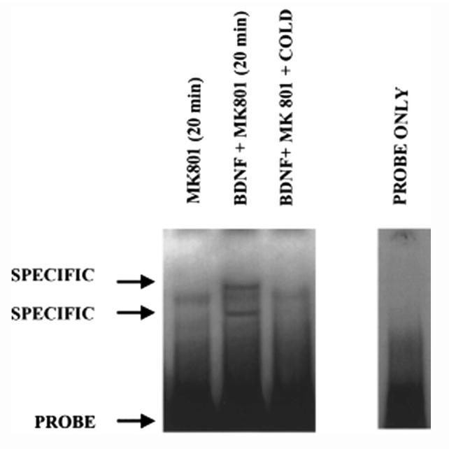
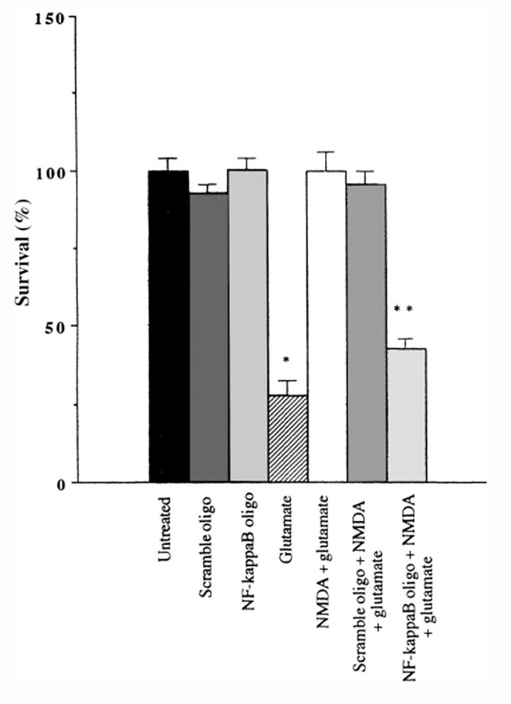
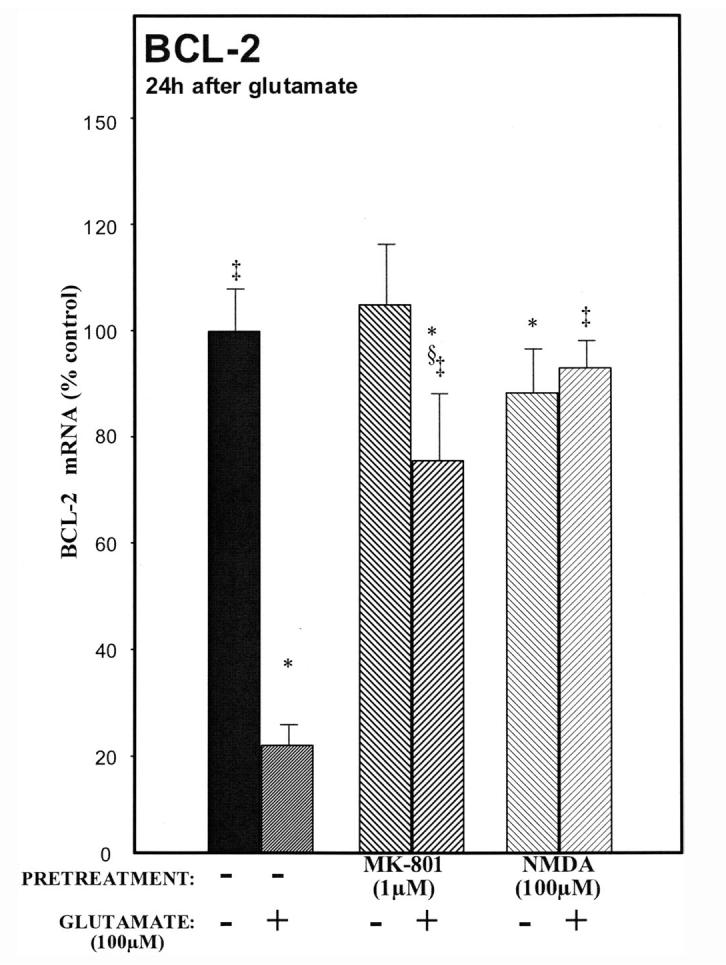
Delineating the mechanisms of survival pathways that exist in neurons will provide important insight into how neurons utilize intracellular proteins as neuroprotectants against the causes of acute neurodegeneration. We have employed cultured rat cerebellar granule cells as a model for determining the mechanisms of these intraneuronal survival pathways. Glutamate has long been known to kill neurons by an N-methyl-d-aspartate (NMDA) receptor-mediated mechanism. Paradoxically, subtoxic concentrations of NMDA protect neurons against glutamate-mediated excitotoxicity. Because NMDA protects neurons in physiologic concentrations of glucose and oxygen, we refer to this phenomenon as physiologic preconditioning. One of the major mechanisms of NMDA neuroprotection involves the activation of NMDA receptors leading to the rapid release of brain-derived neurotrophic factor (BDNF). BDNF then binds to and activates its cognate receptor, receptor tyrosine kinase B (TrkB). The efficient utilization of these two receptors confers remarkable resistance against millimolar concentrations of glutamate that kill more than eighty percent of the neurons in the absence of preconditioning the neurons with a subtoxic concentration of NMDA. Exactly how the neurons mediate neuroprotection by activation of both receptors is just beginning to be understood. Both NMDA and TrkB receptors activate nuclear factor kappaB (NF-kappaB), a transcription factor known to be involved in protecting neurons against many different kinds of toxic insults. By converging on survival transcription factors, such as NF-kappaB, NMDA and TrkB receptors protect neurons. Thus, crosstalk between these very different receptors provides a rapid means of neuronal communication to upregulate survival proteins through release and transcriptional activation of messenger RNA.
Figures


Similar articles
-
The excitoprotective effect of N-methyl-D-aspartate receptors is mediated by a brain-derived neurotrophic factor autocrine loop in cultured hippocampal neurons.J Neurochem. 2005 Aug;94(3):713-22. doi: 10.1111/j.1471-4159.2005.03200.x. Epub 2005 Jul 5. J Neurochem. 2005. PMID: 16000165
-
Co-activation of the phosphatidylinositol-3-kinase/Akt signaling pathway by N-methyl-D-aspartate and TrkB receptors in cerebellar granule cell neurons.Amino Acids. 2002;23(1-3):11-7. doi: 10.1007/s00726-001-0103-9. Amino Acids. 2002. PMID: 12373512
-
N-methyl-D-aspartate and TrkB receptors protect neurons against glutamate excitotoxicity through an extracellular signal-regulated kinase pathway.J Neurosci Res. 2005 Apr 1;80(1):104-13. doi: 10.1002/jnr.20422. J Neurosci Res. 2005. PMID: 15744743 Free PMC article.
-
Preconditioning and neurotrophins: a model for brain adaptation to seizures, ischemia and other stressful stimuli.Amino Acids. 2007;32(3):299-304. doi: 10.1007/s00726-006-0414-y. Epub 2006 Sep 29. Amino Acids. 2007. PMID: 16998712 Review.
-
Toward the development of strategies to prevent ischemic neuronal injury. In vitro studies.Ann N Y Acad Sci. 1997 Oct 15;825:209-19. doi: 10.1111/j.1749-6632.1997.tb48431.x. Ann N Y Acad Sci. 1997. PMID: 9369988 Review.
Cited by
-
Is there a place for cerebral preconditioning in the clinic?Transl Stroke Res. 2010 Jan 14;1(1):4-18. doi: 10.1007/s12975-009-0007-7. Transl Stroke Res. 2010. PMID: 20563278 Free PMC article.
-
Genome instability in Alzheimer disease.Mech Ageing Dev. 2017 Jan;161(Pt A):83-94. doi: 10.1016/j.mad.2016.04.005. Epub 2016 Apr 20. Mech Ageing Dev. 2017. PMID: 27105872 Free PMC article. Review.
-
Pathophysiology and neuroprotection of global and focal perinatal brain injury: lessons from animal models.Pediatr Neurol. 2015 Jun;52(6):566-584. doi: 10.1016/j.pediatrneurol.2015.01.016. Epub 2015 Jan 31. Pediatr Neurol. 2015. PMID: 26002050 Free PMC article. Review.
-
P2X7, NMDA and BDNF receptors converge on GSK3 phosphorylation and cooperate to promote survival in cerebellar granule neurons.Cell Mol Life Sci. 2010 May;67(10):1723-33. doi: 10.1007/s00018-010-0278-x. Epub 2010 Feb 10. Cell Mol Life Sci. 2010. PMID: 20146080 Free PMC article.
-
Toll-like receptors and opportunities for new sepsis therapeutics.Curr Infect Dis Rep. 2012 Oct;14(5):455-61. doi: 10.1007/s11908-012-0273-5. Curr Infect Dis Rep. 2012. PMID: 22805995
References
-
- Kirino T, Tsujita Y, Tamura A. Induced tolerance to ischemia in gerbil hippocampal neurons. J. Cereb. Blood Flow Metab. 1991;11:299–307. - PubMed
-
- Kitagawa K, Matsumoto M, Tagaya M, et al. “Ischemic tolerance” phenomenon found in the brain. Brain Res. 1990;528:21–24. - PubMed
-
- Liu Y, Kato H, Nakata N, Kogure K. Protection of rat hippocampus against ischemic neuronal damage by pretreatment with sublethal ischemia. Brain Res. 1992;586:121–124. - PubMed
-
- Kitagawa K, Matsumoto M, Kuwabara K, et al. “Ischemic tolerance” phenomenon detected in various brain regions. Brain Res. 1991;561:203–211. - PubMed
-
- Abe H, Nowak TS., Jr. Gene expression and induced ischemic tolerance following brief insults. Acta Neurobiol. Exp. 1996;56:3–8. - PubMed
Publication types
MeSH terms
Substances
Grants and funding
LinkOut - more resources
Full Text Sources
