Infection with Mycobacterium bovis BCG diverts traffic of myelin oligodendroglial glycoprotein autoantigen-specific T cells away from the central nervous system and ameliorates experimental autoimmune encephalomyelitis
- PMID: 12853387
- PMCID: PMC164279
- DOI: 10.1128/cdli.10.4.564-572.2003
Infection with Mycobacterium bovis BCG diverts traffic of myelin oligodendroglial glycoprotein autoantigen-specific T cells away from the central nervous system and ameliorates experimental autoimmune encephalomyelitis
Abstract
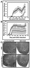
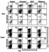
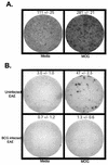
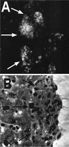
Infectious agents have been proposed to influence susceptibility to autoimmune diseases such as multiple sclerosis. We induced a Th1-mediated central nervous system (CNS) autoimmune disease, experimental autoimmune encephalomyelitis (EAE) in mice with an ongoing infection with Mycobacterium bovis strain bacillus Calmette-Guérin (BCG) to study this possibility. C57BL/6 mice infected with live BCG for 6 weeks were immunized with myelin oligodendroglial glycoprotein peptide (MOG(35-55)) to induce EAE. The clinical severity of EAE was reduced in BCG-infected mice in a BCG dose-dependent manner. Inflammatory-cell infiltration and demyelination of the spinal cord were significantly lessened in BCG-infected animals compared with uninfected EAE controls. ELISPOT and gamma interferon intracellular cytokine analysis of the frequency of antigen-specific CD4(+) T cells in the CNS and in BCG-induced granulomas and adoptive transfer of MOG(35-55)-specific green fluorescent protein-expressing cells into BCG-infected animals indicated that nervous tissue-specific (MOG(35-55)) CD4(+) T cells accumulate in the BCG-induced granuloma sites. These data suggest a novel mechanism for infection-mediated modulation of autoimmunity. We demonstrate that redirected trafficking of activated CNS antigen-specific CD4(+) T cells to local inflammatory sites induced by BCG infection modulates the initiation and progression of a Th1-mediated CNS autoimmune disease.
Figures


Similar articles
-
De novo central nervous system processing of myelin antigen is required for the initiation of experimental autoimmune encephalomyelitis.J Immunol. 2002 Apr 15;168(8):4173-83. doi: 10.4049/jimmunol.168.8.4173. J Immunol. 2002. PMID: 11937578
-
MOG extracellular domain (p1-125) triggers elevated frequency of CXCR3+ CD4+ Th1 cells in the CNS of mice and induces greater incidence of severe EAE.Mult Scler. 2014 Sep;20(10):1312-21. doi: 10.1177/1352458514524086. Epub 2014 Feb 19. Mult Scler. 2014. PMID: 24552747
-
Mycobacterium bovis bacille Calmette-Guérin infection in the CNS suppresses experimental autoimmune encephalomyelitis and Th17 responses in an IFN-gamma-independent manner.J Immunol. 2008 Nov 1;181(9):6201-12. doi: 10.4049/jimmunol.181.9.6201. J Immunol. 2008. PMID: 18941210 Free PMC article.
-
T- and B-cell responses to myelin oligodendrocyte glycoprotein in experimental autoimmune encephalomyelitis and multiple sclerosis.Glia. 2001 Nov;36(2):220-34. doi: 10.1002/glia.1111. Glia. 2001. PMID: 11596130 Review.
-
Myelin oligodendrocyte glycoprotein: a novel candidate autoantigen in multiple sclerosis.J Mol Med (Berl). 1997 Feb;75(2):77-88. doi: 10.1007/s001090050092. J Mol Med (Berl). 1997. PMID: 9083925 Review.
Cited by
-
Virally activated CD8 T cells home to Mycobacterium bovis BCG-induced granulomas but enhance antimycobacterial protection only in immunodeficient mice.Infect Immun. 2007 Mar;75(3):1154-66. doi: 10.1128/IAI.00943-06. Epub 2006 Dec 18. Infect Immun. 2007. PMID: 17178783 Free PMC article.
-
Infections and autoimmunity: a panorama.Clin Rev Allergy Immunol. 2008 Jun;34(3):283-99. doi: 10.1007/s12016-007-8048-8. Clin Rev Allergy Immunol. 2008. PMID: 18231878 Free PMC article. Review.
-
CONTINUOUS REPOPULATION OF LYMPHOCYTE SUBSETS IN TRANSPLANTED MYCOBACTERIAL GRANULOMAS.Eur J Microbiol Immunol (Bp). 2011 Mar;1(1):59-69. doi: 10.1556/EuJMI.1.2011.1.8. Eur J Microbiol Immunol (Bp). 2011. PMID: 22096617 Free PMC article.
-
Immunomodulation with microbial vaccines to prevent type 1 diabetes mellitus.Nat Rev Endocrinol. 2010 Mar;6(3):131-8. doi: 10.1038/nrendo.2009.273. Nat Rev Endocrinol. 2010. PMID: 20173774 Review.
-
Conflicting Role of Mycobacterium Species in Multiple Sclerosis.Front Neurol. 2017 May 19;8:216. doi: 10.3389/fneur.2017.00216. eCollection 2017. Front Neurol. 2017. PMID: 28579973 Free PMC article. Review.
References
-
- Altschul, S. F., W. Gish, W. Miller, E. W. Myers, and D. J. Lipman. 1990. Basic local alignment search tool. J. Mol. Biol. 215:403-410. - PubMed
-
- Andersen, E., H. Isager, and K. Hyllested. 1981. Risk factors in multiple sclerosis: tuberculin reactivity, age at measles infection, tonsillectomy and appendectomy. Acta Neurol. Scand. 63:131-135. - PubMed
-
- Ben-Nun, A., I. Mendel, G. Sappler, and N. Kerlero de Rosbo. 1995. A 12-kDa protein of Mycobacterium tuberculosis protects mice against experimental autoimmune encephalomyelitis. Protection in the absence of shared T cell epitopes with encephalitogenic proteins. J. Immunol. 154:2939-2948. - PubMed
-
- Ben-Nun, A., and S. Yossefi. 1992. Staphylococcal enterotoxin B as a potent suppressant of T lymphocytes: trace levels suppress T lymphocyte proliferative responses. Eur. J. Immunol. 22:1495-1503. - PubMed
Publication types
MeSH terms
Substances
Grants and funding
LinkOut - more resources
Full Text Sources
Medical
Research Materials

