. 2003 Jul 9;23(14):6030-40.
doi: 10.1523/JNEUROSCI.23-14-06030.2003.

Retinoschisin, a photoreceptor-secreted protein, and its interaction with bipolar and muller cells

Affiliations

Retinoschisin, a photoreceptor-secreted protein, and its interaction with bipolar and muller cells

Silvia N M Reid et al. J Neurosci. .

Abstract

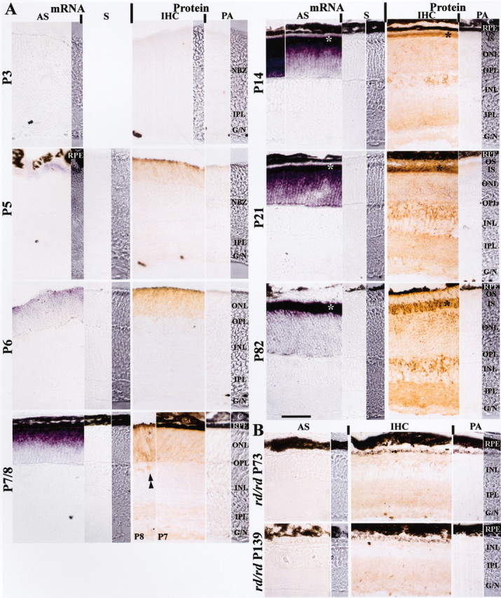
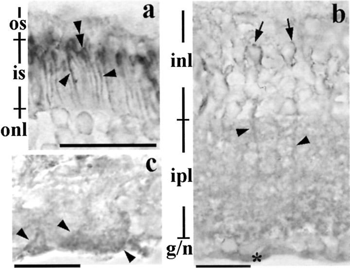
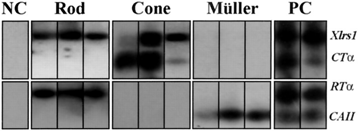
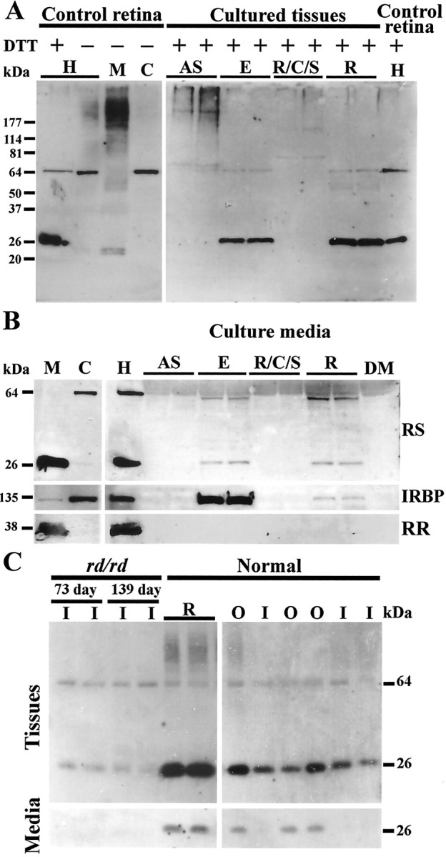
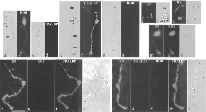
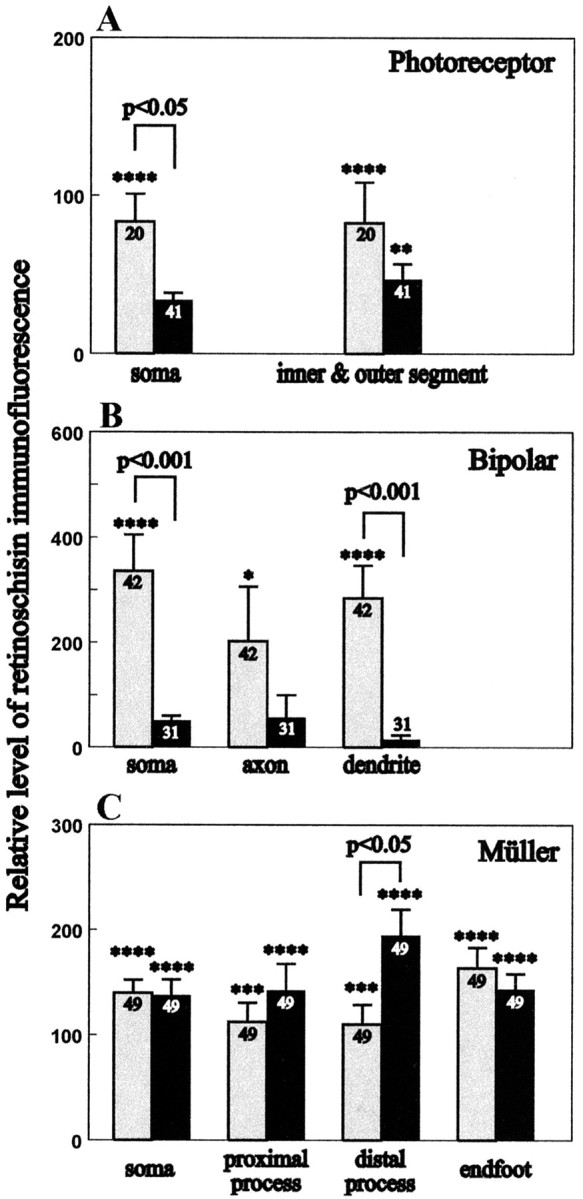
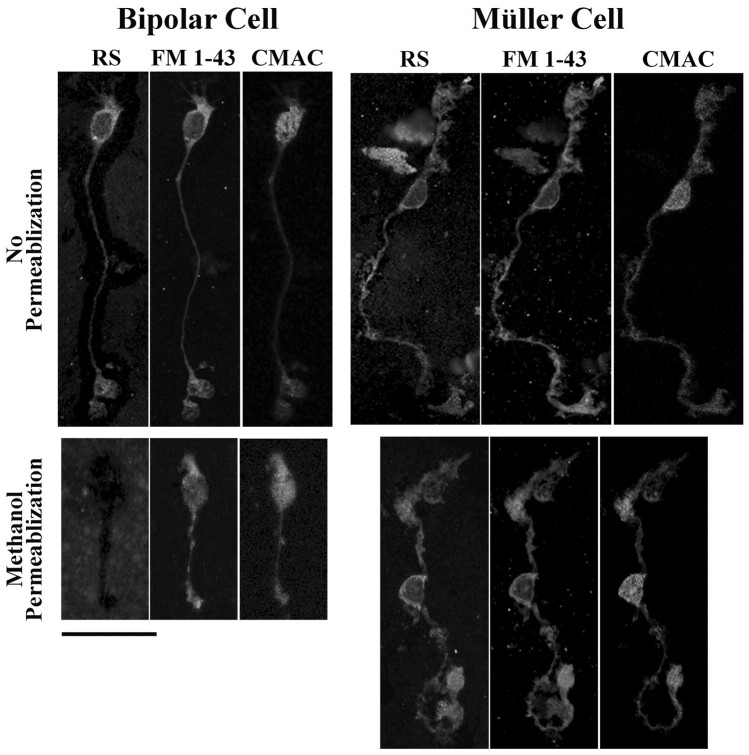
Usually, photoreceptors interact with other retinal cells through the neurotransmitter glutamate. Here we describe a nonsynaptic interaction via a secreted protein, retinoschisin. Using in situ hybridization, we found that from early postnatal life retinoschisin mRNA is present only in the outer retina of the mouse, and with single-cell RT-PCR we demonstrated its localization in rod and cone photoreceptor cells but not in Müller cells. Western blot analyses of proteins from cultured ocular tissues and microdissected outer and inner retinas, as well as from the culture media of these samples, showed that retinoschisin is secreted from the photoreceptor cells. Immunostaining of permeabilized and nonpermeabilized dissociated retinal cells revealed that retinoschisin is mainly inside and outside the photoreceptors, outside bipolar cells, and associated with plasma membranes of Müller cells and inside their distal processes. Because we showed previously that retinoschisin is distributed all over the retina, our current data suggest that after synthesis and secretion by the photoreceptors, retinoschisin reaches the surface of retinal cells and mediates interactions/adhesion between photoreceptor, bipolar, and Müller cells, contributing to the maintenance of the cytoarchitectural integrity of the retina. These interactions may not occur when the gene encoding retinoschisin is mutated, as it occurs in X-linked juvenile retinoschisis, a disease that results in morphological and electrophysiological defects of the retina.


Figures

Figure 1.
Figure 1.
Distribution of retinoschisin mRNA and protein in normal (A) and rd/rd mouse (B) retinas. A, Retinoschisin mRNA was detected with a digoxigenin-labeled antisense-cRNA probe (AS) and visualized by the purple reaction product of the alkaline phosphatase reaction; no staining was observed with sense-cRNA probe (S) used as control. Retinoschisin was detected by immunohistochemistry (IHC); the brown peroxidase-reaction product was observed after immunoreaction with an affinity-purified polyclonal antibody against amino acid residues 24–37 of retinoschisin (aa 24–37). This antibody was preabsorbed with aa 24–37 and used as control (PA). NBZ, Neuroblastic zone; IPL, inner plexiform layer; G/N, ganglion cell and nerve fiber layers; RPE, retinal pigment epithelium; ONL, outer nuclear layer; OPL, outer plexiform layer; INL, inner nuclear layer; C, choroid; OS, outer segment of photoreceptors; IS, inner segment of photoreceptors. Retinoschisin mRNA and protein were not detected in the retina of normal mice at P3 but were found at a very low level at P5. A mismatched mRNA and protein localization started on P7 and persisted into adulthood (P82). Retinoschisin mRNA was in the outer retina throughout postnatal development (outer NBZ on P5; ONL on P6 and P7/8; IS and ONL on P14, 21, and 82). In contrast, retinoschisin (Protein) was initially in the outer retina (outer NBZ on P5 and ONL on P6) and then appeared in the inner retina. Immunopositive cell bodies in the INL (double arrowheads) started appearing at P8 and showed increased intensity as the retina matured. By P21, retinoschisin was abundant in all layers. Both mRNA and protein were most concentrated in the area corresponding to the photoreceptor inner segments (*). Visualization of the mRNA was achieved with a 2 hr alkaline phosphatase reaction (P7/8, 14, and 21) or 10.5 hr (P3, 5, 6, and 82, and the left micrograph in the AS column for P14). B, At P73 and P139, most photoreceptors had degenerated in the rd/rd retinas, and retinoschisin mRNA was not detected even after 10.5 hr of reaction. Faint retinoschisin staining was present in the INL, the IPL, and the G/N. The micrograph strips at the far right of the mRNA and protein panels for each age are high-contrast bright-field micrographs taken from the same section as the adjacent micrographs. High-contrast micrographs were also taken from a portion of the sections in the AS panel at P3, the IHC panel at P3, and the AS panel at P5. Scale bar: for all micrographs, 50 μm.
Figure 2.
Figure 2.
Retinoschisin in the adult retina. a, Retinoschisin-immunolabeling surrounds the inner segment of the photoreceptors (arrowheads) and is concentrated in the area adjacent to where the inner segment joins the outer segment (double arrowheads). b, Immunohistochemical reaction products in the inner retina appear around cell bodies (arrows) in the INL, processes in the INL and IPL (arrowheads), and in the nerve fiber layer (*). c, The immunoreaction products are also associated with the end feet of Müller cells (arrowheads) in the nerve fiber layer. Scale bar: a, b, 20 μm; c, 13 μm. See Figure 1 for abbreviations.
Figure 3.
Figure 3.
RT-PCR detection of Xlrs1 mRNA expression in rod, cone, and Müller cells. Retinoschisin mRNA was found in individual cells expressing either the rod-specific α1-subunit of transducin (RTα) or the cone-specific α2-subunit of transducin (CTα), but not in cells expressing carbonic anhydrase II (CAII) mRNAs. The expression of these cell-specific genes was examined by RT-PCR using gene-specific primers and was used to reveal the identity of the isolated cells (rod, cone, and Müller cells, respectively). Expression of the above genes was not found in cells that were determined by morphology to be neither photoreceptors nor Müller cells [negative control (NC)]. Clusters containing multiple retinal cells were collected as the positive control (PC), and expression of all four genes was detected in these samples.
Figure 4.
Figure 4.
Retinoschisin in normal mouse retinal extracts and its secretion from different ocular tissues analyzed by Western blots. Proteins from all samples were subjected to SDS-PAGE. Western blots were labeled with the RS24–37 antibody. A, Left panel, Retinoschisin from a mouse retinal homogenate(H), with or without the addition of DTT, and nonreduced membrane (M) and cytosol (C) fractions (see Materials and Methods). Right panel, Proteins from cultured eye tissues incubated with DTT. AS, Anterior segment; E, eyecup; R/C/S, retinal pigment epithelium/choroid/sclera; R, retina. B, Left panel, Retinal cellular fractions treated with DTT. Right panel, Retinal homogenate used as reference and the culture media of ocular tissues in A after treatment with DTT. C, Left panel, Cultured adult rd/rd retinas, containing only the inner layers, and normal retina (top panel); culture media of above samples (bottom panel). Right panel, Microdissected inner and outer layers of normal retinas (top panel) and their culture media (bottom panel). The 27 kDa protein was observed only under reducing conditions in retinal samples and their culture media (see Results for details). IRBP, a predominately cytosolic protein, was used as a positive control for secretion and rod rhodopsin (RR), an intrinsic membrane protein, was used as a negative control. RS24–37 antibody also labeled a 65 kDa protein present in the retinal cytosol. DM, DMEM culture medium; RS, retinoschisin; I, inner retina; O, outer retina.
Figure 5.
Figure 5.
Retinoschisin in photoreceptor cells and marker-identified bipolar and Müller cells. The bipolar cell shown (a) was recognized by the ROB antibody when permeabilized with proteinase K (b); bipolar cells were not immunopositive for the Müller cell protein CRALBP (c, d). Müller cells were identified by an antibody against CRALBP after Triton X-100 permeabilization (e, f) and were not labeled by ROB (g, h). Cells in a and b and g and h were immunostained at the same time and so were cells in c and d and e and f. The retinoschisin antibody (RS) labeled photoreceptor cells that were either permeabilized with methanol (i, j) or not permeabilized (k, l). Bipolar cells (mo) and Müller cells (ps and tx) that had been immunostained for retinoschisin were subjected to sequential immunostainings, and the image acquisition after each staining was completed before the next round of staining. The nonpermeabilized bipolar cell in m was also labeled by the ROB antibody (n). The methanol-permeabilized Müller cell in p was not labeled by ROB (q) but was labeled by the antibody against CRALBP (r). The nonpermeabilized Müller cell in t was not labeled by the antibody against the intracellular protein CRALBP (u). After Triton X-100 permeabilization, this Müller cell was not positive for ROB (v), but it was labeled by the CRALBP antibody (w). a, c, e, g, j, l, o, s, and x were taken with bright field under high contrast. b and h, d and f, q and r, and uw were acquired with the same exposure time. dd, Dendrite; sm, soma; ax, axon; dp, distal process; pp, proximal process; ef, end foot; os, outer segment; is, inner segment; my, myoid; nl, nucleus. Scale bar, 25 μm.
Figure 6.
Figure 6.
Relative levels of retinoschisin immunofluorescence in nonpermeabilized and methanol-permeabilized photoreceptor (A), bipolar (B), bipolar, and Müller (C) cells. Immunofluorescent images of retinal cells were analyzed from confocal optical sections recorded under identical conditions. The relative level of retinoschisin immunofluorescence represents the percentage difference from the control. The immunofluorescence of the control was obtained using the anti-retinoschisin antibody preabsorbed with the peptide used to generate it. A lack of difference between the antibody staining of the sample and the control was given a value of 0 for the relative retinoschisin immunofluorescence. Bars represent the averaged relative retinoschisin immunofluorescence ± the corresponding SEM for the number of observations performed (indicated in the bar). Gray bars represent nonpermeabilized cells; black bars represent methanol-permeabilized cells. Statistical comparisons between cells treated with retinoschisin antibody and the peptide preabsorbed antibody are indicated with asterisks only for those that are at a significant p level: *p < 0.5; **p < 0.01; ***p < 0.005; ****p < 0.0001. The relative level of retinoschisin immunofluorescence is higher without than with permeabilization for the soma of photoreceptor cells (p < 0.05) and the soma (p < 0.001) and dendrite (p < 0.001) of bipolar cells. The level was significantly higher after permeabilization at the distal process of Müller cells (p < 0.05).
Figure 7.
Figure 7.
Confocal images of triple-labeled bipolar and Müller cells. Enzymatically dissociated retinal cells were incubated with culture medium containing 7-amino-4-chloromethylcoumarin (CMAC) for 15 min at 37°C to stain cytosol and then under the same conditions with FM 1–43-containing medium to label cell membranes. The labeled cells were fixed in paraformaldehyde and smeared onto glass slides. Immunostaining for retinoschisin was performed immediately before or after permeabilization with methanol. The antigen was visualized with Alexa Fluor 488-conjugated secondary antibody. The retinoschisin immunostaining (RS) pattern of nonpermeabilized bipolar cells was nearly identical to the cell plasma membrane labeling with FM 1–43 but was different from the cytosol labeling with CMAC. Methanol permeabilization abolished retinoschisin immunoreactivity of bipolar cells but not the FM 1–43 and CMAC stainings. These results show that retinoschisin is localized outside bipolar cells. The antibody against retinoschisin immunolabeled nonpermeabilized Müller cells. This staining pattern was nearly identical to that obtained with FM 1–43 and persisted after methanol permeabilization, indicating that retinoschisin is associated with the plasma membrane of Müller cells. Scale bar, 25 μm.

Similar articles

Cited by

References

    1. Alexander KR, Fishman GA, Grover S ( 2000) Temporal frequency deficits in the electroretinogram of the cone system in X-linked retinoschisis. Vision Res 40: 2861–2868. - PubMed
    1. Ando A, Takahashi K, Sho K, Matsushima M, Okamura A, Uyama M ( 2000) Histopathological findings of X-linked retinoschisis with neovascular glaucoma. Graefe's Arch Clin Exp Ophthalmol 238: 1–7. - PubMed
    1. Apweiler R, Attwood TK, Bairoch A, Bateman A, Birney E, Biswas M, Bucher P, Cerutti L, Corpet F, Croning MDR, Durbin R, Falquet L, Fleischmann W, Gouzy J, Hermjakob H, Hulo N, Jonassen I, Kahn D, Kanapin A, Karavidopoulou Y, et al. ( 2001) The InterPro database, an integrated documentation resource for protein families, domains and functional sites. Nucleic Acids Res 29: 37–40. - PMC - PubMed
    1. Baumgartner S, Littleton JT, Broadie K, Bhat MA, Harbecke R, Lengyel JA, Chiquet-Ehrismann R, Prokop A, Bellen HJ ( 1996) A Drosophila neurexin is required for septate junction and blood-nerve barrier formation and function. Cell 87: 1059–1068. - PubMed
    1. Blanks JC, Adinolfi AM, Lolley RN ( 1974) Synaptogenesis in the photoreceptor terminal of the mouse retina. J Comp Neurol 156: 81–94. - PubMed

Publication types

MeSH terms