. 2003 Jul 9;23(14):6041-9.
doi: 10.1523/JNEUROSCI.23-14-06041.2003.

Cav1.4alpha1 subunits can form slowly inactivating dihydropyridine-sensitive L-type Ca2+ channels lacking Ca2+-dependent inactivation

Affiliations

Cav1.4alpha1 subunits can form slowly inactivating dihydropyridine-sensitive L-type Ca2+ channels lacking Ca2+-dependent inactivation

Alexandra Koschak et al. J Neurosci. .

Abstract

The neuronal L-type calcium channels (LTCCs) Cav1.2alpha1 and Cav1.3alpha1 are functionally distinct. Cav1.3alpha1 activates at lower voltages and inactivates more slowly than Cav1.2alpha1, making it suitable to support sustained L-type Ca2+ inward currents (ICa,L) and serve in pacemaker functions. We compared the biophysical and pharmacological properties of human retinal Cav1.4alpha1 using the whole-cell patch-clamp technique after heterologous expression in tsA-201 cells with other L-type alpha1 subunits. Cav1.4alpha1-mediated inward Ba2+ currents (IBa) required the coexpression of alpha2delta1 and beta3 or beta2a subunits and were detected in a lower proportion of transfected cells than Cav1.3alpha1. IBa activated at more negative voltages (5% activation threshold; -39mV; 15 mm Ba2+) than Cav1.2alpha1 and slightly more positive than Cav1.3alpha1. Voltage-dependent inactivation of IBa was slower than for Cav1.2alpha1 and Cav1.3alpha1( approximately 50% inactivation after 5 sec; alpha2delta1 + beta3 coexpression). Inactivation was not increased with Ca2+ as the charge carrier, indicating the absence of Ca2+-dependent inactivation. Cav1.4alpha1 exhibited voltage-dependent, G-protein-independent facilitation by strong depolarizing pulses. The dihydropyridine (DHP)-antagonist isradipine blocked Cav1.4alpha1 with approximately 15-fold lower sensitivity than Cav1.2alpha1 and in a voltage-dependent manner. Strong stimulation by the DHP BayK 8644 was found despite the substitution of an otherwise L-type channel-specific tyrosine residue in position 1414 (repeat IVS6) by a phenylalanine. Cav1.4alpha1 + alpha2delta1 + beta channel complexes can form LTCCs with intermediate DHP antagonist sensitivity lacking Ca2+-dependent inactivation. Their biophysical properties should enable them to contribute to sustained ICa,L at negative potentials, such as required for tonic neurotransmitter release in sensory cells and plateau potentials in spiking neurons.


Figures
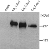
Figure 1.
Figure 1.
Heterologous expression of different LTCC subunits in tsA-201 cells. Cells were transfected with Cav1.2α1, Cav1.3α1, or Cav1.4α1 together with β3 and α2δ1 subunit cDNA as described in Materials and Methods. Expression of α1 subunit proteins was analyzed in immunoblots of membranes prepared from lyzed cells after separation on 8% SDS-PAGE gels (10 μg of membrane protein per lane) using a generic anti-α1 sequence directed antibody (anti-CP1382–1400). No α1 immunoreactivity was present in mock-transfected cells used as a control. One of four experiments yielding similar results is shown.
Figure 2.
Figure 2.
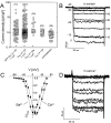
Biophysical properties of IBa and ICa through Cav1.4α1 subunits. Cav1.4α1 subunits were expressed together with β3 + α2δ1 (A—C) or β2a + α2δ1 (A), as described in Materials and Methods, using 15 mm Ba2+ (A—C) or 15 mM Ca2+ (D) as charge carrier. A, Expression density was determined by depolarizing pulses to Vmax.IBa was measured 48 – 82 hr after transfection for the indicated number of cells. Small currents measured in untransfected cells were attributable to endogenous non-L-type currents that were <1.72 pA/pF. Asterisks indicate statistically significant difference to untransfected cells (p < 0.01; Kruskal—Wallis test, followed by Dunn's multiple comparison test). B, Superimposed currents were activated by depolarizing Cav1.4α1-transfected cells during 50 msec pulses from an HP of -90 mV to between -60 and 40 mV in 10 mV steps. C, Normalized I—V curves for Cav1.4α1 coexpressed with β3 and α2δ1 subunits using 15 mm Ba2+ (black squares) or Ca2+ (black circles) as charge carriers. Biophysical parameters are given in Table 1. D, Protocol as in B but with 15 mm Ca2+ in the bath solution.
Figure 3.
Figure 3.
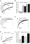
Inactivation properties of Cav1.4α1 Ca2+ channels. A, IBa (black traces) through Cav1.3α1 and Cav1.4α1 subunits coexpressed with β3 and α2δ1 subunits were elicited by 10 sec depolarizing pulses from an HP of -90 mV to Vmax. Representative current traces for Cav1.3α1 (n = 4) and Cav1.4α1 (n = 17) channels are shown. Traces were normalized to the peak current amplitudes. For the experiments shown, inactivation measured during 5 and 10 sec depolarizing pulses was as follows: Cav1.4α1, 41 and 56%; Cav1.3α1, 89 and 97%. A representative trace for current through Cav1.4α1 recorded with 15 mm Ca2+ as charge carrier is illustrated in gray superimposed on the IBa trace indicated in black. B, Percent current inactivation measured after 0.25, 5, and 10 sec depolarizations to Vmax in Cav1.4α1-transfected cells using either 15 mm Ba2+ or 15 mM Ca2+ as the charge carrier. Inactivation of currents during pulses was not significantly different for Ba2+ (black bars) and Ca2+ (gray bars) (n = 7; p > 0.05). C, D, Inactivation for Cav1.2α1 (C) and Cav1.3α1 (D) during 2 sec depolarizing pulses toVmax with 15 mm Ba2+ (black trace) or 15 mM Ca2+ (gray trace) as charge carriers. For Cav1.3α1, a variable noninactivating ICa component was found (9 – 40%; n = 4), whereas remaining Cav1.2α1 currents were always <3.5% (n = 7). E, Inactivation of IBa through Cav1.4α1 cotransfected with β3 (black) or β2a (gray) and α2δ1. Currents were normalized to peak IBa. Currents were elicited by depolarization from an HP of -90 mV to Vmax. F, Percentage of inactivation of IBa through Cav1.4α1 cotransfected with β3 (black; n = 17) or β2a (gray;n = 7), and α2δ1 was determined after 5 and 10 sec during a depolarization from an HP of -90 mV toVmax. Currents were normalized to peakIBa. Inactivation with β2a coexpression was 27.2±4.6% (after 5 sec) and 44.6 ± 6.4% (after 10 sec; n=7), respectively. Asterisks indicate a statistically significant difference to β2a coexpression (p < 0.01).
Figure 8.
Figure 8.
Sequence alignment of L-type Cav1.4α1 with Cav1.2α1 and Cav1.3α1. An amino acid exchange within the proposed DHP-binding domain of LTCCs is indicated by an arrow. A tyrosine residue conserved in all L-type α1 subunits is replaced by phenylalanine in position 1414 of Cav1.4α1. The sequence alignment also illustrates amino acid differences between Cav1.4α1 and Cav1.2α1 or Cav1.3α1, which might explain the differences in Ca2+-dependent inactivation described under our experimental conditions. Sequence stretches previously identified as critical determinants for calmodulin-binding and Ca2+-dependent inactivation (de Leon et al., 1995; Zuhlke et al., 1999; Mouton et al., 2001; Pitt et al., 2001) are indicated.
Figure 4.
Figure 4.
DHP antagonist sensitivity of Cav1.4α1 Ca2+ channels. Cav1.4α1 was coexpressed with β3 and α2δ1 subunits in tsA-201 cells and recorded in bath solution containing 15 mm Ba2+ as charge carrier. A, IBa was elicited from depolarizations to Vmax (filled circles) in the absence or presence (gray bar) of the DHP antagonist isradipine. For the experiment shown,IBa block by 1 μm isradipine was 80%. Corresponding peak current traces elicited by 40 msec depolarizations in the absence (control) and presence of isradipine are shown in the inset. B, Concentration-dependent inhibition was measured from a holding potential of -90 mV for Cav1.4α1 (black triangles) during superfusion of the cell with bath solution containing the indicated concentrations of isradipine. The dose—response relationship was compared with Cav1.3α1 (squares) (Koschak et al., 2001). Pronounced voltage dependence of isradipine block of IBa through Cav1.4α1 subunits was observed by changing the HP from -90 to -50 mV (open triangle). Asterisks indicate statistically significant difference (p < 0.05; data are means ± SE).
Figure 5.
Figure 5.
DHP agonist sensitivity of Cav1.4α1 Ca2+ channels. For all experiments, Cav1.4α1 subunits were coexpressed with β3 and α2δ1 subunits in tsA-201 cells. Charge carrier was 15 mm Ba2+. One representative experiment (of 9) is shown. A, Stimulation of IBa through Cav1.4α1 Ca2+ channels by the Ca2+ channel activator BayK 8644 (BayK). IBa was elicited by depolarizations to Vmax before and after application of BayK 8644-containing solution (5 μm). Maximal IBa is plotted against time. The inset shows representative traces in the absence (control) and presence of BayK 8644. B, Current—voltage relationship for Cav1.4α1 in the absence (black circles) and presence of 5 μm BayK 8644 (BayK; gray circles). V0.5, act was -8 and -21.1 mV for the control and BayK 8644-modulated current, respectively. One representative experiment (of 7) is shown.
Figure 6.
Figure 6.
Voltage-dependent facilitation of Cav1.4α1 subunits. A, Schematic representation of the voltage protocol used to elicit facilitation. Channel activity was recorded in tsA-201 cells transfected with Cav1.4α1, β3, and α2δ1 subunits in 15 mm Ba2+ solution. Test pulses (TPs) of 400 msec were applied with or without a 200 msec PP. A representative current trace (facilitation ratio, 1.11) is shown. Note the differences in activation and inactivation kinetics time courses. B, Comparison of voltage-dependent facilitation of different LTCCs. Facilitation of Cav1.2α1, Cav1.3α1, and Cav1.4α1 IBa is expressed as the amplitude ratio of currents recorded with a prepulse (+PP) over the respective control currents without prepulse (-PP). A similar extent of facilitation was observed for all LTCC subtypes tested (p > 0.05). Facilitation was observed in seven of seven Cav1.2α1-transfected cells, eight of 20 Cav1.3α1-transfected cells, and 10 of 17 Cav1.4α1 (+ β3 + α2δ1)-transfected cells.
Figure 7.
Figure 7.
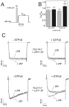
Depolarization-induced facilitation of Cav1.4α1 subunits. A, Pulse protocol used to determine G-protein modulation of Cav1.4α1- and Cav2.1α1-mediated IBa. Facilitation was measured during 50 msec test pulses (TP) to Vmax either with or without a 5 msec PP to 140 mV. B, Facilitation ratios for Cav1.4α1 and Cav2.1α1 Ca2+ channels (coexpressed with β3 and α2δ1) in the absence (control; gray bars) and presence of 300 μm intracellular GTPγS (black bars) using the pulse protocol described in A. Recordings were started after ≥3 min of dialysis with GTPγS. The following facilitation ratios were obtained in the absence and presence of GTPγS, respectively:Cav1.4α1, 1.1±0.02 (n=11 of 17 cells), 1.12±0.02 (n=7 of 14 cells); Cav2.1α1, 0.99 ± 0.01 (n = 7 of 7 cells), 1.3 ± 0.07 (n = 10 of 10 cells). Asterisk indicates statistically significant difference (p < 0.01). C, Representative current traces for the experiments described in B.

Similar articles

Cited by

References

    1. Alaburda A, Perrier JF, Hounsgaard J ( 2002) Mechanisms causing plateau potentials in spinal motoneurones. Adv Exp Med Biol 508: 219–226. - PubMed
    1. Ball SL, Powers PA, Shin HS, Morgans CW, Peachey NS, Gregg RG ( 2002) Role of the beta(2) subunit of voltage-dependent calcium channels in the retinal outer plexiform layer. Invest Ophthalmol Vis Sci 43: 1595–1603. - PubMed
    1. Bech-Hansen NT, Naylor MJ, Maybaum TA, Pearce WG, Koop B, Fishman GA, Mets M, Musarella MA, Boycott KM ( 1998) Loss-of-function mutations in a calcium-channel alpha1-subunit gene in Xp11.23 cause incomplete X-linked congenital stationary night blindness. Nat Genet 19: 264–267. - PubMed
    1. Berntson A, Taylor WR, Morgans CW ( 2003) Molecular identity, synaptic localization, and physiology of calcium channels in retinal bipolar cells. J Neurosci Res 71: 146–151. - PubMed
    1. Bourinet E, Charnet P, Tomlinson WJ, Stea A, Snutch TP, Nargeot J ( 1994) Voltage-dependent facilitation of a neuronal α1C L-type calcium channel. EMBO J 13: 5032–5039. - PMC - PubMed

Publication types

MeSH terms