. 2003 Jul 9;23(14):6132-40.
doi: 10.1523/JNEUROSCI.23-14-06132.2003.

Sequential signaling through Notch1 and erbB receptors mediates radial glia differentiation

Affiliations

Sequential signaling through Notch1 and erbB receptors mediates radial glia differentiation

Brooke A Patten et al. J Neurosci. .

Abstract

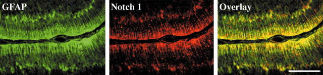
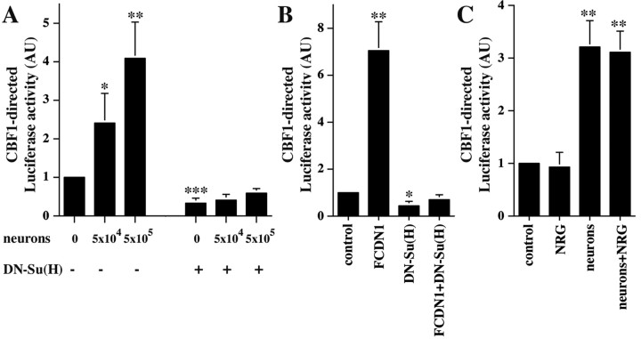
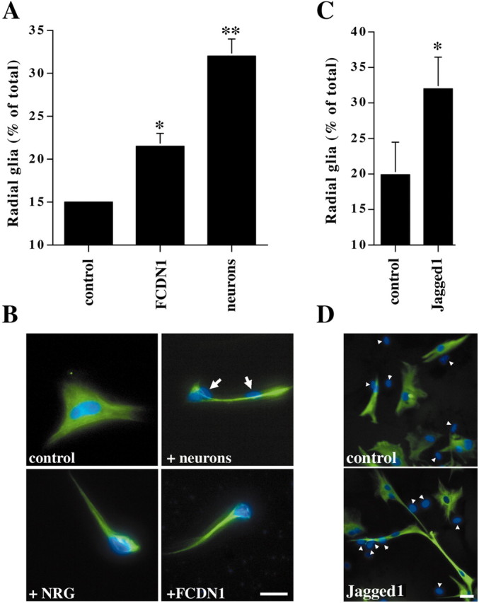
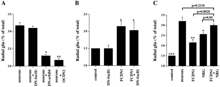
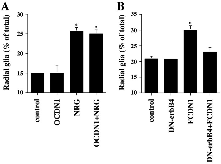
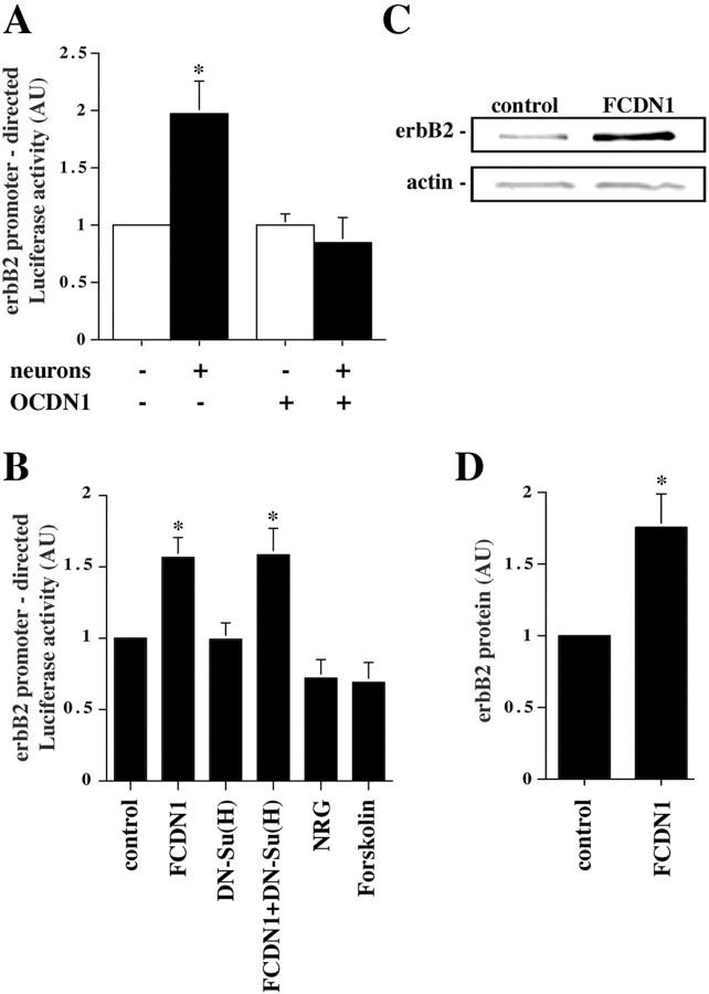
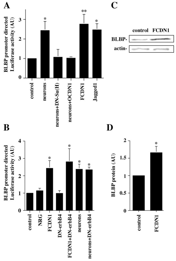
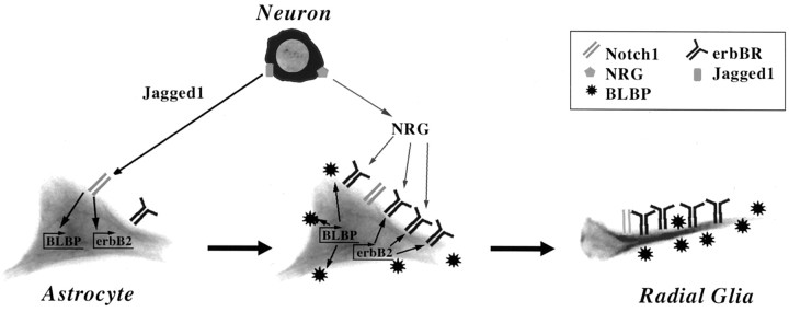
Radial glia cells both generate neurons and physically guide nascent neurons to their target destination in the cortex, and as such they are essential for CNS development. It has been proposed that in the developing cerebellum, neuronal contact induces radial glia formation, however, the mechanisms involved in this process are not well understood. Here we demonstrate that neuronal induction of radial glia formation is the result of sequential signaling through Notch1 and erbB receptors. First, Notch1 activation by neuronal contact induces the glial expression of the brain lipid binding protein (BLBP) and erbB2 genes. Interestingly, two different signaling pathways mediate these effects of Notch1 on transcription, BLBP expression being dependent on Su(H), whereas erbB2 is regulated by a yet unidentified Notch1 pathway. The subsequent increase in erbB2 receptor expression makes the glia more responsive to neuronal NRG, which then induces the morphological transformation into radial glia. Thus, these results unveil some of the mechanisms underlying radial glia formation.


Figures

Figure 1.
Figure 1.
Notch1 is expressed by Bergmann glia fibers in the developing cerebellum. Parasagittal sections through the P6 rat cerebellum stained with antibodies against GFAP (green) and Notch1 (red). The overlay image demonstrates colocalization of GFAP and Notch1 staining in Bergmann radial glia. Scale bar, 250 μm.
Figure 2.
Figure 2.
Contact with granule cells activates Notch signaling in cerebellar glia. A, Purified P6 cerebellar glia were cotransfected with plasmids encoding the CBF1-Luciferase reporter and with either DN-Su(H) or a control plasmid. Twenty-four hours later, purified P6 cerebellar granule cells (5 × 10⁴ or 5 × 10⁵) were added to the culture. The next day cells were lysed, and Luciferase activity was measured. The results represent the mean ± SEM of three independent experiments. Asterisks mark which conditions are significantly different from the control. *p < 0.05; **p < 0.005; ***p < 0.0005. AU, Arbitrary units. B, Purified P6 cerebellar glia were cotransfected with plasmids encoding the CBF1-Luciferase reporter and FCDN1, DN-Su(H), or a control plasmid, or both FCDN1 and DN-Su(H) together. Two days later, cells were lysed, and Luciferase activity was measured. The results represent the mean ± SD of two independent experiments. Asterisks mark which conditions are significantly different from the control. *p < 0.003; **p < 0.0005. C, Purified P6 cerebellar glia were transfected with a plasmid encoding the CBF1-Luciferase reporter. Twenty-four hours later, purified P6 cerebellar granule cells or NRG (1 nm) were added to the culture. The next day cells were lysed, and Luciferase activity was measured. The results represent the mean ± SD of two independent experiments. Asterisks mark which conditions are significantly different from the control, p < 0.005.
Figure 3.
Figure 3.
Activated Notch1 and Jagged1 induce radial formation. A, Purified P6 cerebellar glia were transfected with a plasmid encoding either FCDN1 or a control plasmid. Twenty-four hours later, purified P6 cerebellar granule cells (1 × 10⁵) were added, and the next day glial morphology was examined. Radial glia were identified as GFAP(+) cells with at least one thin process longer than 50 μm. The results represent the mean ± SEM of four independent experiments. Asterisks mark which conditions are significantly different from the control. *p < 0.005; **p < 0.0001. B, Images show representative glia under control conditions, with neurons (white arrows indicate neuronal nuclei), with NRG (1 nm), or transfected with a plasmid encoding FCDN1. Glia were stained with antibodies against GFAP (green), and nuclei were stained with Hoechst (blue). The morphology of the radial glia is similar under all stimuli. Scale bar, 20 μm. C, Jagged1-expressing or parental fibroblasts were added to cultures of purified P6 cerebellar glia. Twenty-four hours later, radial glia morphology was examined. The results represent the mean ± SEM of three independent experiments; p = 0.008. D, Images show representative glia cocultured with parental fibroblasts or Jagged1-expressing fibroblasts (white arrows indicate fibroblast nuclei). Scale bar, 20 μm. Glia were stained with antibodies against GFAP (green), and nuclei were stained with Hoechst (blue).
Figure 4.
Figure 4.
Notch and erbB receptor signaling are both necessary for neuronal induction of radial glia. A, Purified P6 cerebellar glia were transfected with plasmids encoding DN-Su(H), DN-erbB4, OCDN1, or a control plasmid. Twenty-four hours later, purified P6 granule neurons were added, and morphology was examined the next day. The results represent the mean ± SEM of four independent experiments. Asterisks mark which conditions are significantly different from the neuronal treatment. *p < 0.004; **p < 0.0007. B, Purified P6 cerebellar glia were transfected with plasmids encoding DN-Su(H), FCDN1, or a control plasmid. Two days later morphology was examined. The results represent the mean ± SEM of three independent experiments. Asterisks mark which conditions are significantly different from the control. *p < 0.004. C, Purified P6 cerebellar glia were transfected with plasmids encoding either FCDN1 or a control plasmid. Twenty-four hours later, purified P6 granule neurons or NRG (1 nm) was added, and morphology was examined the next day. The results represent the mean ± SEM of three independent experiments. Asterisks mark which conditions are significantly different from the neuronal treatment. *p < 0.02; **p < 0.0005; ***p < 0.0001.
Figure 5.
Figure 5.
Notch1 and erbB act sequentially to induce radial glia formation. A, Purified P6 cerebellar glia were transfected with either a plasmid encoding OCDN1 or a control plasmid. Twenty-four hours later, NRG (1 nm) was added to the culture, and morphology was examined the next day. The results represent the mean ± SEM of three independent experiments. Asterisks mark which conditions are significantly different from the control; p < 0.0003. B, Purified P6 cerebellar glia were transfected with plasmids encoding DN-erbB4, or FCDN1, or both DN-erbB4 and FCDN1 together, or a control plasmid. Twenty-four hours later, glial morphology was examined. The results represent the mean ± SEM of three independent experiments. The asterisk marks which condition is significantly different from the control; p < 0.008.
Figure 6.
Figure 6.
Notch1 signaling mediates the neuronal induction of erbB2 expression in glia. A, Purified P6 cerebellar glia were cotransfected with plasmids encoding the erbB2-Luciferase reporter and either OCDN1 or a control plasmid. Twenty-four hours later, purified P6 granule neurons were added to the culture, and the next day cells were lysed and Luciferase activity measured. The results represent the mean ± SEM of five independent experiments. The asterisk marks which condition is significantly different from the control; p < 0.028. B, Purified P6 cerebellar glia were cotransfected with plasmids encoding the erbB2-Luciferase reporter and FCDN1, DN-Su(H), or a control plasmid. Twenty-four hours later, NRG (1 nm) or forskolin (5 μm) was added to the culture. The next day cells were lysed, and Luciferase activity was measured. The results represent the mean ± SEM of at least three independent experiments. Asterisks mark which conditions are significantly different from the control; p < 0.04. C, Purified P6 cerebellar glia were transfected with either FCDN1 or a control plasmid, lysed 48 hr later, and equal amounts of protein were subjected to quantitative Western blot analysis with rabbit polyclonal anti-erbB2 and mouse monoclonal anti-actin antibodies for normalization. D, Intensities of the erbB2 immunoreactivity normalized to the actin control. The data represent the mean ± SEM of four independent experiments; p < 0.007.
Figure 7.
Figure 7.
Notch1 signaling mediates the neuronal induction of BLBP promoter transcription in glia. A, Purified P6 cerebellar glia were cotransfected with plasmids encoding the BLBP-Luciferase reporter and FCDN1, OCDN1, DN-Su(H), or a control plasmid. Twenty-four hours later, purified P6 granule neurons or Jagged1-expressing fibroblasts were added to the culture, and the next day cells were lysed and Luciferase activity was measured. The results represent the mean ± SEM of at least three independent experiments. Asterisks mark which conditions are significantly different from the control. *p < 0.028; **p < 0.018. B, Purified P6 cerebellar glia were cotransfected with plasmids encoding the BLBP-Luciferase reporter and FCDN1, DN-erbB4, or a control plasmid. Twenty-four hours later, purified P6 granule neurons or NRG (1 nm) was added to the culture. The next day cells were lysed, and Luciferase activity was measured. The results represent the mean ± SEM of three independent experiments. Asterisks mark which conditions are significantly different from the control. *p<0.04. C, Purified P6 cerebellar glia were transfected with either FCDN1 or a control plasmid, lysed 48 hr later, and equal amounts of protein were subjected to quantitative Western blot analysis with rabbit polyclonal anti-BLBP and mouse monoclonal anti-actin antibodies for normalization. D, Intensities of the BLBP immunoreactivity normalized to the actin control. The data represent the mean ± SEM of three independent experiments; p = 0.046.
Figure 8.
Figure 8.
A model for neuronal induction of radial glia by sequential signaling through Notch and erbB pathways. Initial contact by a Jagged1-expressing neuron activates astrocytic Notch receptors. Notch signaling then induces expression of BLBP and erbB2 in the glia. The increase in erbB receptor expression makes the astrocyte more responsive to neuron-derived NRG, which subsequently induces the glia to adopt a radial morphology and to support neuronal migration.

Similar articles

Cited by

References

    1. Alvarez-Buylla A, Theelen M, Nottebohm F ( 1990) Proliferation “hot spots” in adult avian ventricular zone reveal radial cell division. Neuron 5: 101–109. - PubMed
    1. Anton ES, Marchionni MA, Lee KF, Rakic P ( 1997) Role of GGF/neuregulin signaling in interactions between migrating neurons and radial glia in the developing cerebral cortex. Development 124: 3501–3510. s - PubMed
    1. Canoll PD, Musacchio JM, Hardy R, Reynolds R, Marchionni MA, Salzer JL ( 1996) GGF/neuregulin is a neuronal signal that promotes the proliferation and survival and inhibits the differentiation of oligodendrocyte progenitors. Neuron 17: 229–243. - PubMed
    1. Chanas-Sacre G, Rogister B, Moonen G, Leprince P ( 2000) Radial glia phenotype: origin, regulation, and transdifferentiation. J Neurosci Res 61: 357–363. - PubMed
    1. Culican SM, Baumrind NL, Yamamoto M, Pearlman AL ( 1990) Cortical radial glia: identification in tissue culture and evidence for their transformation to astrocytes. J Neurosci 10: 684–692. - PMC - PubMed

Publication types

MeSH terms

LinkOut - more resources