. 2003 Jul 9;23(14):6152-60.
doi: 10.1523/JNEUROSCI.23-14-06152.2003.

GRK1-dependent phosphorylation of S and M opsins and their binding to cone arrestin during cone phototransduction in the mouse retina

Affiliations

GRK1-dependent phosphorylation of S and M opsins and their binding to cone arrestin during cone phototransduction in the mouse retina

Xuemei Zhu et al. J Neurosci. .

Abstract

The shutoff mechanisms of the rod visual transduction cascade involve G-protein-coupled receptor (GPCR) kinase 1 (GRK1) phosphorylation of light-activated rhodopsin (R*) followed by rod arrestin binding. Deactivation of the cone phototransduction cascade in the mammalian retina is delineated poorly. In this study we sought to explore the potential mechanisms underlying the quenching of the phototransduction cascade in cone photoreceptors by using mouse models lacking rods and/or GRK1. Using the "pure-cone" retinas of the neural retina leucine zipper (Nrl) knock-out (KO, -/-) mice (Mears et al., 2001), we have demonstrated the light-dependent, multi-site phosphorylation of both S and M cone opsins by in situ phosphorylation and isoelectric focusing. Immunoprecipitation with affinity-purified polyclonal antibodies against either mouse cone arrestin (mCAR) or mouse S and M cone opsins revealed specific binding of mCAR to light-activated, phosphorylated cone opsins. To elucidate the potential role of GRK1 in cone opsin phosphorylation, we created Nrl and Grk1 double knock-out (Nrl-/-Grk1-/-) mice by crossing the Nrl-/- mice with Grk1-/- mice (Chen et al., 1999). We found that, in the retina of these mice, the light-activated cone opsins were neither phosphorylated nor bound with mCAR. Our results demonstrate, for the first time in a mammalian species, that cone opsins are phosphorylated and that CAR binds to phosphorylated cone opsins after light activation.


Figures
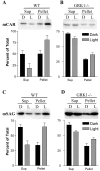
Figure 1.
Figure 1.
Light- and GRK1-dependent membrane association of mCAR. Adult WT or Grk1 -/- mice were killed either at mid-day in the light or dark-adapted overnight and killed in the dark under IR light. The retinas were dissected under room light from light-adapted mice (L) or under IR light from dark-adapted mice (D) and were homogenized. The supernatants (Sup; soluble fraction) and pellets (membrane fraction) were separated by centrifugation, and the pellets were resuspended in the same volume of buffer. Equal volumes of proteins were resolved on replicate 11.5% SDS-PAGE gels and transferred to PVDF membranes, which were detected with the mCAR (LUMIJ) or rod arrestin (mSAG, C10C10) antibodies with an ECL kit. In each panel a representative immunoblot and a histogram representing quantitative data (mean ± SEM) from at least three immunoblots are shown.
Figure 2.
Figure 2.
Characterization of anti-mouse S opsin (A) and M opsin (B) antibodies by Western blotting and immunohistochemistry. Western blot analysis was performed with normal adult C57BL/6J mouse whole retinal homogenate, and immunohistochemistry was done on C57BL/6J mouse retinal frozen sections. Cone photoreceptor cells were labeled with biotinylated peanut agglutinin and visualized with Texas Red-avidin D in red (b,e). S and M opsins were labeled with anti-S and anti-M opsin polyclonal antibodies, respectively, and visualized with fluorescein label in green (a, d). Dual immunofluorescence labeling verified both S and M opsin immunoreactivities localized to cone cells (c, f). OS, Outer segments; IS, inner segments; ONL, outer nuclear layer. Scale bar, 20 μm.
Figure 3.
Figure 3.
Localization of GRK1 in both S and M cone photoreceptors of the normal mouse retina. Adult C57 mouse retinal frozen sections were triple labeled fluorescently with the GRK1-specific monoclonal antibody D11 (A, E, I), the fluorescent nuclear dye PI (red in C, G, K), and the polyclonal anti-S opsin (B), anti-M opsin (F), or anti-mCAR (LUMIJ, J). Overlay of A and B with PI staining (C) or E and F with PI staining (G) reveals colocalization of GRK1 with both S opsins (C) and M opsins (G) in the cone outer segments, and overlay of I and J with PI staining (K) shows colocalization of GRK1 with mCAR also in the cone outer segments. A phase-contrast image (D, H, L) also is shown for each section. Note that the staining of GRK1 and S and M opsins is restricted to the outer segments (OS), whereas the staining of mCAR is diffused throughout the cone photoreceptors but condensed in the cone outer segments and the synaptic terminals. IS, Inner segments; ONL, outer nuclear layer; OPL, outer plexiform layer. Scale bar, 20 μm.
Figure 4.
Figure 4.
Localization of mCAR in both S and M cone photoreceptors of the normal mouse retina. Adult C57 mouse retinal whole mounts were double labeled immunofluorescently with either anti-S (A) or anti-M opsin antibody (D) and the anti-mCAR antibody LUMIJ (B, E). Overlay of A and B (C) or D and E (F) reveals colocalization of mCAR with both S and M opsins in the cone outer segments.
Figure 5.
Figure 5.
GRK1 and mCAR both are expressed in all photoreceptors of the Nrl -/- mouse retina. Adult Nrl-/- mouse retinal frozen sections were double labeled fluorescently with D11 (A) + anti-S opsin (B), D11 (E) + anti-M opsin (F), LUMIJ (I) + anti-S opsin (J), and LUMIJ (M) + anti-M opsin (N). Overlay of A and B (C) or E and F (G) reveals colocalization of GRK1 with both S opsins (C) and M opsins (G) in the shortouter segments, and overlay of I and J (K) or M and N (O) shows colocalization of mCAR with both S and M opsins also in the outer segments. Note that the staining of GRK1 and S and M opsins is restricted to the outer segment layer (OS), whereas the staining of mCAR is diffused throughout the photoreceptors but condensed in the outer segments and the synaptic terminals. A phase-contrast image (D, H, L, P) is shown for each section. IS, Inner segments; ONL, outer nuclear layer; OPL, outer plexiform layer. Scale bar, 20 μm.
Figure 6.
Figure 6.
In situ light-dependent phosphorylation of opsins ex vivo. A, In situ phosphorylation of opsins. WT, Nrl -/-, and Nrl -/-Grk1 -/- mice were dark-adapted overnight and killed. The retinas were dissected under IR light and incubated in phosphate-free Krebs' buffer containing 1.25 mCi/ml 32P orthophosphate for 30 min at RT in the dark. One retina of each strain was homogenized in SDS sample buffer in the dark (D) while the other retina was exposed to bright sunlight (L) for 10 min before homogenization. The proteins were resolved on an 11.5% SDS-PAGE gel and transferred to a PVDF membrane. Opsin phosphorylation was detected by autoradiography on a PhosphorImager screen. B, Immunoblot analysis of the same membrane in A, using polyclonal antibodies to mouse S or M opsin or the rhodopsin monoclonal antibody 1D4, sequentially, to observe the quantity and location of the opsins relative to the radioactive phosphate (32P) identified in A. Note the slightly higher molecular weight of the phosphorylated as compared with the unphosphorylated species of S and M opsin and rhodopsin.
Figure 7.
Figure 7.
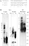
Multiple phosphorylation of S and M opsins ex vivo. A, Alignment of C-terminal sequences for mouse rhodopsin and S and M opsins. The conserved amino acids among all three opsins are indicated with an asterisk. Potential phosphorylation sites (serine and threonine residues) are underlined. B, Separation of S and M opsin and rhodopsin and their phosphorylated species by isoelectric focusing (IEF). Retinas from Nrl -/- (S and M opsins) and WT mice (rhodopsin) were dissected under infrared light, exposed to direct bright sunlight (∼8000 fc) for 10 min (L), or kept in total darkness (D). Samples were processed as described in Materials and Methods, and a fraction corresponding to one-tenth of a retina was applied per lane. The pH gradient of the gel is indicated on the right. Phosphorylated S and M opsin and rhodopsin species with an increasing number of phosphates per opsin (P/O) yielded bands at increasingly acidic pH.
Figure 8.
Figure 8.
Coimmunoprecipitation of mCAR with light-activated, phosphorylated cone opsins. A, Coimmunoprecipitation of mCAR with phosphorylated S and M opsins from light-exposed Nrl -/- mouse retinas. Nrl -/- mouse retinas were dissected under IR light from dark-adapted mice and incubated in phosphate-free Krebs' buffer containing 1.25 mCi/ml 32P orthophosphate for 30 min. Four retinas were exposed to bright sunlight (∼8000 fc) for 10 min while another four retinas remained in the dark. The retinas were homogenized in lysis buffer, and the supernatant was immunoprecipitated with rabbit polyclonal antibodies against mCAR (α-mCAR), Sopsin (α-Sopsin), Mopsin (α-Mopsin), or CRX (α-CRX). The precipitated proteins were resolved on SDS-PAGE gels and transferred to PVDF membranes. Phosphorylated opsins (32P) were detected by exposing the membranes to a PhosphorImager screen. Then the membranes were subjected to Western blot analysis with antibodies against mCAR and S and M opsins. B, Immunoprecipitation with the same antibodies as in A from the Nrl -/-Grk1 -/- mouse retinas. Note the lack of phosphorylation of either S or M opsin even after light exposure. Also, mCAR is not coimmunoprecipitated with either of the cone opsins.

Similar articles

Cited by

References

    1. Abdulaeva G, Hargrave PA, Smith WC ( 1995) The sequence of arrestins from rod and cone photoreceptors in the frogs Rana catesbeiana and Rana pipiens Localization of gene transcripts by reverse-transcription polymerase chain reaction on isolated photoreceptors. Eur J Biochem 234: 437–442. - PubMed
    1. Arshavsky V, Antoch MP, Philippov PP ( 1987) On the role of transducin GTPase in the quenching of a phosphodiesterase cascade of vision. FEBS Lett 224: 19–22. - PubMed
    1. Baylor D ( 1996) How photons start vision. Proc Natl Acad Sci USA 93: 560–565. - PMC - PubMed
    1. Baylor DA, Burns ME ( 1998) Control of rhodopsin activity in vision. Eye 12: 521–525. - PubMed
    1. Baylor DA, Lamb TD, Yau KW ( 1979) The membrane current of single rod outer segments. J Physiol (Lond) 288: 589–611. - PMC - PubMed

Publication types

MeSH terms

LinkOut - more resources