. 2003 Jul 15;22(14):3503-13.
doi: 10.1093/emboj/cdg356.

Mechanism of the electron transfer catalyst DsbB from Escherichia coli

Affiliations

Mechanism of the electron transfer catalyst DsbB from Escherichia coli

Ulla Grauschopf et al. EMBO J. .

Abstract

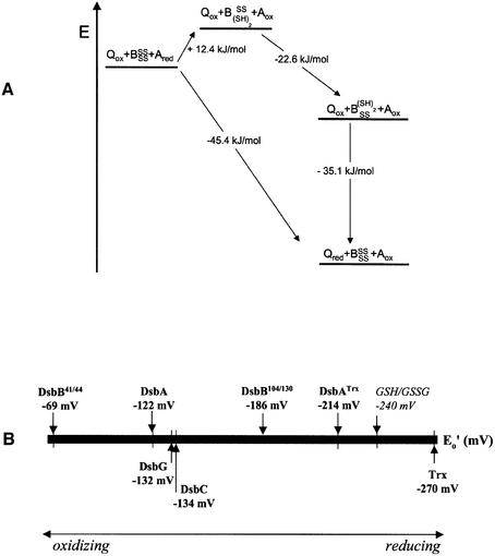
The membrane protein DsbB from Escherichia coli is essential for disulfide bond formation and catalyses the oxidation of the periplasmic dithiol oxidase DsbA by ubiquinone. DsbB contains two catalytic disulfide bonds, Cys41-Cys44 and Cys104-Cys130. We show that DsbB directly oxidizes one molar equivalent of DsbA in the absence of ubiquinone via disulfide exchange with the 104-130 disulfide bond, with a rate constant of 2.7 x 10 M(-1) x s(-1). This reaction occurs although the 104-130 disulfide is less oxidizing than the catalytic disulfide bond of DsbA (E(o)' = -186 and -122 mV, respectively). This is because the 41-44 disulfide, which is only accessible to ubiquinone but not to DsbA, is the most oxidizing disulfide bond in a protein described so far, with a redox potential of -69 mV. Rapid intramolecular disulfide exchange in partially reduced DsbB converts the enzyme into a state in which Cys41 and Cys44 are reduced and thus accessible for reoxidation by ubiquinone. This demonstrates that the high catalytic efficiency of DsbB results from the extreme intrinsic oxidative force of the enzyme.


Figures

None
Fig. 1. Spectroscopic characterization of DsbB at pH 7.0 and 25°C. (A) UV spectrum of DsbB purified according to Bader et al. (2000), before (broken line) and after (solid line) reduction of bound ubiquinone Q8 by NaBH4. (B) UV spectrum of DsbB purified according to the present protocol, which includes an additional detergent wash step to remove ubiquinone Q8, before (broken line) and after (solid line) the addition of NaBH4. (Inset) Coomassie Brilliant Blue-stained SDS gel: lane 1, purified DsbB; lane 2, molecular mass standard. (C) Fluorescence spectrum of ubiquinone-free, wild-type DsbB (2 µM) in 0.1% (w/v) DM at pH 7.0 before (solid line) and after (broken line) reduction of both disulfide bonds by DTT (170 µM).
None
Fig. 2. Redox equilibria with glutathione of wild-type DsbB, the CCSS variant, the SSCC variant, and wild-type DsbA in 0.1% DM at pH 7.0 and 25°C. Proteins (1 µM) were incubated in the presence of 1 mM GSSG and varying concentrations of GSH (0.2 µM to 11 mM), and tryptophan fluorescence at 330 nm was recorded. (A) Original fluorescence data obtained for wild-type DsbB (squares), CCSS (circles) and SSCC (diamonds). Data for wild-type DsbB and the CCSS variant were fitted according to equation 1 (see Materials and methods) (solid lines). (B) Normalized equilibrium data, showing the fractions of partially and fully reduced wild-type DsbB (squares), reduced CCSS (circles) and reduced wild-type DsbA (triangles) under different redox conditions.
None
Fig. 3. Fluorescence titrations in 0.1% (w/v) DM at pH 7.0 and 25°C, establishing the stoichiometry of the oxidation of DsbA by wild-type DsbB, and the DsbB-catalysed oxidation of DsbA by ubiquinone Q1. (A) Titration of oxidized DsbB (1 µM) with reduced DsbA. Fluorescence at 330 nm (circles) is plotted against the DsbA:DsbB ratio. The solid line describes a fit according to equation 3 (see Materials and methods) with an equilibrium constant of 62, corresponding to the difference in redox potential between DsbA and the 41–44 disulfide of DsbB. (B) Reduced DsbA (1 µM) was mixed with catalytic amounts (20 nM) of oxidized wild-type DsbB and titrated with a solution of ubiquinone Q1. Fluorescence at 330 nm (circles) is plotted against the UQ1:DsbAred ratio. The arrow at UQ1:DsbAred = 0.96 indicates the equivalence point of the titration.
None
Fig. 4. Determination of the redox potential of the 104–130 disulfide bond in the SSCC variant of DsbB at pH 7.0 and 25°C in 0.1% (w/v) DM by fluorescence titration. (A) Wild-type DsbA oxidizes the 104–130 disulfide in the SSCC variant. The reduced SSCC variant (1 µM) was titrated with oxidized wild-type DsbA, and the fluorescence at 330 nm (circles) was plotted against the DsbA:SSCC ratio. Data were fitted (solid line) according to equation 3 and were consistent with a calculated equilibrium constant of 146 (Supplementary table I). (B) Redox equilibrium between the SSCC variant and DsbA–Trx. The oxidized SSCC variant (1 µM) was titrated with reduced DsbA–Trx. The fluorescence at 330 nm (circles) was plotted against the DsbA:SSCC ratio. Data were fitted according to equation 3 (solid line) and yielded an equilibrium constant of 9.2 ± 5.2. (Inset) Fast oxidation of DsbA–Trx (0.5 µM) by equimolar amounts of the oxidized SSCC variant, measured by stopped-flow fluorescence. The solid line corresponds to a second-order fit, yielding a rate constant of 3.3 × 105 M–1 s–1. (C) Wild-type DsbB oxidizes two molar equivalents of DsbA–Trx. Oxidized wild-type DsbB (1 µM) was titrated with reduced DsbA–Trx, and the fluorescence at 330 nm (circles) was plotted against the DsbA–Trx:DsbB ratio.
None
Fig. 5. Rapid oxidation of wild-type DsbA by wild-type DsbB at pH 7.0 in 0.1% (w/v) DM, measured by stopped-flow fluorescence. (A) Oxidized DsbB and reduced DsbA were mixed at a 1:1 ratio (final concentrations: 1 µM each). The increase in fluorescence at 330 nm was recorded and data were fitted according to second-order kinetics (continuous line), yielding a rate constant of 2.7 ± 0.2 × 106 M–1 s–1. (B) Oxidation of DsbA by DsbB does not stop at the level of a DsbB–DsbA mixed disulfide. A Coomassie Brilliant Blue-stained, non-reducing SDS gel showns proteins after TCA-precipitation and modification with AMS under the same conditions that were used previously for redox potential determination and identification of intramolecular disulfide bonds between DsbA and DsbB (Inaba and Ito, 2002; Kadokura and Beckwith, 2002; Regeimbal and Bardwell, 2002). (Lanes 1–4) Reaction between oxidized DsbB and reduced wild-type DsbA; (lanes 5–8) reaction between the oxidized SSCC variant and reduced DsbA–Trx. Lane 1, oxidized DsbA; lane 2, reduced DsbA; lane 3, 1:1 mixture of reduced DsbA and oxidized wild-type DsbB; lane 4, oxidized wild-type DsbB; lane 5, oxidized DsbA–Trx; lane 6, reduced DsbA–Trx; lane 7, mixture between reduced DsbA–Trx and oxidized SSCC; lane 8, oxidized SSCC. Note that Coomassie Brilliant Blue staining of DsbB is significantly weaker compared with equimolar amounts of DsbA.
None
Fig. 6. Energy diagram of the proposed catalytic mechanism of DsbB (A). The free enthalpies (ΔG) of the individual reaction steps were calculated from the standard redox potentials according to ΔG = –2·F·ΔE, where F is the Faraday constant and ΔE corresponds to the redox potential difference between the respective species (see Supplementary table I). BSSSS corresponds to fully oxidized DsbB, BSS(SH)2 represents partially reduced DsbB in which the Cys41–44 disulfide bond is formed and Cys104 and Cys130 are reduced, and B(SH)2SS is partially reduced DsbB in which the Cys104–130 disulfide is formed and Cys41 and Cys44 are reduced. A redox potential of +0.113 V was used for ubiquinone/ubiquinol (Gennis and Stewart, 1996). (B) Scale of redox potentials of enzymes involved in disulfide bond formation in E.coli (see Sevier and Kaiser, 2002, and references therein).

Similar articles

Cited by

References

    1. Bader M., Muse,W., Zander,T. and Bardwell,J. (1998) Reconstitution of a protein disulfide catalytic system. J. Biol. Chem., 273, 10302–10307. - PubMed
    1. Bader M., Muse,W., Ballou,D.P., Gassner,C. and Bardwell,J.C. (1999) Oxidative protein folding is driven by the electron transport system. Cell, 98, 217–227. - PubMed
    1. Bader M.W., Xie,T., Yu,C.A. and Bardwell,J.C. (2000) Disulfide bonds are generated by quinone reduction. J. Biol. Chem., 275, 26082–26088. - PubMed
    1. Bardwell J.C., McGovern,K. and Beckwith,J. (1991) Identification of a protein required for disulfide bond formation in vivo. Cell, 67, 581–589. - PubMed
    1. Bardwell J.C., Lee,J.O., Jander,G., Martin,N., Belin,D. and Beckwith,J. (1993) A pathway for disulfide bond formation in vivo. Proc. Natl Acad. Sci. USA, 90, 1038–1042. - PMC - PubMed

Publication types

LinkOut - more resources