. 2003 Jul 15;22(14):3514-23.
doi: 10.1093/emboj/cdg363.

14-3-3s regulate fructose-2,6-bisphosphate levels by binding to PKB-phosphorylated cardiac fructose-2,6-bisphosphate kinase/phosphatase

Affiliations

14-3-3s regulate fructose-2,6-bisphosphate levels by binding to PKB-phosphorylated cardiac fructose-2,6-bisphosphate kinase/phosphatase

Mercedes Pozuelo Rubio et al. EMBO J. .

Abstract

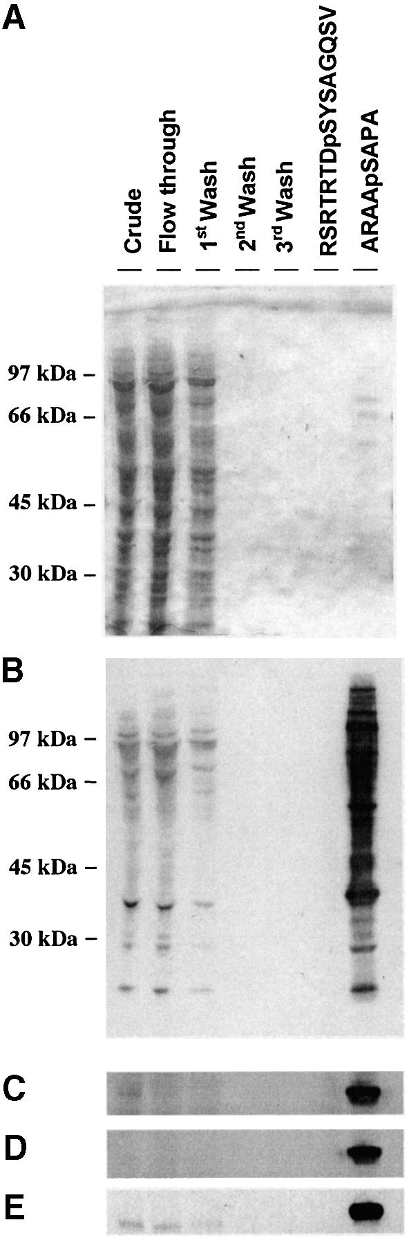
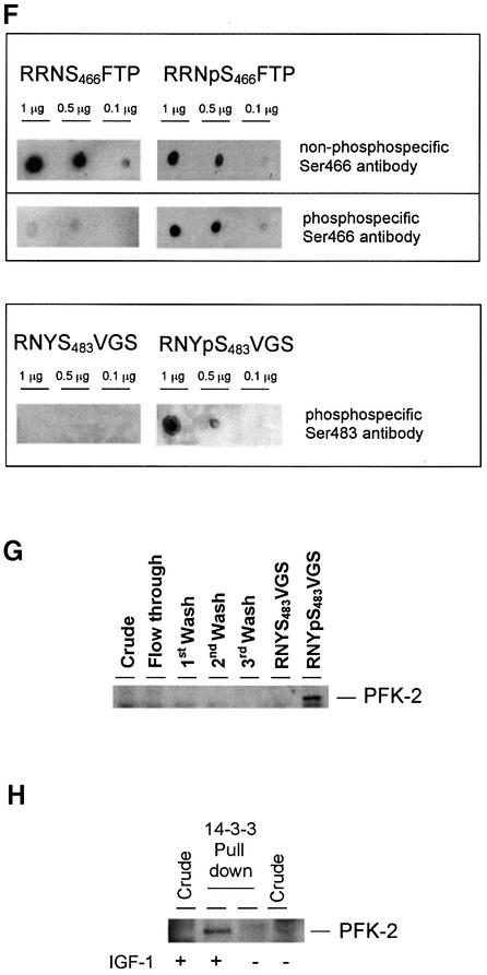
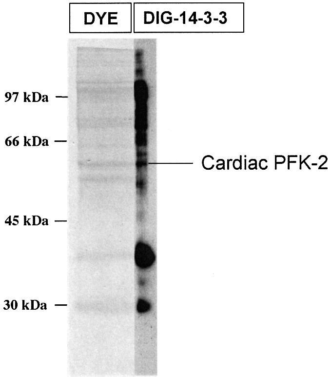
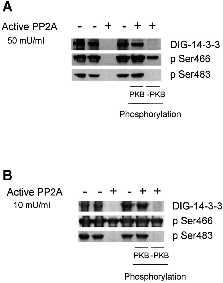
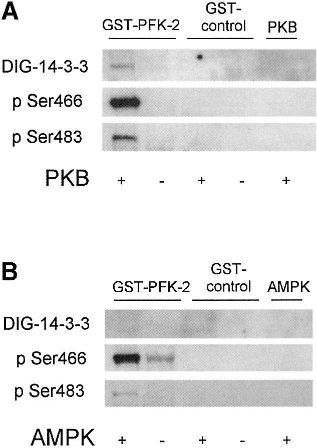
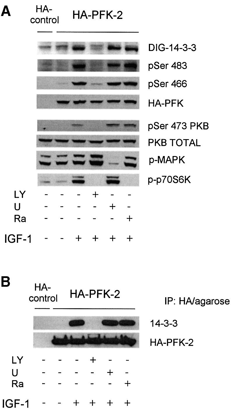
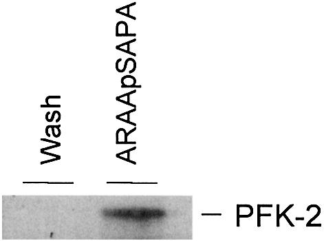
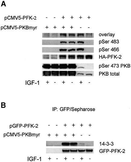
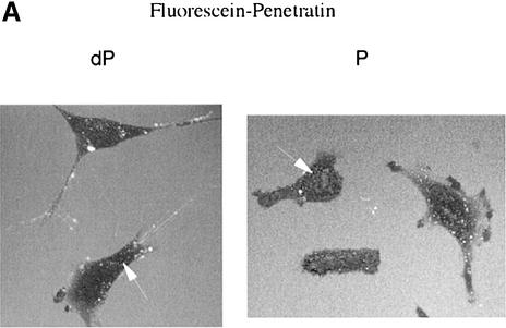
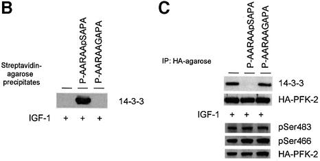
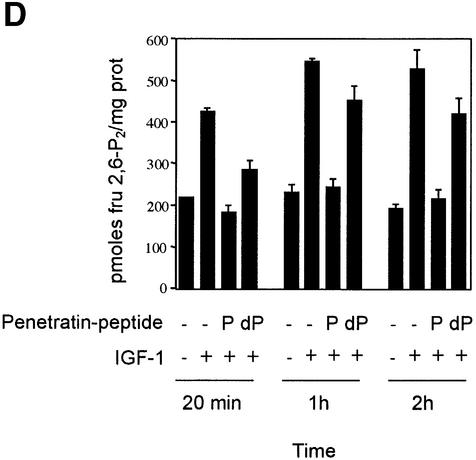
The cardiac isoform of 6-phosphofructo-2-kinase/ fructose-2,6-bisphosphatase (PFK-2), regulator of the glycolysis-stimulating fructose-2,6-bisphosphate, was among human HeLa cell proteins that were eluted from a 14-3-3 affinity column using the phosphopeptide ARAApSAPA. Tryptic mass fingerprinting and phospho-specific antibodies showed that Ser466 and Ser483 of 14-3-3-affinity-purified PFK-2 were phosphorylated. 14-3-3 binding was abolished by selectively dephosphorylating Ser483, and 14-3-3 binding was restored when both Ser466 and Ser483 were phosphorylated with PKB, but not when Ser466 alone was phosphorylated by AMPK. Furthermore, the phosphopeptide RNYpS(483)VGS blocked binding of PFK-2 to 14-3-3s. These data indicate that 14-3-3s bind to phosphorylated Ser483. When HeLa cells expressing HA-tagged PFK-2 were co-transfected with active PKB or stimulated with IGF-1, HA-PFK-2 was phosphorylated and bound to 14-3-3s. The response to IGF-1 was abolished by PI 3-kinase inhibitors. In addition, IGF-1 promoted the binding of endogenous PFK-2 to 14-3-3s. When cells were transduced with penetratin-linked AARAApSAPA, we found that this reagent bound specifically to 14-3-3s, blocked the IGF-1-induced binding of HA-PFK-2 to 14-3-3s, and completely inhibited the IGF-1-induced increase in cellular fructose-2,6-bisphosphate. These findings suggest that PKB-dependent binding of 14-3-3s to phospho-Ser483 of cardiac PFK-2 mediates the stimulation of glycolysis by growth factor.


Figures

None
Fig. 1. Phosphorylated cardiac PFK-2 is highly enriched in the specific phosphopeptide elution pool from a 14-3-3 column. An extract of serum-grown HeLa cells was chromatographed on 14-3-3–Sepharose (see Materials and methods). The following amounts of protein were run on 10% Bis-Tris SDS–PAGE and transferred to nitrocellulose: extract, flow through, and start of salt wash (30 µg); middle and end of salt wash (protein undetectable); control peptide mock elution pool (<1 µg); and ARAApSAPA pool (2 µg). Blots were analysed for protein with Ponceau S (A) and DIG-14-3-3 overlay (B). Western blotting was performed with an antibody recognizing both the seryl-phosphorylated and unphosphorylated forms of the peptide RRNSFTP, corresponding to residues 463–469 of human cardiac PFK-2 (C), phospho-specific cardiac PFK-2 (pSer466) antibody (D), and phospho-specific cardiac PFK-2 (pSer483) antibody (E). DIG-14-3-3 overlays were performed as for western blots, except that DIG-labelled 14-3-3 was used instead of primary antibody, followed by anti-DIG horseradish peroxidase-secondary antibody (Moorhead et al., 1999). (F) Different amounts of synthetic peptides were tested by dot-blot using affinity-purified antibodies that bound to both phosphorylated and unphosphorylated RRN(p)S466TFP peptides (top panel), phospho-specific antibodies against RRNpS466FTP (pSer466 antibody, middle panel), and phospho-specific antibodies against RNYpS483VGS (pSer483 antibody, bottom panel). (G) Fractions from a 14-3-3–Sepharose column where 1 mM RNYS483VGS was used as the control peptide, and 1 mM RNYpS483VGS (corresponding to the pSer483 phosphorylation site on PFK-2) was used to elute proteins. PFK-2 was identified using the same antibody as in (C). (H) Untransfected HeLa cells were serum-starved for 12 h, then stimulated for 20 min with or without 100 ng/ml IGF-1, as indicated. Cell extracts (3 mg of protein) were incubated for 1 h with 50 µl 14-3-3–Sepharose, and washed pellets were extracted with SDS sample buffer, run on SDS–PAGE and probed with the same anti-PFK-2 antibody used in (C).
None
Fig. 2. Cardiac PFK-2 is among 14-3-3 affinity-purified HeLa proteins. 14-3-3 affinity-purified proteins (200 µg) were fractionated further by Mono Q anion-exchange chromatography. Fractions that were eluted between 300 and 400 mM NaCl were run on a 10% Bis-Tris SDS–polyacrylamide gel, blotted onto a Fluorotrans membrane, and stained with sulphorhodamine B. A narrow slice of the stained lane was processed in an overlay (labelled DIG-14-3-3) to identify 14-3-3-binding proteins. The remainder of the protein bands were analysed by MALDI-TOF tryptic mass fingerprinting (see Supplementary data).
None
Fig. 3. Effects of dephosphorylation and rephosphorylation of cardiac PFK-2 in the 14-3-3 column elution pool. (A) Lanes 1 and 2 contain 2 µg of 14-3-3-binding proteins eluted with ARAApSAPA from a 14-3-3 affinity column [plus or minus 5 µM microcystin-LR (MC-LR), respectively]. For lanes 3, 5 and 6, 14-3-3-binding proteins (2 µg) were dephosphorylated with 50 mU/ml PP2A for 30 min at 30°C, and dephosphorylation was stopped with 5 µM MC-LR. Lane 4 is a control where the MC-LR and PP2A had been pre-mixed. In lanes 5 and 6, the dephosphorylated protein was rephosphorylated with 100 µM ATP/10 mM MgCl2, with (lane 5) or without (lane 6) 1 U/ml PKB for 30 min at 30°C, and the reaction was stopped with sample buffer. Samples were analysed by DIG-14-3-3 overlay and western blotting using phospho-specific cardiac PFK-2 (pSer466) and phospho-specific cardiac PFK-2 (pSer483) antibodies as indicated. (B) Identical experiment to (A), except that PP2A was used at 10 mU/ml.
None
Fig. 4. Direct binding to 14-3-3s after phosphorylation of GST-tagged cardiac PFK-2 with PKB, but not after phosphorylation with AMPK. (A) Purified GST–PFK2, GST control or no substrate were incubated in the presence and absence of PKB (1 U/ml) with MgATP for 30 min at 30°C, and analysed by DIG-14-3-3 overlays, phospho-specific cardiac PFK-2 (pSer466) antibody, and phospho-specific cardiac PFK-2 (pSer483) antibody, as indicated. (B) As for A, expect that AMPK (10 U/ml) was used in place of PKB.
None
Fig. 5. Binding of 14-3-3s to HA-PFK-2 in IGF-1-treated HeLa cells. HeLa cells were transfected with a plasmid expressing HA-PFK-2. After 16 h, cells were serum-starved for a further 12 h, then stimulated for 20 min with or without 100 ng/ml IGF-1. Where indicated, cells were incubated with LY294002 (LY; 100 µM for 1 h), U0126 (U; 10 µM for 1 h) or rapamycin (Ra; 100 nM for 30 min) prior to stimulation with IGF-1. (A) Cell extracts (30 µg) were probed for binding to DIG-14-3-3s (14-3-3 overlay), phospho-specific antibodies that recognize pSer483 and pSer466 on cardiac PFK-2, anti-HA antibodies, phospho-specific antibodies against pSer473 on PKBα, and anti-PKB-total. Only the part of the 14-3-3 overlay in the region of HA-PFK-2 is shown. (B) HA-PFK-2 and associated proteins were precipitated from cell extracts (500 µg of lysate protein) with 20 µl of anti-HA affinity matrix (Roche). The washed immunoprecipitates were resolved using SDS–PAGE, transferred to nitrocellulose and probed for binding to the K19 antibodies that recognize all seven human 14-3-3 isoforms and anti-HA antibodies. (C) The washed immunoprecipitates were probed for binding to antibodies specific for individual 14-3-3 isoforms (see Materials and methods) and anti-HA antibodies.
None
Fig. 6. Binding of endogenous 14-3-3s to endogenous PFK-2. An extract of serum-grown HeLa cells (500 mg) was passed through anti-14-3-3 antibody column (1 mg K-19 antibody bound to agarose). After washing with 500 column volumes of 0.5 M NaCl, 14-3-3-binding proteins were eluted with 1 mM ARAApSAPA. Samples of salt wash and the ARAApSAPA elution pool were concentrated, run on SDS–PAGE and transferred to nitrocellulose. Western blotting (same antibody as in Figure 1C) was used to identify endogenous PFK-2.
None
Fig. 7. Binding of 14-3-3s to HA-PFK-2 in myristylated PKB-transfected HeLa cells. HeLa cells were transfected with plasmid pCMV5-PFK-2 to express HA-PFK-2 (A), or pGFPPFK2 to express GFP–HA-PFK-2 (B). Where indicated, cells were co-transfected with a plasmid expressing PKB with a myristylation consensus site (pCMV5-PKBmyr). (A) Cell extracts (30 µg) were analysed for binding to DIG-14-3-3s (14-3-3 overlay), phospho-specific antibodies that recognize pSer483 and pSer466 on cardiac PFK-2 and anti-HA antibodies, and phospho-specific antibodies against pSer473 on PKBα and anti-PKB. (B) GFP–HA-PFK-2 and associated proteins were precipitated from cell extracts (500 µg) with 3 µg anti-GFP antibodies (20 µl of protein G–Sepharose). Washed anti-GFP immunoprecipitates were subjected to SDS–PAGE, transferred to nitrocellulose and blotted with K19 antibodies that recognize all seven human 14-3-3 isoforms and anti-GFP antibodies.
None
Fig. 8. Use of penetratin-ARAApSAPA to test the effects of disrupting 14-3-3 binding to cellular PFK-2. (A) HeLa cells were incubated with 30 µM of fluorescein-penetratin-AARAASAPA (dP) or 30 µM of fluorescein-penetratin-AARAApSAPA (P) for 1 h. Live cells were observed by fluorescence microscopy. Arrows indicate examples of green fluorescent spots in cells. (B–D) HeLa cells were transfected with the plasmid expressing HA-PFK-2, and after 16 h were serum-starved for a further 12 h, then stimulated for 20 min, 1 h or 2 h with 100 ng/ml IGF-1. Where indicated, the cells were incubated with 100 µg/ml (30 µM) biotin-penetratin-AARAApSAPA or biotin-penetratin-AAR AAGAPA for 1 h prior to stimulation with IGF-1. (B) Biotinylated peptides and associated proteins were precipitated from cell extracts (500 µg of lysate protein) with 20 µl streptavidin-agarose (Amersham-Pharmacia Biotech). Washed pellets were extracted in SDS sample buffer, subjected to SDS–PAGE, transferred to nitrocellulose and blotted with the K19 pan-14-3-3 antibodies. (C) HA-PFK-2 and associated proteins were precipitated from cell extracts (500 µg of lysate protein) with 20 µl anti-HA-agarose. Washed precipitates were subjected to SDS–PAGE, transferred to nitrocellulose and blotted with the K19 pan-14-3-3 and anti-HA antibodies (upper two panels). Lysates from each set of cells (30 µg of protein) were probed with phospho-specific antibodies that recognize pSer483 and pSer466 on cardiac PFK-2 and anti-HA antibodies (lower three panels). (D) Effect of penetratin-AARAApSAPA and penetratin-AARAAGAPA on the IGF-1-induced increase in cellular fru-2,6-P2 levels. Cells were washed, extracted and assayed for fru-2,6-P2 at the times indicated. Results represent means ± standard deviations for three separate experiments, with each assayed in duplicate.

Similar articles

Cited by

References

    1. Andjelkovic M. et al. (1997) Role of translocation in the activation and function of protein kinase B. J. Biol. Chem., 272, 31515–31524. - PubMed
    1. Bertrand L., Alessi,D.R., Deprez,J., Deak,M., Viaene,E., Rider,M.H. and Hue,L. (1999) Heart 6-phosphofructo-2-kinase activation by insulin results from Ser-466 and Ser-483 phosphorylation and requires 3-phosphoinositide-dependent kinase-1, but not protein kinase B. J. Biol. Chem., 274, 30927–30933. - PubMed
    1. Campbell D.G. and Morrice,N.A. (2002) Identification of protein phosphorylation sites by a combination of mass spectrometry and solid phase Edman sequencing. J. Biomol. Technol., 13, 119–130. - PMC - PubMed
    1. Chesney J., Mitchell,R., Benigni,F., Bacher,M., Spiegel,L., Al-Abed,Y., Han,J.H., Metz,C. and Bucala,R. (1999) An inducible gene product for 6-phosphofructo-2-kinase with an AU-rich instability element: role in tumor cell glycolysis and the Warburg effect. Proc. Natl Acad. Sci. USA, 96, 3047–3052. - PMC - PubMed
    1. Dale S., Wilson,W.A., Edelman,A.M. and Hardie,D.G. (1995) Similar substrate recognition motifs for mammalian AMP-activated protein kinase, higher plant HMG-CoA reductase kinase-A, yeast SNF1 and mammalian calmodulin-dependent protein kinase I. FEBS Lett., 361, 191–195. - PubMed

Publication types

MeSH terms