. 2003 Jul 15;22(14):3546-56.
doi: 10.1093/emboj/cdg342.

T1alpha/podoplanin deficiency disrupts normal lymphatic vasculature formation and causes lymphedema

Affiliations

T1alpha/podoplanin deficiency disrupts normal lymphatic vasculature formation and causes lymphedema

Vivien Schacht et al. EMBO J. .

Abstract

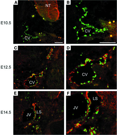
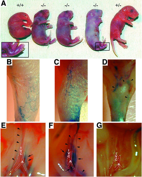
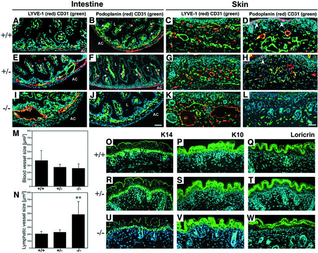
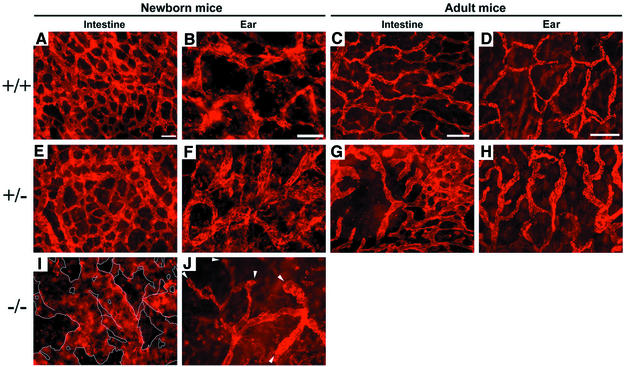
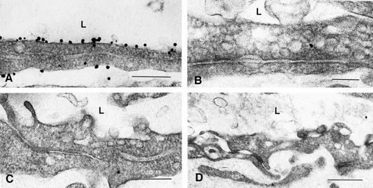
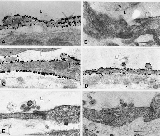
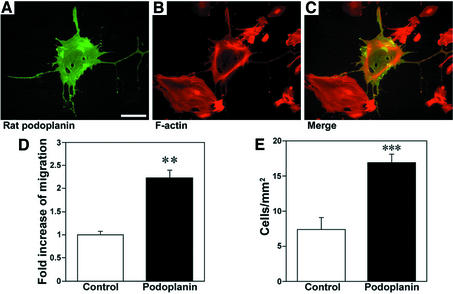
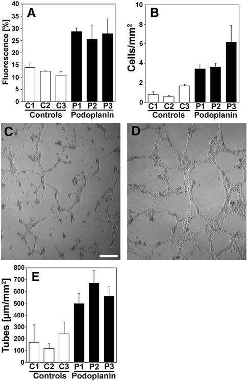
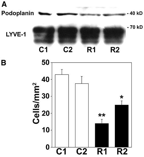
Within the vascular system, the mucin-type transmembrane glycoprotein T1alpha/podoplanin is predominantly expressed by lymphatic endothelium, and recent studies have shown that it is regulated by the lymphatic-specific homeobox gene Prox1. In this study, we examined the role of T1alpha/podoplanin in vascular development and the effects of gene disruption in mice. T1alpha/podoplanin is first expressed at around E11.0 in Prox1-positive lymphatic progenitor cells, with predominant localization in the luminal plasma membrane of lymphatic endothelial cells during later development. T1alpha/podoplanin(-/-) mice die at birth due to respiratory failure and have defects in lymphatic, but not blood vessel pattern formation. These defects are associated with diminished lymphatic transport, congenital lymphedema and dilation of lymphatic vessels. T1alpha/podoplanin is also expressed in the basal epidermis of newborn wild-type mice, but gene disruption did not alter epidermal differentiation. Studies in cultured endothelial cells indicate that T1alpha/podoplanin promotes cell adhesion, migration and tube formation, whereas small interfering RNA-mediated inhibition of T1alpha/podoplanin expression decreased lymphatic endothelial cell adhesion. These data identify T1alpha/podoplanin as a novel critical player that regulates different key aspects of lymphatic vasculature formation.


Figures

None
Fig. 1. T1α/podoplanin is expressed by Prox1-positive lymphatic progenitor cells during embryogenesis. (A and B) At day 10.5 of embryonic mouse development, Prox1 (green) is already expressed by endothelial cells predominantly located on one side of the anterior cardinal vein (CV), whereas the expression of T1α/podoplanin (red) is still restricted to the neural tube (NT). (C and D) At E12.5, T1α/podoplanin-positive endothelial cells are present throughout the anterior cardinal vein. Budding Prox1-positive lymphatic progenitor cells also express T1α/podoplanin. (E and F) At E14.5, the expression of T1α/podoplanin becomes restricted to Prox1-positive lymphatic endothelial cells of the lymph sac (LS), whereas no or only low-level expression is detected on endothelial cells of the jugular vein (JV). Bars = 50 µm.
None
Fig. 2. Congenital skin lymphedema and impaired lymphatic transport in homozygous T1α/podoplanin knockout mice. (A) Newborn wild-type (+/+) and heterozygous mice (+/–) showed no phenotypic abnormalities, whereas T1α/podoplanin-null mice (–/–) died immediately after birth due to respiratory failure. Examination of T1α/podoplanin–/– mice revealed smoothened skin texture, thickened wrinkles, particularly in the neck area, and swelling of the lower extremities (inset). (BD) Intradermal lymphatic capillaries and larger collecting lymphatic vessels of wild-type (B) and T1α/podoplanin+/– (C) mice were filled with dye after injection of Evan’s blue dye into the dorsum of the paws. In contrast, only enlarged subcutaneous lymphatic collectors (D, arrowheads) were detected in T1α/podoplanin–/– mice. After intradermal injection of Evan’s blue dye into the paws, retroperitoneal lymphatic vessels (arrowheads) and lymph nodes (arrows) were stained blue in wild-type (E) and T1α/podoplanin+/– (F) mice, but no dye was detected in the retroperitoneal lymphatics of T1α/podoplanin–/– mice (G). IVC = inferior vena cava; A = aorta.
None
Fig. 3. T1α/podoplanin deficiency leads to dilation of lymphatics, but not of blood vessels, in the skin and intestine. (A–L) Immunofluorescence stains of the ileum (A, B, E, F, I and J; L = lumen; AC = abdominal cavity) and the skin (C, D, G, H, K and L) for CD31 (green) and LYVE-1 (red, C, G and K) or T1α/podoplanin (red, B, F, J, D, H and L) revealed slightly enlarged lymphatic vessels of the submucosal plexus of T1α/podoplanin+/– mice (E), whereas T1α/podoplanin–/– mice have greatly enlarged lymphatics (I). Lacteals of wild-type and T1α/podoplanin+/– mice were LYVE-1 positive (asterisks, A and E), whereas no LYVE-1-positive lacteals were detected in T1α/podoplanin–/– mice (I). Lymphatic expression of T1α/podoplanin was confirmed in the ileum and the skin of wild-type (B and D) and T1α/podoplanin+/– (F and H) mice (asterisks = lacteals), but was not detected in T1α/podoplanin–/– mice (J and L). Staining for CD31 revealed no differences of the size of blood vessels in all of the mice. These findings were confirmed by computer-assisted image analysis which revealed a significant increase in the size of lymphatics (N), but not of blood vessels (M), in T1α/podoplanin–/– mice. Because some T1α/podoplanin-expressing basal keratinocytes were found in the epidermis of wild-type and T1α/podoplanin+/– mice (D and H; arrowheads), the comparative expression of the epidermal differentiation markers K14, K10 and loricrin was investigated (OW). No differences in the expression of these markers were seen in the three genotypes. Bars = 50 µm.
None
Fig. 4. Abnormal patterning of lymphatic capillaries in T1α/podoplanin-deficient mice. (A–H) LYVE-1 whole-mount stains of lymphatic capillaries in the ileum (A, C, E, G and I) and ear (B, D, F, H and J) of newborn (A, B, E, F, I and L) and adult mice (C, D, G and H) revealed a regular network of lymphatics (A and E) in the intestine of newborn wild-type and T1α/podoplanin+/– mice. The network patterning was completely irregular and the diameter of lymphatics was strikingly increased in T1α/podoplanin–/– mice (I). The lymphatic vessels in the ear of T1α/podoplanin–/– mice also developed an irregular network (J) with a higher number of blind beginnings of lymphatics (arrowheads) compared with the lymphatic networks in the ears of newborn wild-type and T1α/podoplanin+/– mice (B and F). In the intestine and ears of adult wild-type mice, regular networks of lymphatic vessels were found (C and D), whereas areas of enlarged lymphatics (G and H) and incomplete network patterning (G) were seen in adult T1α/podoplanin+/– mice. Bars for (A), (B), (E), (F) and (J) = 100 µm, (C) and (G) = 200 µm, (D) and (H) = 300 µm.
None
Fig. 5. Ultrastructural localization of T1α/podoplanin in murine intestinal lymphatic vessels, but not in blood vessels, by immuno-nanogold staining. (A) In newborn wild-type mice, membrane-bound T1α/podoplanin was detected at the luminal (L) side of the lymphatic endothelium in the intestine (ileum). Fewer immuno-nanogold particles were observed at the abluminal plasma membrane and no T1α/podoplanin was detected within lymphatic endothelial cells. (B) T1α/podoplanin expression was completely absent from blood vascular endothelial cells in the intestine of newborn wild-type mice. (C) Absence of specific labeling of wild-type lymphatic endothelium after omission of the primary anti-T1α/podoplanin antibody. (D) Lymphatic endothelial cells of a newborn T1α/podoplanin–/– mouse do not react with the anti-T1α/podoplanin antibody. Bars for (A) = 0.4 µm, (B) = 0.2 µm, (C) = 0.3 µm, (D) = 0.5 µm.
None
Fig. 6. Comparable ultrastructural localization of LYVE-1 in intestinal lymphatic vessels of wild-type and T1α/podoplanin–/– mice. (A) High levels of immuno-nanogold labeling for LYVE-1 were detected at both the luminal (L) and the abluminal plasma membrane of lymphatic endothelium in the ileum of newborn wild-type mice. Some labeling of the lateral plasma membranes was also observed, whereas LYVE-1 was absent from the cytoplasm. (B) LYVE-1 expression was absent from blood vascular endothelium. (C and D) Lymphatic endothelial cells in the intestine of T1α/podoplanin–/– mice also had high levels of LYVE-1 immuno-nanogold labeling of the luminal and abluminal plasma membranes. The lateral plasma membranes were also labeled, with the exception of punctate contact areas between adjacent cells (A, C and D, arrows). (E and F) Replacement of the primary LYVE-1 antibody with an unrelated rabbit IgG control resulted in the absence of lymphatic endothelial cell labeling in the ileum of wild-type (E) and of T1α/podoplanin-null mice (F). Bars for (A), (C), (D) and (E) = 0.5 µm, (B) and (F) = 0.4 µm.
None
Fig. 7. Overexpression of T1α/podoplanin in HMEC-1 cells enhances endothelial cell migration and adhesion in vitro. (AC) Stable transfection of HMEC-1 cells with rat T1α/podoplanin cDNA resulted in the production of the rat T1α/podoplanin protein (green; A and C) and in the formation of long filopodia, which were not seen in cells that did not overexpress T1α/podoplanin. Some of the rat T1α/podoplanin-expressing cells showed a marginal accumulation of F-actin bundles (red, B and C). Bar = 150 µm. (D and E) Overexpression of T1α/podoplanin significantly stimulated cell migration (D) and adhesion (E) of HMEC-1 cells. **P < 0.01; ***P < 0.001.
None
Fig. 8. Overexpression of T1α/podoplanin in EOMA cells enhances cell migration, adhesion and tube formation in vitro. (A and B) Stable transfection of EOMA cells with rat T1α/podoplanin cDNA resulted in significantly enhanced haptotactic cell migration and adhesion to type I collagen in all three clones tested (P1–P3), as compared with control clones (C1–C3). (C and D) Enhanced formation of tube-like structures by T1α/podoplanin-overexpressing EOMA cells after seeding onto Matrigel (24 h; D), as compared with control vector-transfected cells (C). Bar = 50 µm. (E) Overexpression of T1α/podoplanin significantly enhanced the formation of tube-like structures in all three T1α/podoplanin-overexpressing clones (P1–P3) as compared with control clones (C1–C3).
None
Fig. 9. Inhibition of T1α/podoplanin expression by siRNA transfection reduces human lymphatic endothelial cell adhesion to type I collagen. (A) Four days after siRNA transfection of human primary lymphatic endothelial cells, endogenous T1α/podoplanin protein levels, but not LYVE-1 levels, were decreased by two different siRNA oligonucleotides (R1 and R2), as compared with control cells (C1, control vector cDNA; C2, sham-transfected cells). (B) Human dermal lymphatic endothelial cells showed significantly reduced adhesion to type I collagen 4 days after siRNA transfection (R1 and R2), as compared with control cells (C1 and C2). *P < 0.05; **P < 0.01.

Similar articles

Cited by

References

    1. Bartos V., Kolc,J., Vanecek,R. and Malek,P. (1979) Lymph and blood enzymes and pathologic alterations in canine experimental pancreatitis after administration of benzo-pyrones. Scand. J. Gastroenterol., 14, 343–347. - PubMed
    1. Breiteneder-Geleff S., Matsui,K., Soleiman,A., Meraner,P., Poczewski,H., Kalt,R., Schaffner,G. and Kerjaschki,D. (1997) Podoplanin, novel 43-kd membrane protein of glomerular epithelial cells, is down-regulated in puromycin nephrosis. Am. J. Pathol., 151, 1141–1152. - PMC - PubMed
    1. Carmeliet P. (2000) Mechanisms of angiogenesis and arteriogenesis. Nat. Med., 6, 389–395. - PubMed
    1. Detmar M. and Hirakawa,S. (2002) The formation of lymphatic vessels and its importance in the setting of malignancy. J. Exp. Med., 196, 713–718. - PMC - PubMed
    1. Dobbs L.G., Williams,M.C. and Gonzalez,R. (1988) Monoclonal antibodies specific to apical surfaces of rat alveolar type I cells bind to surfaces of cultured, but not freshly isolated, type II cells. Biochim. Biophys Acta, 970, 146–156. - PubMed

Publication types

MeSH terms