. 2003 Jul 15;22(14):3580-90.
doi: 10.1093/emboj/cdg343.

Induction of cell death by the BH3-only Bcl-2 homolog Nbk/Bik is mediated by an entirely Bax-dependent mitochondrial pathway

Affiliations

Induction of cell death by the BH3-only Bcl-2 homolog Nbk/Bik is mediated by an entirely Bax-dependent mitochondrial pathway

Bernhard Gillissen et al. EMBO J. .

Abstract

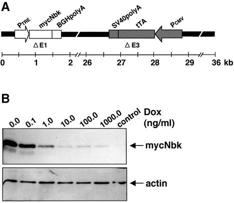
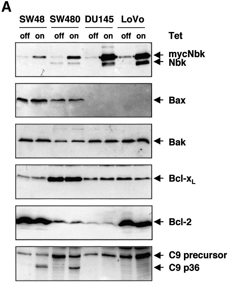
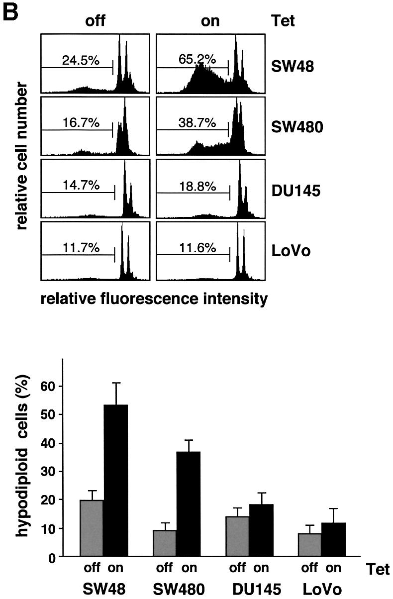
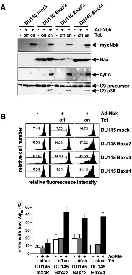
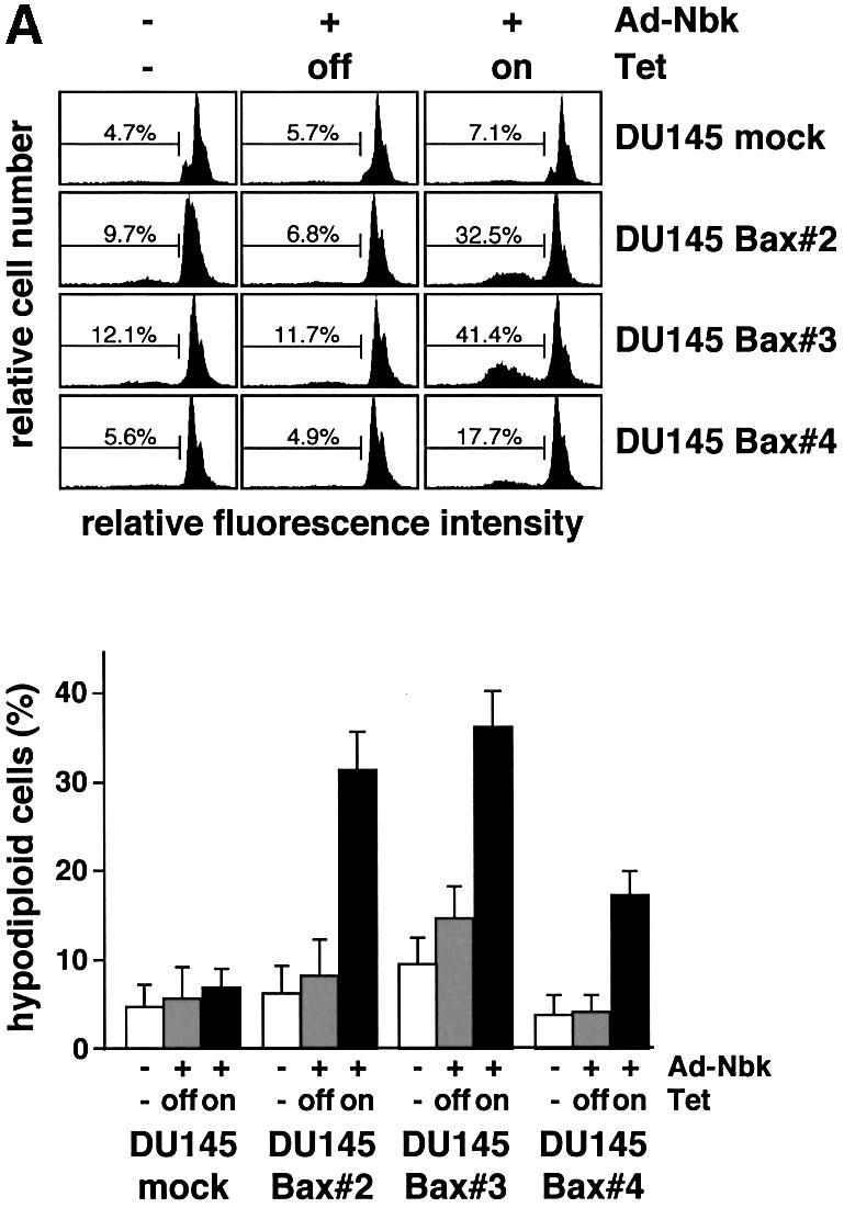
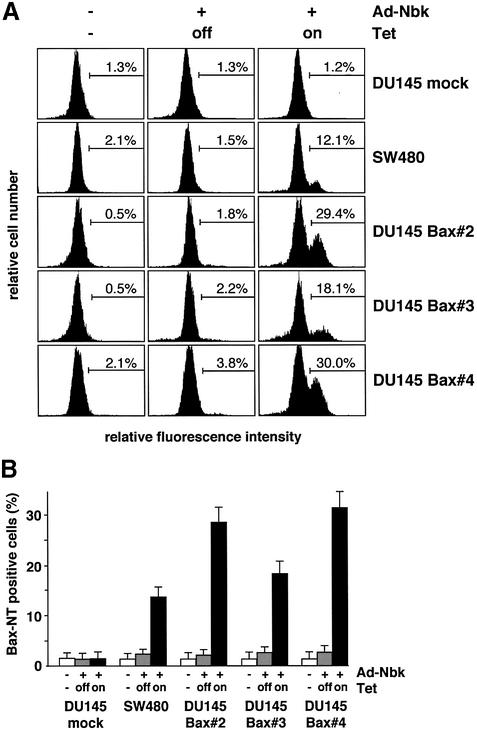
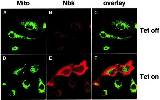
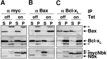
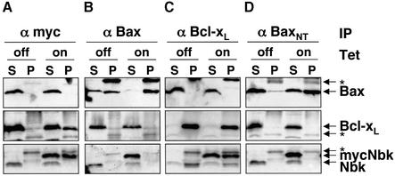
Nbk/Bik (natural born killer/Bcl-2-interacting killer) is a tissue-specific BH3-only protein whose molecular function is still largely unknown. To investigate the mechanism of Nbk action, we established a single- vector adenoviral system based on the Tet-off conditional expression of Nbk. Upon Nbk expression, only Bax-positive, but not Bax-deficient cells were found to undergo apoptosis. Interestingly, Nbk failed to induce apoptosis in the absence of Bax, even despite expression of the related molecule Bak. Re-expression of Bax restored the sensitivity to Nbk. Similarly, Bax wild-type HCT116 cells were highly susceptible, whereas HCT116 Bax knock-out cells remained resistant to Nbk-induced apoptosis. In Bax-positive cells, Nbk induced a conformational switch in the Bax N-terminus coinciding with cytochrome c release, mitochondrial permeability transition and caspase-9 processing. Immunoprecipitation studies revealed that Nbk interacts with Bcl-x(L) and Bcl-2 but not with Bax. Since, in addition, Nbk did not localize to the mitochondria, our data suggest a model in which Nbk acts as an indirect killer to trigger Bax-dependent apoptosis, whereas Bak is not sufficient to confer sensitivity to Nbk.


Figures

None
Fig. 1. Inducible Nbk expression mediated by Ad5-myc-Nbk-tTA. (A) Genomic structure of recombinant adenovirus Ad5-myc-Nbk-tTA. Ad5 sequences are indicated by black dashes. E1 and E3 regions of Ad5 are replaced by the myc-NBK expression cassette (white boxes) and tTA expression cassette (shaded boxes), respectively. [PCMV, immediate-early promoter of cytomegalovirus; tTA, tetracyclin- controlled (Tet-off) transactivator; PTRE, tetracyclin-responsive element located 5′ of the minimal immediate-early CMV promoter.] (B) Western blot analysis of Nbk expression in DU145 cells. Cells were infected with Ad5-myc-NBK-tTA at an m.o.i. of 25 (except control) and treated with increasing concentrations of doxycyclin (Dox) for 24 h. Equal protein loading was confirmed by immunoblotting using an anti-actin antibody. Strong induction of Nbk protein can be detected after doxycyclin withdrawal (Tet-on condition) whereas, in the presence of doxycyclin (Tet-off condition), Nbk expression is repressed to a weak background signal.
None
Fig. 2. Nbk expression induces apoptosis in Bax-positive but not in Bax-negative cells. Lovo, DU145, SW480 and SW48 cells were infected with Ad5-myc-NBK-tTA and cultured for 24 h in the presence (Tet-off condition) or absence of doxycyclin (Tet-on condition). (A) Western blot analysis for Nbk, Bax, Bak, Bcl-xL and Bcl-2 expression and processing of procaspase-9. (B) Flow cytometric detection of apoptotic cells based on measurement of the cellular DNA content. Upper panel: representative experiment. The percentage of hypodiploid, apoptotic cells is indicated between markers. Lower panel: means ± SD of the percentage of hypodiploid cells from three independent experiments.
None
Fig. 3. Apoptotic alterations induced by Nbk in Bax-expressing DU145 clones. DU145 clones expressing exogenous Bax were generated by retroviral transfer of the Bax cDNA under control of the CMV promoter. The DU145-mock clone was generated by transduction with control HyTK retrovirus. Stable clones were transduced with Ad5-myc-Nbk-tTA and cultured for 24 h in the presence (Tet-off condition) or absence of doxycyclin (Tet-on condition). Control cells were mock treated and grown in the absence of doxycyclin. (A) Western blot analysis of Bax and Nbk expression, cytochrome c release and procaspase-9 processing in DU145-Bax clones 24 h after infection with Ad5-myc-NBK-tTA. (B) Disruption of mitochondrial membrane potential (Δψm) by mycNbk in DU145-Bax cells. Cells were incubated with JC-1, a cationic dye that exhibits potential-dependent accumulation in mitochondria, and fluorescence intensity was measured by flow cytometry. Upper panel: representative experiment. The percentage of cells with Δψm loss is indicated between markers. Lower panel: means ± SD of the percentages of cells with Δψm loss from three independent experiments.
None
Fig. 4. Bax is required for Nbk-induced apoptosis. Stable Bax-expressing clones were transduced with Ad5-myc-Nbk-tTA as described in Figure 3 and cultured in the presence (Tet-off condition) or absence of doxycyclin (Tet-on condition). Control cells were mock treated and grown in the absence of doxycyclin. (A) Flow cytometric measurement of hypodiploid DNA. Upper panel: representative experiment. The percentage of apoptotic cells in a representative experiment was measured 24 h after adenoviral tranduction and is indicated between markers. Lower panel: means ± SD from three independent experiments. (B) Detection of phosphatidlyserine exposure: flow cytometric detection of apoptotic cells was performed by staining with annexin V–FITC to detect exposure of phosphatidylserine onto the cell surface 18 h after transduction. Cells were counterstained with propidium iodide (PI) to detect membrane damage. PI-positive cells were considered as necrotic and excluded from the analysis. The percentage of apoptotic cells is indicated between markers. Upper panel: representative experiment. Lower panel: means ± SD from three independent experiments.
None
Fig. 5. Induction of apoptosis by Nbk is entirely Bax dependent in HCT116 cells. HCT116 Bax wild-type (WT) and HCT116 Bax knock-out (k.o.) cells were transduced with Ad5-myc-Nbk-tTA and cultured for 24 h in the presence (Tet-off condition) or absence of doxycyclin (Tet-on condition). (A) Morphology of HCT116 cells. (B) Western blot analyses of Nbk, Bax, Bak expression, PARP and procaspase-9 cleavage. (C) Breakdown of the mitochondrial membrane potential. Loss of Δψm was measured on the single-cell level by the use of the cationic dye JC-1. Means ± SD were calculated from three independent experiments. (D) Apoptosis induction by Nbk. Genomic DNA fragmentation was measured on a single-cell level by determination of cells with a hypodiploid DNA content. Means ± SD were calculated from three independent experiments.
None
Fig. 6. Bax undergoes a conformational change in response to Nbk expression. SW480 cells, Bax-negative DU145 mock transfectants and DU145-Bax clones 2, 3 and 4 were transduced with Ad5-myc-NBK-tTA and cultured for 24 h in the presence (Tet-off condition) or absence of vector and doxycyclin (Tet-on condition). Control cells were grown in the absence of doxycyclin. (A) Cells were stained with a conformation-specific antibody against the Bax N-terminus and analyzed by flow cytometry. The percentage of immunostained cells is indicated between markers. (B) Bar chart showing mean ± SD of cells expressing activated Bax. Data were obtained from three independent experiments.
None
Fig. 7. Subcellular localization pattern of Nbk. Bax-expressing DU145 cells (clone 3) were transduced with Ad5-myc-Nbk-tTA. Cells were stained for Nbk protein expression after 16 h culture in the presence (Tet-off condition, AC) or absence of doxycyclin (Tet-on condition, DF). Nbk was visualized by goat-anti-Nbk followed by Alexa Fluor 594-conjugated chicken anti-goat IgG (red fluorescence; B and E). Cells were counterstained with MitoTracker Green (A and D). (C and F) The overlay of the Nbk and MitoTracker Green signals.
None
Fig. 8. Co-immunoprecipitation analyses show Nbk binding to Bcl-xL but not to Bax. SW480 cells were transduced with Ad5-myc-Nbk-tTA in the presence (Tet-off condition) or absence of doxycyclin (Tet-on condition) for 24 h. Lysates of cells solubilized in Triton X-100 buffer were immunoprecipitated with an anti-pan-Bax, anti-Bcl-xL or anti-c-Myc antibody. Immune complexes were resolved by SDS–PAGE and the presence of the respective protein was detected by immunoblotting, as indicated by arrows. Migration of the immunoglobulin light chain is indicated by an asterisk. S, supernatant; P, immunoprecipitate.
None
Fig. 9. Conformation-specific immunoprecipitation in CHAPS buffer. SW480 cells were infected with Ad5-myc-Nbk-tTA in the presence (Tet-off condition) or absence of doxycyclin (Tet-on condition) for 24 h, followed by cell lysis in CHAPS buffer. Immunoprecipitation was performed with antibodies against pan-Bax, activated Bax (conformation-specific antibody against the N-terminal epitope Bax-NT), Bcl-xL or c-myc. Immune complexes were resolved by SDS–PAGE, and the presence of each protein was detected by immunoblotting as indicated by arrows. Migration of the immunoglobulin light chain is indicated by an asterisk. S, supernatant; P, immunoprecipitate.
None
Fig. 10. Interaction between Nbk and Bcl-2. SW48 cells expressing high levels of endogenous Bcl-2 were transduced with Ad5-myc-Nbk-tTA in the presence (Tet-off condition) or absence of doxycyclin (Tet-on condition) for 24 h, followed by cell lysis in (A) Triton X-100 or (B) CHAPS buffer. Immunoprecipitation was performed with antibodies against pan-Bax, Bcl-xL, Bcl-2 or c-myc. Immune complexes were resolved by SDS–PAGE, and the presence of each protein was detected by immunoblotting as indicated by arrows. Migration of the immunoglobulin light chain is indicated by an asterisk. S, supernatant; P, immunoprecipitate.

Similar articles

Cited by

References

    1. Antonsson B., Montessuit,S., Lauper,S., Eskes,R. and Martinou,J.C. (2000) Bax oligomerization is required for channel-forming activity in liposomes and to trigger cytochrome c release from mitochondria. Biochem. J., 345, 271–278. - PMC - PubMed
    1. Borner C. (2003) The Bcl-2 protein family: sensors and checkpoints for life-or-death decisions. Mol. Immunol., 39, 615–647. - PubMed
    1. Boyd J.M. et al. (1995) Bik, a novel death-inducing protein shares a distinct sequence motif with Bcl-2 family proteins and interacts with viral and cellular survival-promoting proteins. Oncogene, 11, 1921–1928. - PubMed
    1. Cheng E.H., Wei,M.C., Weiler,S., Flavell,R.A., Mak,T.W., Lindsten,T. and Korsmeyer,S.J. (2001) Bcl-2, Bcl-xL sequester BH3 domain-only molecules preventing Bax- and Bak-mediated mitochondrial apoptosis. Mol. Cell, 8, 705–711. - PubMed
    1. Daniel P.T., Pun,K.T., Ritschel,S., Sturm,I., Holler,J., Dörken,B. and Brown,R. (1999) Expression of the death gene Bik/Nbk promotes sensitivity to drug-induced apoptosis in corticosteroid-resistant T-cell lymphoma and prevents tumor growth in severe combined immunodeficient mice. Blood, 94, 1100–1107. - PubMed

Publication types

MeSH terms