. 2003 Jul 15;22(14):3613-23.
doi: 10.1093/emboj/cdg362.

Cofactor Tpr2 combines two TPR domains and a J domain to regulate the Hsp70/Hsp90 chaperone system

Affiliations

Cofactor Tpr2 combines two TPR domains and a J domain to regulate the Hsp70/Hsp90 chaperone system

Alexander Brychzy et al. EMBO J. .

Abstract

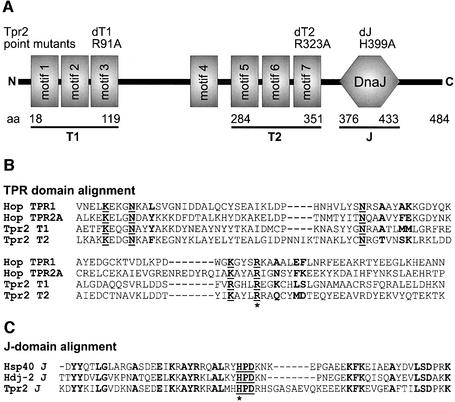
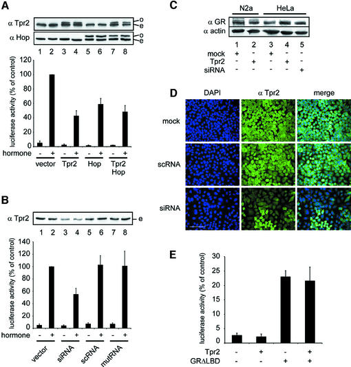
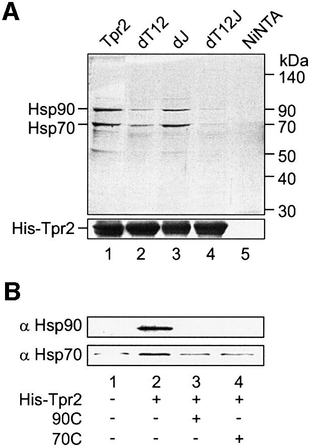
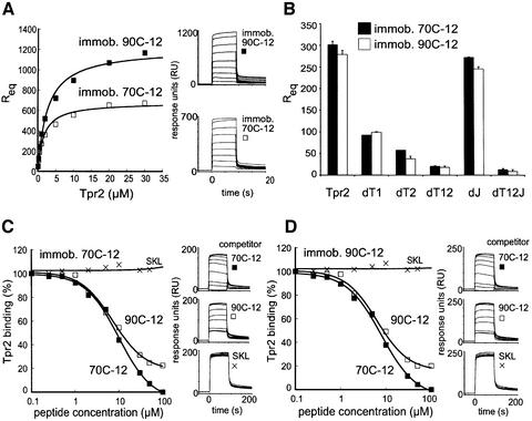
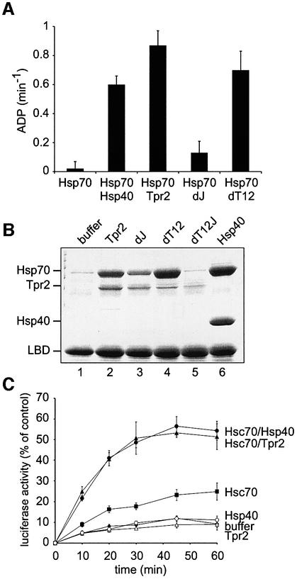
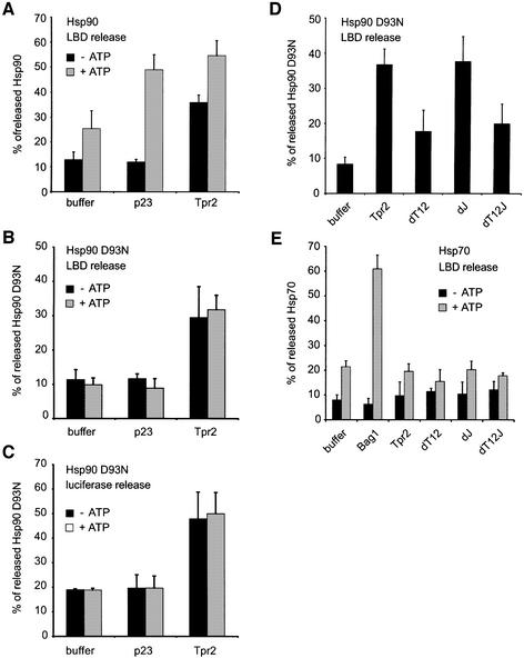
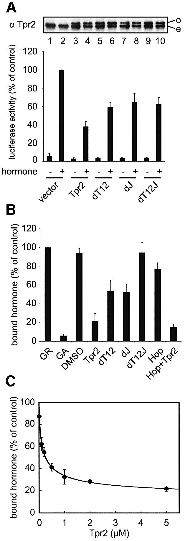
In the eukaryotic cytosol, Hsp70 and Hsp90 cooperate with various co-chaperone proteins in the folding of a growing set of substrates, including the glucocorticoid receptor (GR). Here, we analyse the function of the co-chaperone Tpr2, which contains two chaperone-binding TPR domains and a DnaJ homologous J domain. In vivo, an increase or decrease in Tpr2 expression reduces GR activation, suggesting that Tpr2 is required at a narrowly defined expression level. As shown in vitro, Tpr2 recognizes both Hsp70 and Hsp90 through its TPR domains, and its J domain stimulates ATP hydrolysis and polypeptide binding by Hsp70. Furthermore, unlike other co-chaperones, Tpr2 induces ATP-independent dissociation of Hsp90 but not of Hsp70 from chaperone-substrate complexes. Excess Tpr2 inhibits the Hsp90-dependent folding of GR in cell lysates. We propose a novel mechanism in which Tpr2 mediates the retrograde transfer of substrates from Hsp90 onto Hsp70. At normal levels substoichiometric to Hsp90 and Hsp70, this activity optimizes the function of the multichaperone machinery.


Figures

None
Fig. 1. Prediction of Tpr2 structural domains. (A) Domain prediction revealed seven tetratricopeptide motifs in human Tpr2 (motifs 1–7). Two sets of three motifs were predicted to form dicarboxylate clamp domains (T1 and T2). A DnaJ homology domain (J) near the C-terminus was also identified. The boundaries of the predicted domains are indicated as amino acid residue numbers. Introduced point mutations (dT1, dT2 and dJ) are listed with the respective amino acid exchange. (B) Sequences of the predicted T1 and T2 domains in Tpr2 were aligned against the Hsp70- and Hsp90-binding dicarboxylate clamp domains of Hop, TPR1 and TPR2A, respectively. Conserved residues that participate in the formation of the dicarboxylate clamp are underlined in boldface. Residues in boldface alone determine the specificity of chaperone binding. The arginine residues mutated to alanine in the dT1 and dT2 point mutants are marked with an asterisk. (C) The sequence of the predicted J domain in Tpr2 was aligned against the J domains of Hsp40 and Hdj-2. The functional HPD motif is underlined in boldface. Conserved residues are marked in boldface. The histidine residue mutated to alanine in the dJ point mutant is marked with an asterisk.
None
Fig. 2. Perturbation of Tpr2 expression levels reduce glucocorticoid receptor (GR) activation in vivo. Cells were transfected with a plasmid encoding a luciferase reporter gene downstream of a GR response element (GRE) and a control plasmid encoding β-galactosidase. Cells were treated for 24 h with 1 µM dexamethasone where indicated and harvested. Cell lysates were tested for luciferase activity and normalized against β-galactosidase activity. Samples were immunoblotted with antibodies against Tpr2, Hop or GR. In all figures, error bars show standard deviations from the mean of at least three independent experiments. (A) Empty vector (lanes 1–2) or vectors encoding myc-tagged Tpr2 (lanes 3–4), myc-tagged Hop (lanes 5–6) or both Tpr2 and Hop (lanes 7–8) were co-transfected into N2A cells together with the reporter and control plasmids. Top, immunoblot with antibodies against Tpr2 or Hop; transfected overexpressed myc-tagged proteins (o) are visible as bands above endogenous species (e). Bottom, GR-activated normalized luciferase expression in cells under conditions indicated. Columns correspond to the above immunoblot. (B) A double-stranded siRNA oligomer against the Tpr2 RNA was co-transfected into HeLa cells with the reporter and control plasmids (lanes 3–4). Control experiments included either an empty vector (lanes 1–2) or double-stranded RNA oligomers with a scrambled sequence (scRNA, lanes 5–6) or mutated sequence (mutRNA, lanes 7–8). Top, immunoblot against endogenous Tpr2 (e). Bottom, normalized GR-activated luciferase expression under indicated conditions. Columns correspond to the above immunoblot. (C) The indicated total cell lysates were resolved on SDS–PAGE and immunoblotted for GR, and for actin as a loading control. (D) Immunofluorescence of cells treated with siRNA against Tpr2 or control cells. Nuclei are stained with DAPI. Scale bar represents 100 µm. (E) Empty vector or vectors encoding Tpr2, GRΔLBD or both vectors were co-transfected with the reporter and control plasmids. Normalized GR-activated luciferase expression under indicated conditions are plotted.
None
Fig. 3. Hsp90 and Hsp70 are the two major interaction partners of Tpr2. (A) Purified His-tagged wild-type (WT) Tpr2, Tpr2 point mutated in the TPR clamp domains (dT12) or J-domain (dJ), or with the combination of point mutations (dT12J) were tested for binding to reticulocyte lysate (RL) proteins. The indicated proteins at 10 µM concentration were incubated with RL, in parallel with a control reaction with no added protein (Ni-NTA). Complexes were recovered with Ni-NTA agarose and bound proteins eluted with 500 mM NaCl (top). Tpr2 proteins were re-eluted with SDS sample buffer containing 25 mM EDTA (bottom). Samples were resolved by SDS–PAGE and visualized by Coomassie blue staining. The two major bands eluting from WT Tpr2 were identified by immunoblotting as Hsp90 and Hsp70. The position of molecular weight standards is marked on the right. (B) 10 µM Tpr2 together with 50 µM of the C-terminal fragments of Hsp90 (90C, lane 3) or Hsp70 (70C, lane 4) were present during the binding reaction. After recovery with Ni-NTA agarose and elution with 500 mM NaCl, eluted proteins were resolved on SDS–PAGE and detected by immunoblotting against Hsp90 and Hsp70.
None
Fig. 4. Quantitative analysis of the Tpr2–chaperone interactions. 12mer peptides containing the C-terminal sequence of either Hsp70 or Hsp90 (70C-12 and 90C-12 respectively) were covalently coupled to a Biacore chip. Various concentrations of Tpr2 and its mutants were injected and the association and dissociation monitored by the surface plasmon resonance (SPR) signal. (A) Binding kinetics of Tpr2 in the concentration range of 0.1–30 µM were monitored (right panel) with immobilized 90C-12 or 70C-12. The relative response units during the equilibrium phase of binding to 90C-12 or 70C-12 were plotted against Tpr2 concentrations (left panel). (B) Binding efficiency to 70C-12 or 90C-12 of wild-type (WT) or indicated point mutants of Tpr2 at a constant protein concentration of 1 µM was tested. The relative response units during equilibrium binding were plotted. (C and D) Increasing concentrations (0.1–100 µM) of 70C-12 or 90C-12 in solution were used to compete for binding of Tpr2 to immobilized 70C-12 (C) or 90C-12 (D). A control peptide terminating in SKL, which is recognized by the TPR domain of Pex5p, but not by Hop, was also tested. Binding kinetics were monitored (right panels) and Tpr2 binding as a percentage of the control without soluble peptides was plotted against soluble peptide concentration (left panels).
None
Fig. 5. The Tpr2 J domain regulates Hsp70. (A) 1 µM Hsc70/Hsp70, 2 µM Hsp40 and 2 µM wild-type or mutant Tpr2 in the indicated combinations were incubated at 30°C in the presence of [α-32P]ATP and 0.1 mM ATP. Aliquots of each reaction were stopped at different time points with 25 mM EDTA, resolved by thin layer chromatography and evaluated by PhosphorImager scanning. Steady-state ATPase rates were calculated from the linear range of the reactions. (B) Purified, partially-folded GR ligand binding domain (LBD) was bound to Ni-NTA agarose and beads were incubated for 10 min at room temperature with 5 µM Hsc70/Hsp70 and either no added protein (buffer), 5 µM wild-type (WT) or point mutated Tpr2, or Hsp40. Beads were recovered and bound proteins eluted with SDS sample buffer. Samples were resolved by SDS–PAGE and visualized by Coomassie blue staining. (C) Guanidine-denatured luciferase was diluted 100-fold into reactions containing 3% RL and ATP, supplemented with buffer or 1 µM Hsc70/Hsp70 with 2 µM Hsp40 or Tpr2 where indicated. Luciferase refolding at 30°C was monitored over time and plotted as a percentage of the activity of native luciferase.
None
Fig. 6. Tpr2 dissociates Hsp90 but not Hsp70 from substrate polypeptide complexes. Partially-folded myc-His-tagged ligand binding domain (LBD) or guanidine-denatured myc-tagged luciferase were pre-bound to anti-myc antibodies covalently coupled to protein G–Sepharose. Radiolabelled wild-type Hsp90, or the Hsp90 D93N point mutant unable to bind ATP, or Hsp70 was generated by in vitro translation. The translation reactions were added to reticulocyte lysate (RL) containing ATP and chaperone–substrate complexes were formed for 10 min at 25°C and then immune-isolated. The dissociation of radiolabelled chaperones was assayed under different conditions. After 10 min of dissociation at 25°C, beads and supernatants were separated and proteins in the supernatants precipitated. Released and bound proteins were resolved by SDS–PAGE and quantified by PhosphorImager analysis. The fraction of radiolabelled chaperones released from the complexes was plotted as a percentage of the total. (A) Dissociation of wild-type Hsp90 from LBD was measured in the presence of buffer alone, or 5 µM p23 or Tpr2, without or with 2 mM ATP. (B) Dissociation of Hsp90 D93N from LBD was measured in the presence of buffer alone, or 5 µM p23 or Tpr2, without or with 2 mM ATP. (C) Dissociation of Hsp90 D93N from luciferase was measured in the presence of buffer alone, or 5 µM p23 or Tpr2, without or with 2 mM ATP. (D) Dissociation of Hsp90 D93N from LBD was measured in the presence of buffer alone, or 5 µM wild-type or point mutated Tpr2. (E) Dissociation of Hsp70 was measured in the presence of buffer alone, or 5 µM Bag-1, or 5 µM wild-type or point mutated Tpr2.
None
Fig. 7. The different domains of Tpr2 cooperate to regulate the Hsp70/Hsp90 machinery. (A) Glucocorticoid receptor (GR) activation in N2A cells with or without dexamethasone was determined as in Figure 2A. Empty vector, vectors encoding wild-type or mutant Tpr2 were co-transfected with the reporter and control plasmids. Top, immunoblot against overexpressed (o) and endogenous (e) Tpr2 forms. Bottom, normalized GR-activated luciferase expression under indicated conditions. (B) Full-length GR was added to reticulocyte lysate (RL) supplemented with various purified proteins and 2 mM ATP. Reactions were incubated at 42°C for 5 min to unfold the GR, then at 30°C for 15 min to allow chaperone-mediated refolding of the GR. [3H]Dexamethasone was allowed to bind the GR, unbound hormone was removed by fast gel filtration and bound hormone quantified by scintillation counting. Hormone binding by GR refolded in RL with no additions was set to 100%. As a negative control, RL was treated with 40 µM GA to inhibit Hsp90, or with an equivalent volume of DMSO as a solvent control. Other reactions were supplemented with 2 µM wild-type, mutant Tpr2 or Hop. (C) Hormone binding by GR after refolding in RL supplemented with increasing concentrations of wild-type Tpr2 was measured and plotted against the concentrations of Tpr2 added.

Similar articles

Cited by

References

    1. Brinker A., Scheufler,C., Von Der Mulbe,F., Fleckenstein,B., Herrmann,C., Jung,G., Moarefi,I. and Hartl,F.U. (2002) Ligand discrimination by TPR domain. Relevance and selectivity of EEVD-recognition in Hsp70–Hop–Hsp90 complexes. J. Biol. Chem., 277, 19265–19275. - PubMed
    1. Buchner J. (1999) Hsp90 & Co.—a holding for folding. Trends Biochem. Sci., 24, 136–141. - PubMed
    1. Bukau B. and Horwich,A. (1998) The Hsp70 and Hsp60 chaperone machines. Cell, 92, 351–366. - PubMed
    1. Chen S. and Smith,D.F. (1998) Hop as an adaptor in the heat shock protein 70 (Hsp70) and Hsp90 chaperone machinery. J. Biol. Chem., 273, 35194–35200. - PubMed
    1. Dittmar K.D., Demady,D.R., Stancato,L.F., Krishna,P. and Pratt,W.B. (1997) Folding of the glucocorticoid receptor by the heat shock protein (hsp) 90-based chaperone machinery. The role of p23 is to stabilize receptor–hsp90 heterocomplexes formed by hsp90–p60–hsp70. J. Biol. Chem., 272, 21213–21220. - PubMed

Publication types

MeSH terms