. 2003 Jul 15;22(14):3705-14.
doi: 10.1093/emboj/cdg347.

Stimulation of poly(A) polymerase through a direct interaction with the nuclear poly(A) binding protein allosterically regulated by RNA

Affiliations

Stimulation of poly(A) polymerase through a direct interaction with the nuclear poly(A) binding protein allosterically regulated by RNA

Yvonne Kerwitz et al. EMBO J. .

Abstract

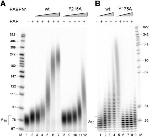
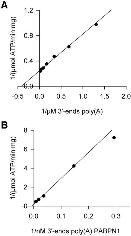
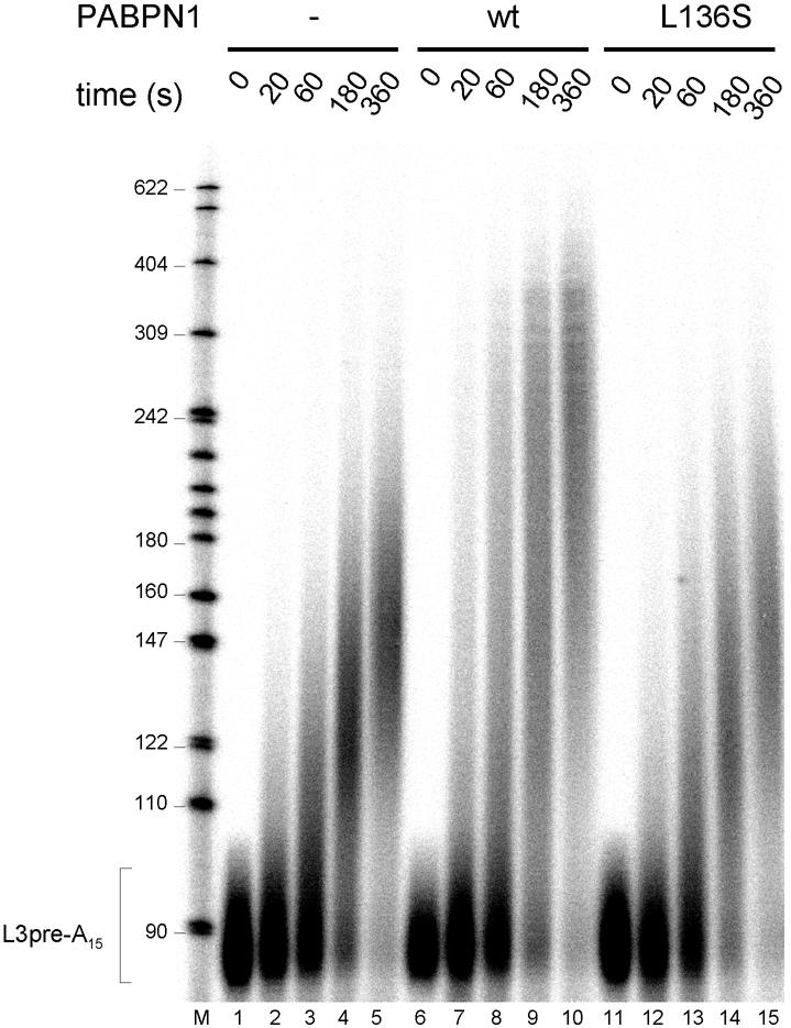
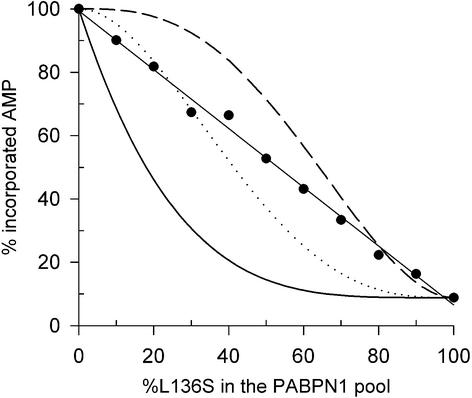
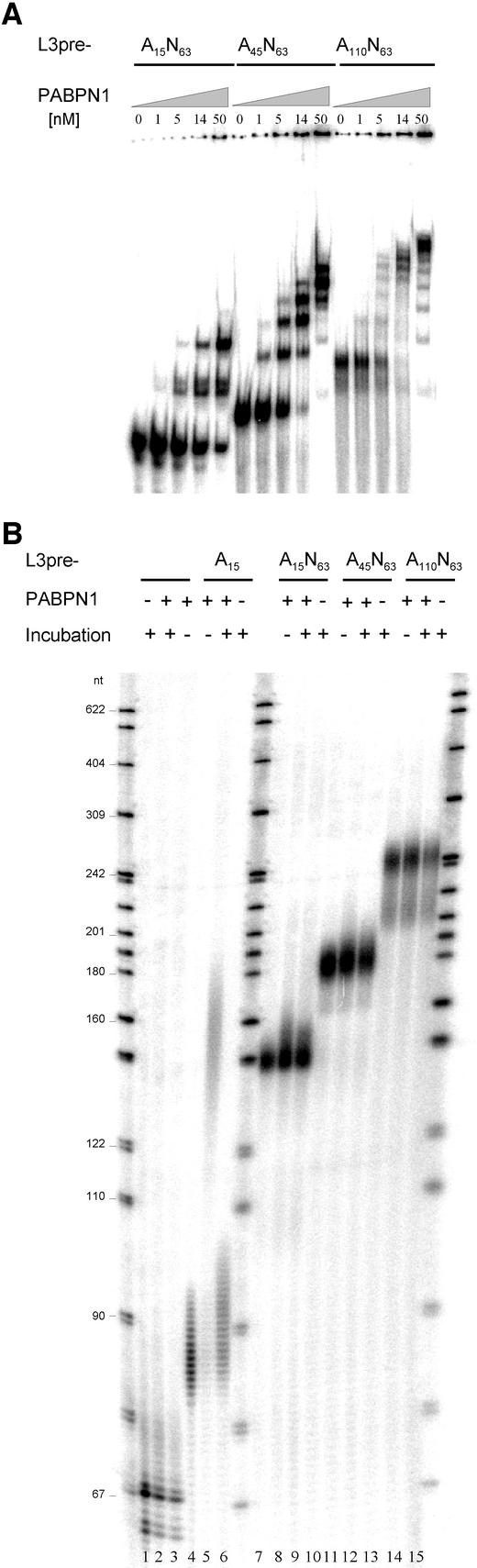
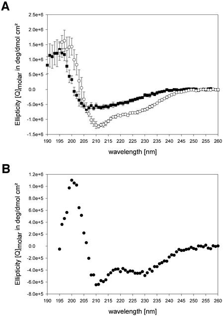
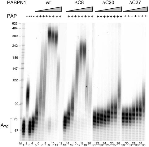
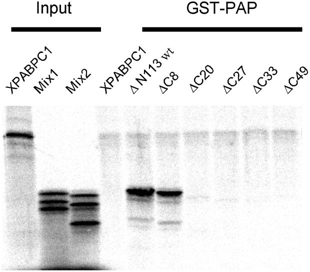
During polyadenylation of mRNA precursors in metazoan cells, poly(A) polymerase is stimulated by the nuclear poly(A) binding protein PABPN1. We report that stimulation depends on binding of PABPN1 to the substrate RNA directly adjacent to poly(A) polymerase and results in an approximately 80-fold increase in the apparent affinity of poly(A) polymerase for RNA without significant effect on catalytic efficiency. PABPN1 associates directly with poly(A) polymerase either upon allosteric activation by oligo(A) or, in the absence of RNA, upon deletion of its N-terminal domain. The N-terminal domain of PABPN1 may function to inhibit undesirable interactions of the protein; the inhibition is relieved upon RNA binding. Tethering of poly(A) polymerase is mediated largely by the C-terminal domain of PABPN1 and is necessary but not sufficient for stimulation of the enzyme; an additional interaction dependent on a coiled-coil structure located within the N-terminal domain of PABPN1 is required for a productive interaction.


Figures

None
Fig. 1. Stimulation of poly(A) polymerase by PABPN1 mutants with reduced affinity for RNA. (A) Eighty fmol of 5′-labeled poly(A) (average chain length 80 nucleotides) were elongated by 50 fmol of poly(A) polymerase for 15 min in the presence of wild-type PABPN1 or the F215A mutant as indicated and analyzed on a denaturing polyacrylamide gel. Lane 1, incubation without poly(A) polymerase. Lane 2, reaction in the absence of PABPN1, lanes 3–7 and 8–12, reactions containing 100, 200, 400, 800 and 1600 fmol of PABPN1. Sizes of DNA markers (lane M) are indicated on the left. PAP, poly(A) polymerase. (B) 5′-labeled oligo(A) (∼25 nucleotides) was elongated in the presence of 200, 400, 800 and 1600 fmol of wild-type PABPN1 or Y174A mutant. Incubation was for 30 min.
None
Fig. 2. Kinetic constants of poly(A) polymerase. Reaction rates of poly(A) polymerase were determined by measuring the incorporation of [α-32P]ATP into RNA as described in Materials and methods. (A) Double-reciprocal plot of reaction rates in the presence of increasing amounts of unfractionated poly(A). (B) Double-reciprocal plot of reaction rates in the presence of increasing amounts of unfractionated poly(A) saturated with PABPN1.
None
Fig. 3. The helical region in the N-terminus of PABPN1 is necessary for poly(A) polymerase stimulation. (A) Heptad repeat representation of the helical region (amino acids 119–147). Hydrophobic amino acids are shaded gray, amino acids in positions a and d are boxed. (B) Polyadenylation with N-terminal truncation mutants of PABPN1: radioactively labeled A80 was incubated for 15 min with poly(A) polymerase (15 fmol) in the presence of PABPN1 as indicated and analyzed on a denaturing polyacrylamide gel. Lane 1, incubation without poly(A) polymerase (PAP); lanes 2–6, incubations with 0, 100, 200, 400 and 800 fmol of wild-type PABPN1; lanes 7–9, incubations with 200, 400 and 800 fmol of ΔN160; lanes 10–15, incubations with 50, 100, 200, 400, 800 and 1600 fmol of ΔN113. Sizes of DNA markers (lane M) are indicated on the left. A reduction of the extent of stimulation at high concentrations of PABPN1 (lanes 5, 6 and 13–15) is usually seen in such assays (for example, Wahle, 1995) and may reflect covering of the RNA to the extent that poly(A) polymerase cannot get access. (C) Polyadenylation of radioactively labeled A80 by 9 fmol of poly(A) polymerase for 15 min. PABPN1 wt, and the substitution mutants A133S, V143A and L136S were added as indicated. Lane 1, poly(A) substrate; lane 2, incubation with poly(A) polymerase; lanes 3–6, 8–12, 13–17, 18–21, incubations with 100, 200, 400, 800 and 1600 fmol of the respective PABPN1 variant. Products were analyzed on a denaturing polyacrylamide gel. Sizes of DNA markers (lane M) are indicated on the left.
None
Fig. 4. PABPN1 L136A is inactive in processive polyadenylation. Reaction mixtures contained 80 fmol of L3pre-A15, 12 fmol of poly(A) polymerase and 120 fmol of CPSF. Wild-type PABPN1 or L136S mutant (300 fmol) was added to the reactions shown in lanes 6–10 and 11–15. After preincubation for 2 min at 37°C, polyadenylation was initiated by the addition of ATP. Reactions were stopped at the time points indicated. RNAs were recovered and analyzed by denaturing gel electrophoresis.
None
Fig. 5. Activity of poly(A) polymerase at different ratios of wild-type PABPN1 to L136S mutant. A95 (1.7 pmol, 3′-ends) were incubated with 0.2 pmol of poly(A) polymerase and 8.5 pmol of wild-type PABPN1 and L136S in different ratios as indicated. Reactions were pre-warmed, started by addition of [α-32P]ATP and incubated for 30 min. The reactions were stopped by TCA precipitation, and the incorporated radioactivity was determined. The data are averaged from two independent experiments. Incorporation in the presence of only wild-type PABPN1 was set to 100%. Statistical model: poly(A) polymerase activity was calculated using the Bernoulli distribution for three different models in which random replacement of one (black line), two (dotted line) or three (dashed line) PABPN1 molecules by L136S reduces the activity of poly(A) polymerase to 8.9% of that in the presence of only wild-type PABPN1.
None
Fig. 6. PABPN1 cannot stimulate poly(A) polymerase from internal binding sites. (A) Binding of PABPN1 to different RNA substrates (80 fmol each) in the presence of 1.25 µg of tRNA was measured by native gel shift assays. RNA was briefly heated to 95°C and chilled on ice before it was added to the binding reaction. Circumstantial evidence indicates that the double band observed in lanes 2–5 is due to conformational heterogeneity of the RNA. It is almost certainly not due to the binding of different numbers of proteins. (B) Specific polyadenylation assays using the same RNA substrates as in (A). Reactions contained 80 fmol of RNA, 20 fmol of poly(A) polymerase and 350 fmol of PABPN1, as indicated. RNA was heated as in (A). Incubation time was 30 min. Products were analyzed on a denaturing polyacrylamide gel.
None
Fig. 7. CD spectra of PABPN1 fragments. (A) The CD spectra of the RNP domain (rectangles) and the α-RNP variant (circles) are shown. (B) Difference spectrum calculated from (A).
None
Fig. 8. The C-terminus of PABPN1 is necessary for stimulation of poly(A) polymerase. Polyadenylation reactions containing radioactively labeled A70 (40 fmol of 3′-ends) and poly(A) polymerase (30 fmol) were incubated with increasing amounts of wild-type or C-terminal truncation variants of PABPN1 as indicated. Lane 1, untreated A70; lane 2, incubation with a 10-fold higher amount of poly(A) polymerase; lanes 3–11, 13–20, 21–28, 29–35, reactions with 0, 100, 200, 500, 1000, 2000, 5000 and 10000 fmol of PABPN1 variants, as indicated. The reaction in the presence of 100 fmol of PABPN1-ΔC27 was left out. Products were analyzed on a denaturing polyacrylamide gel.
None
Fig. 9. The C-terminus of PABPN1 is necessary for poly(A) polymerase binding. GST–PAP was immobilized on GST–Sepharose beads and incubated with 35S-labeled PABPN1-ΔN113 and variants in which the ΔN113 mutation was combined with progressive C-terminal truncations. Labeled cytoplasmic poly(A) binding protein from Xenopus laevis was included as a negative control. Bound proteins as well as 10% of the protein input (mix1: ΔN113, ΔN113-ΔC20, ΔN113-ΔC33 and mix2: ΔN113-ΔC8, ΔN113-ΔC27, ΔN113-ΔC49) were analyzed on a 13% SDS–polyacrylamide gel.

References

    1. Bienroth S., Wahle,E., Suter-Crazzolara,C. and Keller,W. (1991) Purification and characterisation of the cleavage and polyadenylation specificity factor involved in the 3′ processing of messenger RNA precursors. J. Biol. Chem., 266, 19768–19776. - PubMed
    1. Bienroth S., Keller,W. and Wahle,E. (1993) Assembly of a processive messenger RNA polyadenylation complex. EMBO J., 12, 585–594. - PMC - PubMed
    1. Böhm G., Muhr,R. and Jaenicke,R. (1992) Quantitative analysis of protein far UV circular dichroism spectra by neural networks. Protein Eng., 5, 191–195. - PubMed
    1. Bradford M.M. (1976) A rapid and sensitive method for the quantitation of microgram quantities of protein utilizing the principle of protein-dye binding. Anal. Biochem., 72, 248–254. - PubMed
    1. Chen Z., Li,Y. and Krug,R.M. (1999) Influenza A virus NS1 protein targets the poly(A) -binding protein II of the cellular 3′-end processing machinery. EMBO J., 18, 2273–2283. - PMC - PubMed

Publication types