. 2003 Jul 15;22(14):3715-24.
doi: 10.1093/emboj/cdg340.

Early mitotic degradation of the homeoprotein HOXC10 is potentially linked to cell cycle progression

Affiliations

Early mitotic degradation of the homeoprotein HOXC10 is potentially linked to cell cycle progression

Davide Gabellini et al. EMBO J. .

Abstract

Hox proteins are transcription factors involved in controlling axial patterning, leukaemias and hereditary malformations. Here, we show that HOXC10 oscillates in abundance during the cell cycle, being targeted for degradation early in mitosis by the ubiquitin-dependent proteasome pathway. Among abdominal-B subfamily members, the mitotic proteolysis of HOXC10 appears unique, since the levels of the paralogous HOXD10 and the related homeoprotein HOXC13 are constant throughout the cell cycle. When two destruction box motifs (D-box) are mutated, HOXC10 is stabilized and cells accumulate in metaphase. HOXC10 appears to be a new prometaphase target of the anaphase-promoting complex (APC), since its degradation coincides with cyclin A destruction and is suppressed by expression of a dominant-negative form of UbcH10, an APC-associated ubiquitin-conjugating enzyme. Moreover, HOXC10 co-immunoprecipitates the APC subunit CDC27, and its in vitro degradation is reduced in APC-depleted extracts or by competition with the APC substrate cyclin A. These data imply that HOXC10 is a homeoprotein with the potential to influence mitotic progression, and might provide a link between developmental regulation and cell cycle control.


Figures

None
Fig. 1. HOXC10 oscillates during the cell cycle. (A) Asynchronous HeLa cells (A) were synchronized at the G1/S border by double thymidine block (G1/S) and released for 3 h into S phase (S) or synchronized by nocodazole and shaken to collect mitotic (M) and adherent (G2) cells or released in G1 phase for ∼3 h (G1). An anti-HOXC10 immunoblot is shown, whereas the anti-hnRNP A1 immunoblot is to control the loading. The Mr in kDa of pre-stained protein standards is shown alongside. (B) Asynchronous HeLa or C2C12 cells were harvested by shaking off (SO) mitotic or residual adherent (Ad) cells. The corresponding total lysates were immunoblotted with the indicated antibodies. (C) Kinetics (in hours) of HOXC10 during re-entry into G1 phase upon release of nocodazole block. On the same extracts, the levels of cyclin B1 and cyclin D1 were analysed to mark the M/G1 transition of the cell cycle. The hnRNP A1 immunoblot is to check the loading. (D) Time course (in hours) of HoxC10 in serum-stimulated quiescent C2C12 cells. MyoD and cyclin A mark the G1 progression and the beginning of S phase, respectively. The actin immunoblot is to control the loading.
None
Fig. 2. HOXC10 becomes unstable in mitosis at the same time as cyclin A and in quiescent cells, and is targeted for degradation by the ubiquitin–proteasome pathway. (A) The stability of HOXC10 decreases in mitotic-enriched cell fractions. HeLa cells were synchronized at G1/S by a double thymidine block and released in nocodazole medium. At each time point, adherent and shaken off cells were harvested and lysed together. The kinetics of disappearance of cyclin A and the parallel appearance of cyclin B1 (8–12) mark the progressive accumulation of mitotic cells; nevertheless, a fraction of at least 10–20% residual adherent cells was always present in this synchronization procedure. Lower panels: aliquots of cells were collected 2 or 7 h after the release of the G1/S block, and incubated with cycloheximide (chx) for up to 120 min to inhibit neosynthesis of proteins. The indicated immunoblots show the stability of HOXC10, cyclin A and hnRNP A1 under these conditions. (B) The 26S proteasome inhibitor, lactacystin, impairs mitotic or G0 degradation of HOXC10. Asynchronous HeLa cells were cultured without any treatment (lane 1) or incubated with nocodazole for 14 h (lanes 2–6). Lactacystin (lanes 2 and 5) or a mixture of common lysosomal protease inhibitors (lanes 3 and 6) was added to the culture during the last 4 h of nocodazole treatment (Noco). Mitotic (Shaken Off) or residual adherent cells (Adherent) were lysed and analysed in immunoblots with the indicated antibodies. C2C12 quiescent cells (lane 7) were also incubated with lactacystin (lane 8) as above. An aliquot of cells (lane 9) was serum stimulated in S phase to check the expression of the indicated proteins. (CIn vivo ubiquitylation assay on human 293 cells transfected with different combinations of His6-tagged ubiquitin (UbiHis) and HA-tagged Hoxc10 (HOXC10HA) expression vectors. Ni-NTA affinity-purified ubiquitinHis conjugates (Ni) or SDS total lysates (T) of each transfection points were fractionated on an SDS–polyacrylamide gel and immunoblotted with mouse monoclonal anti-HA antibodies. Free HOXC10HA (arrowhead) and ubiquitinHis intermediates (Ubiquitin conjugates) are indicated. The Mr in kDa of pre-stained protein standards is shown next to the panel.
None
Fig. 3. In cycling cells, HOXC10 is specifically targeted for degradation in mitosis, whereas other members of the Hox family show constant levels. (A) HeLa cells transiently transfected with Hoxc13, Hoxd10HA, Hoxc10HA or control expression vector were nocodazole synchronized. Lysates of mitotic (M) or adherent G2 phase (G2) cells were then immunoblotted with anti-HOXC13, anti-HA (HOXC10 and HOXD10), anti-cyclin B1 or anti-hnRNP A1 antibodies. (B) An in vivo ubiquitin assay (see legend of Figure 2D) was applied to members of the Hox family.
None
Fig. 4. In vivo ubiquitylation of HOXC10 requires two functional destruction boxes and is suppressed by a dominant-negative form of UbcH10. (A) An in vivo ubiquitylation assay was carried out as in Figure 2. Note that the overexpression of the UbcH10:C(114)S mutant abolishes the accumulation of ubiquitinHis conjugates of HOXC10 (lane 8), but not that of c-Jun (lane 10). (B) Vectors expressing GFP alone (GFP), GFP-HOXC10 wt (GFP-HOXC10), or a set of GFP–HOXC10 fusion proteins containing the first 164 (T1-164), 188 (T1-188) or lacking the first 121 (Δ1–121) or 186 (Δ1–186) N-terminal residues were subjected to an in vivo ubiquitylation assay. (C) Comparison of D-box sequences identified in APC substrates. The conserved arginine at position 1 and leucine at position 4 are highlighted by grey boxes. Potential HOXC10 D-boxes are located at positions 177–185 (D-box 1) and 320–328 (D-box 2). Alanine replacements in the N-terminal (D-box1AA), C-terminal (D-box2AA) or in both [D-box(1+2)AA] D-box mutants are indicated by arrows. (D) D-box mutants of HOXC10 were subjected to in vivo ubiquitylation assays.
None
Fig. 5. APC may be involved in HOXC10 destruction. (A) HOXC10 is degraded in vitro by mitotic extracts, whereas D-box mutations enhance its stability. In vitro translated 35S-labelled cyclin A and D-box or wild-type HOXC10 proteins were incubated with HeLa mitotic extracts. At the indicated times, in hours, equal aliquots of each reaction were removed, resolved by SDS–PAGE and autoradiographed. Representative kinetics of at least four independent experiments are shown. Mitotic extracts were also prepared from shaken off HeLa cells incubated with lactacystin during the last 4 h of nocodazole treatment. Note that under these conditions, the in vitro degradation of HOXC10 and cyclin A is impaired. (B) HOXC10 is ubiquitylated in mitotic extracts. 35S-labelled HOXC10 was incubated in mitotic extracts supplemented with in vitro translated His6-tagged ubiquitin and processed as above. Longer (upper panel) and shorter (s.e.) exposures are shown. (C) In the same degradation assay, an ∼100-fold excess of in vitro translated cyclin A or bacterial Ni-NTA-purified GST-HOXC10His (0.5 µg) blocks HOXC10 or cyclin A degradation, respectively. Unprogrammed reticulocyte lysate (Control) and GST were assayed as controls. (D) APC immunodepletion of HeLa mitotic extracts. Shaken-off cells were lysed in low salt buffer and incubated for 45 min at room temperature with irrelevant IgG or with anti-CDC27 antibody. The anti-CDC27 immunoblot shows a significant depletion of the APC subunit CDC27 from the supernatant (compare lanes 1 and 2). The corresponding control IgG and anti-CDC27 immunoprecipitations are also shown (lanes 3 and 4). (E) Mitotic lysates depleted as in (D) were incubated with the in vitro translated HOXC10 or cyclin A. Equal aliquots were analysed as above, and exposure time was the same for all gels. (F) Upon transfection of HeLa cells, the recombinant HOXC10 was immunoprecipitated with anti-HOXC10 or irrelevant IgG antibodies. The immunoprecipitations were resolved by SDS–PAGE and analysed with anti-CDC27 antibody (lower panel) or anti-HOXC10 antibody (middle panel). The level of HOXC10 was checked in the supernatant before and after immunoprecipitation (upper panel).
None
Fig. 6. A stable mutant of HOXC10 can delay the metaphase–anaphase transition. (A) Chromatin binding of the D-box(1+2)AA mutant, endogenous or transfected wild-type HOXC10 and the control USF1. Nuclear pellet was extracted in buffer containing the indicated concentration of salt, and the solubilized nuclear proteins were separated from the chromatin-bound proteins by centrifugation. The chromatin nuclear matrix-bound fraction is shown. (B) CAT assay for analysing the transactivation activity of D-box (1+2)AA or wild-type HOXC10 on the LamB2-ori+ CAT reporter gene. (C) HeLa cells were transiently transfected with the GFP-based bicistronic vectors for D-box(1+2)AA or wild-type Hoxc10, synchronized as described in Figure 2B. Anti-HOXC10 immunoblots show that D-box(1+2)AA is more stable than the wild-type in mitotic-enriched extracts. (D) HeLa cells were transiently transfected with the indicated GFP-based bicistronic vectors. The percentage of mitosis (mitotic index) of untransfected HeLa or GFP-positive cells was measured 42–48 h after transfection. The mitotic index (+SD) was calculated from five independent experiments in which at least 350 cells were counted in each experiment. (E) Confocal and transmitted light image of a D-box(1+2)AA-expressing GFP cell. Left panel: GFP fluorescence. Middle panel: DAPI nuclear staining. Right panel: phase contrast. (F) Wild-type HOXC10- or D-box(1+2)AA-expressing GFP-positive cells transfected as above or untransfected HeLa cells were analysed by time-lapse and fluorescence microscopy as shown in Figure 7. A horizontal bar indicates the period of time (in minutes) from a visible chromosome condensation to metaphase–anaphase transition of each cell indicated by a number.
None
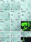
Fig. 7. The time-lapse and fluorescence microscopy show the delay of metaphase–anaphase transition by the D-box(1+2)AA mutant. Living D-box(1+2)AA (AI) or wild-type HOXC10 (JO) GFP-positive cells (numbered arrows) were photographed for GFP fluorescence (I and O) and in phase contrast at an interval of 5 min. The photograms show the indicated cells during entry into mitosis, when chromosome condensation becomes visible (*); at the metaphase to anaphase transition (#); and in late anaphase/telophase (**). The most representative photograms are shown.

Similar articles

Cited by

References

    1. Abate-Shen C. (2002) Deregulated homeobox gene expression in cancer: cause or consequence? Nat. Rev. Cancer, 2, 777–785. - PubMed
    1. Abdurashidova G., Riva,S., Biamonti,G., Giacca,M. and Falaschi,A. (1998) Cell cycle modulation of protein–DNA interactions at a human replication origin. EMBO J., 17, 2961–2969. - PMC - PubMed
    1. Abdurashidova G., Deganuto,M., Klima,R., Riva,S., Biamonti,G., Giacca,M. and Falaschi,A. (2000) Start sites of bidirectional DNA synthesis at the human lamin B2 origin. Science, 287, 2023–2026. - PubMed
    1. Bastians H., Topper,L.M., Gorbsky,G.L. and Ruderman,J.V. (1999) Cell cycle-regulated proteolysis of mitotic target proteins. Mol. Biol. Cell, 10, 3927–3941. - PMC - PubMed
    1. Boncinelli E. (1997) Homeobox genes and disease. Curr. Opin. Genet. Dev., 7, 331–337. - PubMed

Publication types

MeSH terms