. 2003 Jul 22;100(15):8933-8.
doi: 10.1073/pnas.1537685100. Epub 2003 Jul 9.

The ErbB2/ErbB3 heterodimer functions as an oncogenic unit: ErbB2 requires ErbB3 to drive breast tumor cell proliferation

Affiliations

The ErbB2/ErbB3 heterodimer functions as an oncogenic unit: ErbB2 requires ErbB3 to drive breast tumor cell proliferation

Thomas Holbro et al. Proc Natl Acad Sci U S A. .

Abstract

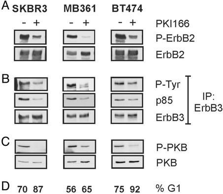
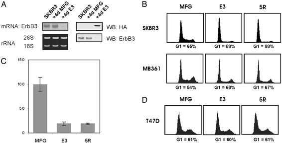
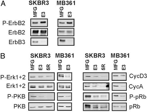
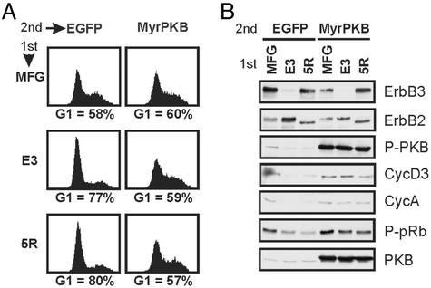
ErbB2 is a receptor tyrosine kinase whose activity in normal cells depends on dimerization with another ligand-binding ErbB receptor. In contrast, amplification of c-erbB2 in tumors results in dramatic overexpression and constitutive activation of the receptor. Breast cancer cells overexpressing ErbB2 depend on its activity for proliferation, because treatment of these cells with ErbB2-specific antagonistic antibodies or kinase inhibitors blocks tumor cells in the G1 phase of the cell cycle. Intriguingly, loss of ErbB2 signaling is accompanied by a decrease in the phosphotyrosine content of ErbB3. On the basis of these results, it has been proposed that ErbB3 might be a partner for ErbB2 in promoting cellular transformation. To test this hypothesis and directly examine the role of the "kinase dead" ErbB3, we specifically ablated its expression with a designer transcription factor (E3). By infection of ErbB2-overexpressing breast cancer cells with a retrovirus expressing E3, we show that ErbB3 is an essential partner in the transformation process. Loss of functional ErbB2 or ErbB3 has similar effects on cell proliferation and cell cycle regulators. Furthermore, expression of constitutively active protein kinase B rescues the proliferative block induced as a consequence of loss of ErbB2 or ErbB3 signaling. These results demonstrate that ErbB2 overexpression and activity alone are insufficient to promote breast tumor cell division. Furthermore, we identify ErbB3's role, which is to couple active ErbB2 to the phosphatidylinositol 3-kinase/protein kinase B pathway. Thus, the ErbB2/ErbB3 dimer functions as an oncogenic unit to drive breast tumor cell proliferation.


Figures

Fig. 1.
Fig. 1.
An ErbB2 kinase inhibitor blocks ErbB3 signaling and activity of the PI3K pathway. SKBR3, MB361, and BT474 breast cancer cells were treated for 30 min (A–C) or 24 h (D) with medium containing 5 μM PKI166 (+) or DMSO vehicle control (–). Protein lysates were probed for phospho-ErbB2 (P-ErbB2) (A) or phospho-PKB (P-PKB) (C). Membranes were stripped and reprobed to control for ErbB2 and PKB levels. (B) ErbB3 was immunoprecipitated from 500 μg of protein lysates, immunoprecipitates were resolved by SDS/PAGE, transferred onto a poly(vinylidene difluoride) membrane, and probed for phosphotyrosine (P-Tyr) or p85PI3K. Membranes were stripped and reprobed to control for ErbB3 levels. (D) Cells were harvested by trypsinization, nuclei were stained with propidium iodide, and a flow cytometric analysis was performed. The percentage of cells in the G1 phase of the cell cycle is indicated.
Fig. 2.
Fig. 2.
ErbB3 expression is required for ErbB2-dependent proliferation. (A) Four days after infection of SKBR3 cells, total RNA or protein was prepared from noninfected cells (SKBR3) or cells infected with the MFG control virus (MFG) or the E3 virus (E3). (Left) An ErbB3 Northern analysis was carried out on 3 μg of total RNA; ribosomal RNA levels are shown below. (Right) Fifty micrograms of protein lysates were probed for HA or ErbB3. (B and D) Cultures were infected with the indicated viruses and collected 4 days later. Cells were harvested by trypsinization, and the nuclei were stained with propidium iodide. The percentage of cells in G1 is indicated. (C) Proliferation assay of SKBR3 cells was performed as described in Materials and Methods. The bars indicate standard deviations.
Fig. 3.
Fig. 3.
The antiproliferative effect of E3 is due to loss of ErbB3. Stable pools of SKBR3 cells transfected with empty pcDNA3 vector (SKBR3-neo) or pcDNA3-ErbB3 (SKBR3-ErbB3) were infected with control (MFG), E3 or 5R viruses. Four days after infection parallel cultures were used to prepare cell lysates (A) or analyzed for cell cycle distribution (B). (A) Fifty micrograms of protein lysates were probed with the indicated antibodies. Reprobing with an ERK1+2 antiserum was used to control for loading. (B) Cells were harvested by trypsinization, and nuclei were stained with propidium iodide. The percentage of cells in G1 is indicated.
Fig. 4.
Fig. 4.
E3 down-regulates cytoplasmic and nuclear signaling molecules but not ErbB2 activity in breast tumor cells. Cultures of SKBR3 or MB361 cells were infected with the indicated viruses, and 4 days later, protein lysates were prepared. Fifty micrograms of protein lysates were resolved by SDS/PAGE and transferred onto poly(vinylidene difluoride) membranes. (A) The same membrane was sequentially probed for phospho-ErbB2 (P-ErbB2), ErbB2, and ErbB3. (B) Membranes were probed for phospho-ERK1+2, phospho-PKB, cyclin D3, cyclin A, phospho-pRb, pRb, ERK1+2, and PKB.
Fig. 5.
Fig. 5.
Expression of constitutively active PKB rescues the G1 block of SKBR3 cells lacking functional ErbB2 or ErbB3. SKBR3 cells were infected at day 0 with either MFG, E3, or 5R viruses (1st). Thirty-six hours later, cultures were infected with a second virus (2nd) expressing either EGFP or myristoylated PKB-IRES-EGFP (MyrPKB). Parallel cultures were collected 4 days after the first infection and stained for DNA content (A) or used to prepare protein lysates (B). In A, cells were harvested by trypsinization and fixed, and the cell cycle distribution of EGFP-positive cells was determined. The percentage of cells in the G1 phase of the cycle is indicated. In B, 15 μg of protein lysates was resolved by SDS/PAGE, transferred onto poly(vinylidene difluoride) membranes, and probed for the indicated proteins.

References

    1. Olayioye, M. A., Neve, R. M., Lane, H. A. & Hynes, N. E. (2000) EMBO J. 19, 3159–3167. - PMC - PubMed
    1. Yarden, Y. & Sliwkowski, M. X. (2001) Nat. Rev. Mol. Cell Biol. 2, 127–137. - PubMed
    1. Pinkas-Kramarski, R., Soussan, L., Waterman, H., Levkowitz, G., Alroy, I., Klapper, L., Lavi, S., Seger, R., Ratzkin, B. J., Sela, M., et al. (1996) EMBO J. 15, 2452–2467. - PMC - PubMed
    1. Riese, D. J., van Raaij, T. M., Plowman, G. D., Andrews, G. C. & Stern, D. F. (1995) Mol. Cell. Biol. 15, 5770–5776. - PMC - PubMed
    1. Graus-Porta, D., Beerli, R. R., Daly, J. M. & Hynes, N. E. (1997) EMBO J. 16, 1647–1655. - PMC - PubMed

Publication types

MeSH terms

Substances