. 2003 Jul 22;100(15):8856-61.
doi: 10.1073/pnas.1431057100. Epub 2003 Jul 9.

Human inhibitory receptors Ig-like transcript 2 (ILT2) and ILT4 compete with CD8 for MHC class I binding and bind preferentially to HLA-G

Affiliations

Human inhibitory receptors Ig-like transcript 2 (ILT2) and ILT4 compete with CD8 for MHC class I binding and bind preferentially to HLA-G

Mitsunori Shiroishi et al. Proc Natl Acad Sci U S A. .

Abstract

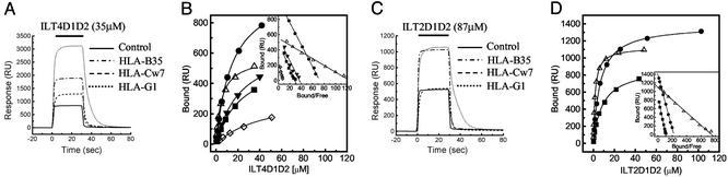
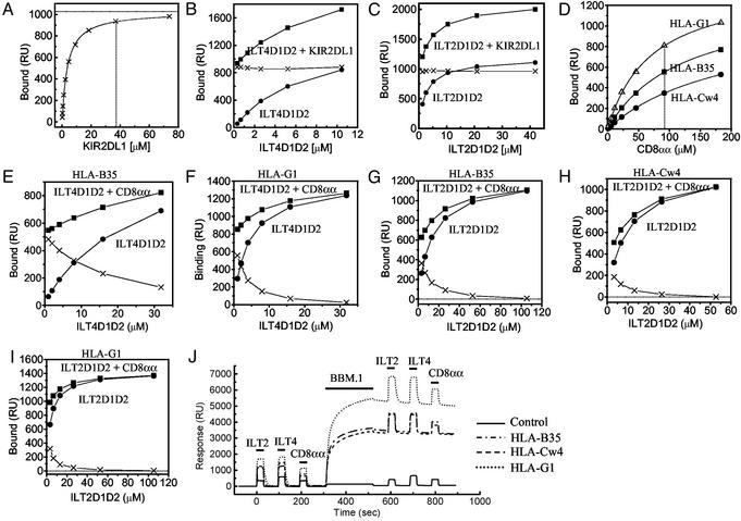
Ig-like transcript 4 (ILT4) (also known as leukocyte Ig-like receptor 2, CD85d, and LILRB2) is a cell surface receptor expressed mainly on myelomonocytic cells, whereas ILT2 (also known as leukocyte Ig-like receptor 1, CD85j, and LILRB1) is expressed on a wider range of immune cells including subsets of natural killer and T cells. Both ILTs contain immunoreceptor tyrosine-based inhibitory receptor motifs in their cytoplasmic tails that inhibit cellular responses by recruiting phosphatases such as SHP-1 (Src homology 2 domain containing tyrosine phosphatase 1). Although these ILTs have been shown to recognize a broad range of classical and nonclassical human MHC class I molecules (MHCIs), their precise binding properties remain controversial. We have used surface plasmon resonance to analyze the interaction of soluble forms of ILT4 and ILT2 with several MHCIs. Although the range of affinities measured was quite broad (Kd = 2-45 microM), some interesting differences were observed. ILT2 generally bound with a 2- to 3-fold higher affinity than ILT4 to the same MHCI. Furthermore, ILT2 and ILT4 bound to HLA-G with a 3- to 4-fold higher affinity than to classical MHCIs, suggesting that ILT/HLA-G recognition may play a dominant role in the regulation of natural killer, T, and myelomonocytic cell activation. Finally, we show that ILT2 and ILT4 effectively compete with CD8 for MHCI binding, raising the possibility that ILT2 modulates CD8+ T cell activation by blocking the CD8 binding as well as by recruiting inhibitory molecules through its immunoreceptor tyrosine-based inhibitory receptor motif.


Figures

Fig. 1.
Fig. 1.
Equilibrium binding of ILT2 and ILT4 to MHCIs. (A and C) ILT4D1D2 (35 μM) (A) and ILT2D1D2 (87 μM) (C) were injected for 30 s through flow cell 1 with control (BSA, solid line), flow cell 2 with HLA-B35 (broken and dotted line), flow cell 3 with HLA-Cw7 refolded by using chemically biotinylated β2m (broken line), and flow cell 4 with HLA-G1 (dotted line). Biotinylated BSA was used as a control. (B and D) Plots of the equilibrium binding responses of ILT4D1D2 (B) and ILT2D1D2 (D) versus concentration. Diamonds, HLA-A11; squares, HLA-B35; circles, HLA-Cw4; downward triangles, HLA-Cw7; upward triangles, HLA-G1. The solid lines represent direct nonlinear fits of the 1:1 Langmuir binding isoform to the data. (Insets) Scatchard plots of the same data are shown. The solid lines are linear fits. RU, response units.
Fig. 2.
Fig. 2.
The effect of KIR2DL1, anti-β2m mAb (BBM.1), or CD8αα on the binding of a soluble ILT2 and ILT4 to MHCIs. (A) Equilibrium binding analysis of KIR2DL1 against HLA-Cw*0401 (Kd ≈ 3.3 μM) on the same sensor chip used in the experiments shown in B and C. The estimated saturation level of KIR2DL1 was calculated by nonlinear curve fitting [1,026 response units (RU)]. (B and C) Binding of ILT4D1D2 (B) and ILT2D1D2 (C) (filled circles) alone or mixed with KIR2DL1 (filled squares). The concentration of KIR2DL1 was 38 μM (A, dotted line). The difference in the binding seen with or without KIR2DL1 was plotted (crosses). (D) Equilibrium binding analysis of CD8αα against HLA-B*3501 (squares), HLA-Cw*0401 (circles), and HLA-G1 (upward triangles) on the same sensor chip used in the experiments shown in EI. The Kd values are listed in Table 1. (E and F) Binding of ILT4D1D2 with or without CD8αα to HLA-B*3501 and HLA-G1, respectively. The concentration of CD8αα was 92 μM(D, dotted line). The difference in the binding seen with or without CD8αα was plotted (crosses) in the experiments shown in EI.(GI) Binding of ILT2D1D2 with or without CD8αα to HLA-B*3501 (G), HLA-Cw*0401 (H), and HLA-G1 (I). The concentration of CD8αα was 92 μM. (J) Binding of ILT2D1D2 (105 μM), ILT4D1D2 (33 μM), and CD8αα (92 μM) before and after injection of anti-β2m BBM.1 mAb to saturation level.
Fig. 3.
Fig. 3.
The putative ILT-binding site of MHCI. (A) Amino acid sequence alignment of α3 domains of HLA class I alleles (183–276). (B) Amino acid sequence alignment of the D1 and D2 domains of ILT2 and ILT4. (C Upper Left) Surface and ribbon diagram of HLA class I molecule with solid (α3 domain) and transparent surface. The putative residues playing an important role in strong binding of ILT2 and ILT4 to HLA-G1 (see Results and Discussion) are shown in orange. The residues of the α3 domain involved in MHCI–CD8αα binding are shown in cyan (35). The putative residues playing a part in the differentiation of ILT binding to different MHCI alleles are shown in pink. The residues interacting with KIR2DL1 are shown in red. The important epitopes of anti-β2m antibody BBM.1 (residues 38, 44, and 45) are shown in blue (34). (Upper Right) The reverse side view of that shown in the Upper Left. The residues of HLA-G1, which differ from those of the other MHCI alleles on the G strand, are shown in green. (Lower) Surface and ribbon diagram of ILT2D1D2. The residues playing an important role in binding to UL18 are shown in yellow (18). The colors used in this figure correspond to those of alignment shown in A and B. The diagrams in C were created with weblab viewer lite (Accelrys, San Diego).

Similar articles

Cited by

References

    1. Martin, A. M., Kulski, J. K., Witt, C., Pontarotti, P. & Christiansen, F. T. (2002) Trends Immunol. 23, 81–88. - PubMed
    1. Samaridis, J. & Collona, M. (1997) Eur. J. Immunol. 27, 660–665. - PubMed
    1. Cosman, D., Fanger, N., Borges, L., Kubin, M., Chin, W., Peterson, L. & Hsu, M.-L. (1997) Immunity 7, 273–282. - PubMed
    1. Borges, L., Hsu, M.-L., Fanger, N., Kubin, M. & Cosman, D. (1997) J. Immunol. 159, 5192–5196. - PubMed
    1. Wagtmann, N., Rojo, S., Eichler, E., Mohrenweiser, H. & Long, O. E. (1997) Curr. Biol. 7, 615–618. - PubMed

Publication types

MeSH terms