Comparative Study
. 2003 Jul 22;100(15):8682-7.
doi: 10.1073/pnas.1133380100. Epub 2003 Jul 9.

X-ray crystal structures of the WT and a hyper-accurate ribosome from Escherichia coli

Affiliations
Comparative Study

X-ray crystal structures of the WT and a hyper-accurate ribosome from Escherichia coli

Antón Vila-Sanjurjo et al. Proc Natl Acad Sci U S A. .

Abstract

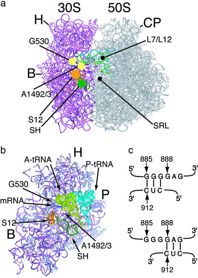
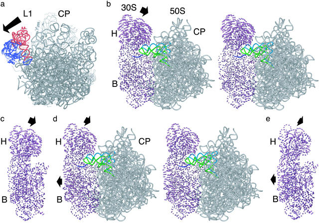
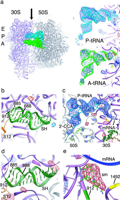
Protein biosynthesis on the ribosome requires accurate reading of the genetic code in mRNA. Two conformational rearrangements in the small ribosomal subunit, a closing of the head and body around the incoming tRNA and an RNA helical switch near the mRNA decoding site, have been proposed to select for complementary base-pairing between mRNA codons and tRNA anticodons. We determined x-ray crystal structures of the WT and a hyper-accurate variant of the Escherichia coli ribosome at resolutions of 10 and 9 A, respectively, revealing that formation of the intact 70S ribosome from its two subunits closes the conformation of the head of the small subunit independent of mRNA decoding. Moreover, no change in the conformation of the switch helix is observed in two steps of tRNA discrimination. These 70S ribosome structures indicate that mRNA decoding is coupled primarily to movement of the small subunit body, consistent with previous proposals, whereas closing of the head and the helical switch may function in other steps of protein synthesis.


Figures

Fig. 1.
Fig. 1.
Components of the ribosome involved in mRNA decoding. (a) View of the ribosome from the A-site side. Elements of the small (30S) subunit and large (50S) subunit that contribute to binding of the anticodon of aa-tRNA and EF-Tu are marked: G530 and A1492/3, nucleotides in 16S rRNA; S12, protein S12; SH, switch helix; L7/L12, proteins L7/L12; SRL, sarcin-ricin loop in 23S rRNA. Other features of the ribosome are marked as follows: H, head of the small subunit; B, small subunit body; P, small subunit platform; CP, central protuberance of the large subunit. (b) View of the interface side of the small subunit, with the large subunit removed for clarity. Nucleotides G530, A1492, and A1493 (yellow) contact both the mRNA (blue) and A-site tRNA (light green). Protein S12 (orange) and the switch helix (dark green) are located adjacent to the decoding site. (c) Proposed switch in base-pairing in helix 27 of 16S rRNA. The 885 conformation (Upper) involves base-pairing between nucleotides 885–887 and 910–912. In the 888 conformation (Lower), nucleotides 888–890 base-pair with nucleotides 910–912.
Fig. 2.
Fig. 2.
Superpositions of ribosomal subunit structures. (a) Position of the L1 stalk in the smD E. coli ribosome structure (blue) compared with that in the T. thermophilus 70S ribosome (red). The 50S subunit is shown from the 30S interface side. The arrow indicates the direction of motion required to move from the closed to open position of the L1 stalk. (b) Stereoview comparing the open 30S subunit conformation to the small subunit in the E. coli smD ribosome. Difference vectors between all conserved phosphorus atoms in the small subunit are shown. Arrows indicate the direction of movement in going from the open conformation to that in the intact ribosome. The large subunit (gray), mRNA (blue), A-site tRNA (light green), and P-site tRNA (light blue) bound to the E. coli smD ribosome are shown for reference. Other components of the ribosome are marked as in Fig. 1. (c) Comparison of the open 30S subunit conformation to the small subunit in the T. thermophilus 70S ribosome structure. Difference vectors are shown as described above. The orientation is the same as in b.(d) Stereoview comparing the closed 30S subunit conformation to the small subunit in the E. coli smD ribosome. (e) Comparison of the closed 30S subunit conformation to the small subunit in the T. thermophilus 70S ribosome structure. The orientation is the same as in d.
Fig. 3.
Fig. 3.
Conformation of the 70S ribosome in the switch helix region. (a) Difference electron density map comparing WT and smD 70S ribosomes from E. coli. The difference map was calculated by using observed diffraction amplitudes from both crystal forms, as described in Materials and Methods. The smD ribosomes contain mRNA, P-site tRNAfMet (P-tRNA, Upper Right) and noncognate A-site tRNAfMet at 50% occupancy (A-tRNA, Lower Right). (Left) A top view of the ribosome is shown with the A site, P site, and E site indicated to the left of the small subunit. The tRNAs are viewed from the perspective of the E site, as indicated by the arrow. The color scheme of the ribosome complex is the same as in Fig. 1. (b) The same difference electron density map in the switch helix region. (c) Difference electron density map comparing the smD 70S ribosome complex to a model lacking ligands throughout refinement. The positive electron density corresponds to P-site tRNA and mRNA bound to the ribosome. The perspective is the same as in a. (d) The same difference electron density map in the switch helix region. (e) Difference electron density map comparing the smD 70S ribosome complex to a ribosome model that contained streptomycin throughout refinement. The streptomycin binding pocket (sm) is shown. Positive electron density (blue, left) appeared after refinements of the model both in the absence and presence of the antibiotic, whereas negative density appeared only when streptomycin was included in the model. In all panels, difference electron density is contoured at 3.0 and –3.0 SDs from the mean (blue and red, respectively).
Fig. 4.
Fig. 4.
Chemical probing of E. coli ribosome decoding complexes. (a) Kethoxal modification of 16S rRNA showing P-site tRNA dependent protection of G926 (4) and the absence of reactivity in the 885–887 stretch in the switch helix as expected for the error-prone conformation (15). Sequencing lanes are marked U, G, C, and A. Lanes for both WT and smD ribosomes were as follows: 1, ribosomes with no kethoxal treatment; 2, ribosomes with kethoxal treatment; 3, ribosomes plus mRNA and tRNAfMet with kethoxal; 4, ribosomes plus mRNA, tRNAfMet, EF-Tu/GTP/Phe-tRNAPhe with kethoxal; 5, ribosomes plus mRNA, tRNAfMet, EF-Tu/GMP-PNP/Phe-tRNAPhe with kethoxal. (b) Protections from DMS modification at nucleotides A1492–A1493 of 16S rRNA dependent on A-site tRNA binding (4). Lanes were loaded as in a, replacing kethoxal with DMS. (c) Protection of A2602 in 23S rRNA from DMS dependent on release of Phe-tRNAPhe after GTP hydrolysis by EF-Tu (EFTu/GTP/Phe-tRNAPhe lanes), and modification of A2602 in codon recognition complexes (EFTu/GMPPNP/Phe-tRNAPhe lanes) (28). Lanes were loaded as in b. (d) Reactivity of A889 in 16S rRNA to DMS, in agreement with the error-prone conformation (15). Lanes were loaded as in b.

Similar articles

Cited by

References

    1. Rodnina, M. V. & Wintermeyer, W. (2001) Annu. Rev. Biochem. 70, 415–435. - PubMed
    1. Ogle, J. M., Carter, A. P. & Ramakrishnan, V. (2003) Trends Biochem. Sci. 28, 259–266. - PubMed
    1. Pettersson, I. & Kurland, C. G. (1980) Proc. Natl. Acad. Sci. USA 77, 4007–4010. - PMC - PubMed
    1. Moazed, D. & Noller, H. F. (1986) Cell 47, 985–994. - PubMed
    1. Moazed, D. & Noller, H. F. (1987) Nature 327, 389–394. - PubMed

Publication types

MeSH terms

LinkOut - more resources