. 2003 Jul 22;100(15):8862-7.
doi: 10.1073/pnas.1133324100. Epub 2003 Jul 9.

Survey of naturally occurring CD4+ T cell responses against NY-ESO-1 in cancer patients: correlation with antibody responses

Affiliations

Survey of naturally occurring CD4+ T cell responses against NY-ESO-1 in cancer patients: correlation with antibody responses

Sacha Gnjatic et al. Proc Natl Acad Sci U S A. .

Abstract

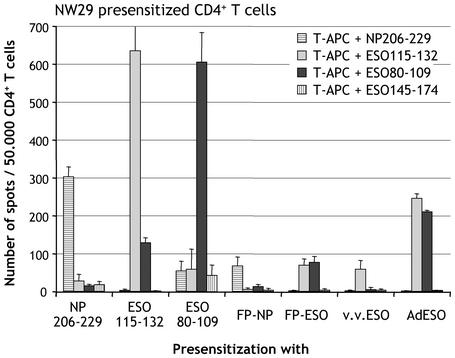
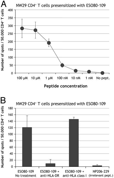
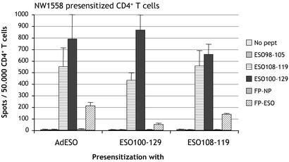
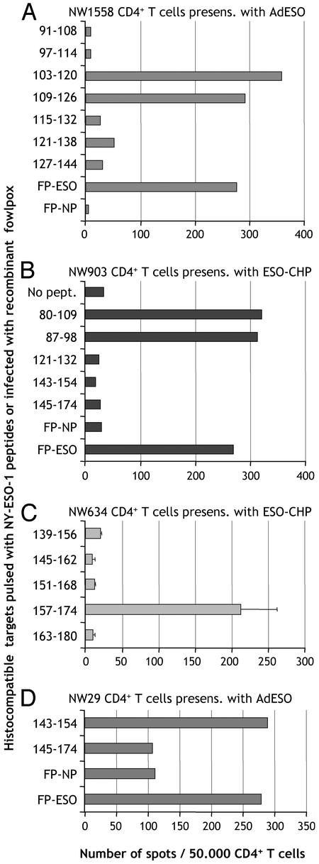
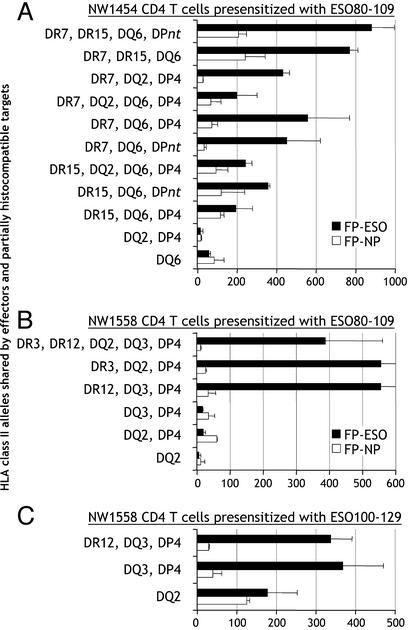
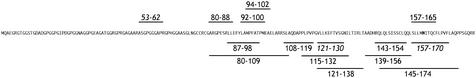
NY-ESO-1 is one of the most immunogenic proteins described in human cancers, based on its capacity to elicit simultaneous antibody and CD8+ T cell responses in vivo. Although HLA class II restricted epitopes from NY-ESO-1 have been identified, no broad survey has yet established the status of natural CD4+ T cell responses in cancer patients in relation to CD8+ and antibody responses. We used a recently developed general strategy for monitoring CD4+ responses that overcomes the need for prior knowledge of epitope or HLA restriction to analyze a series of 31 cancer patients and healthy donors for the presence of CD4+ T cells to NY-ESO-1, and related this response to NY-ESO-1 expression in tumor cells and serum antibodies to NY-ESO-1. None of the 18 patients that tested seronegative for NY-ESO-1 had detectable CD4+ T cell responses. On the contrary, 11 of 13 cancer patients with serum antibodies to NY-ESO-1 had polyclonal CD4+ T cell responses directed against various known and previously undescribed NY-ESO-1 epitopes. NY-ESO-1 peptide 80-109 was the most immunogenic, with 10 of 11 patients responding to this peptide. We show here that 12-mer determinants from NY-ESO-1 eliciting a CD4+ T cell response were peptide 87-98 with promiscuous HLA class II presentation, peptide 108-119 restricted by HLA-DP4, and peptides 121-132 and 145-156, both shorter epitopes from previously described HLA-DR4 peptides, also presented by HLA-DR7. This study represents the next step in compiling a comprehensive picture of the adaptive immune response to NY-ESO-1, and provides a general strategy for analyzing the CD4+ T cell response to other tumor antigens eliciting a humoral immune response.


Figures

Fig. 1.
Fig. 1.
Specificity controls of CD4+ T cell presensitization from melanoma patient NW29. CD4+ T cells were presensitized with indicated peptides or recombinant vectors and tested by ELISPOT at d23 against autologous T-APC pulsed with peptides NP206-229, ESO115-132, ESO80-109, or ESO145-174. Results represent the mean number of spots per 50,000 effector CD4+ T cells in duplicate wells, with error bars indicating standard deviation.
Fig. 2.
Fig. 2.
(A) Titration of peptide ESO80-109 recognized by CD4+ T cells from patient NW29. CD4+ T cells were presensitized with peptide ESO80-109 and tested by ELISPOT against autologous T-APC pulsed with peptide ESO80-109 in concentrations ranging from 100 μM to 1 nM, or not pulsed. (B) Blocking of ESO80-109 recognition by anti-HLA-DR antibodies. The same effectors were tested by ELISPOT against targets pulsed with ESO80-109 or irrelevant control peptide NP206-229 in the presence or absence of anti-HLA-DR (10 μg/ml) or anti-HLA class I (10 μg/ml) monoclonal antibodies. Results represent the mean number of spots per 50,000 effector CD4+ T cells in duplicate wells, with error bars indicating standard deviation.
Fig. 3.
Fig. 3.
Presensitization of CD4+ T cells from ovarian cancer patient NW1558. CD4+ T cells were presensitized with AdESO or with peptides ESO100-129 or ESO108-119 and tested by ELISPOT at d19 against autologous T-APC pulsed with ESO98-105, ESO108-119, or ESO100-129, or infected with FP-NP or FP-ESO. Results represent the mean number of spots per 50,000 effector CD4+ T cells in duplicate wells, with error bars indicating standard deviation.
Fig. 4.
Fig. 4.
Mapping of epitopes recognized by CD4+ T cells from different cancer patients. (A) CD4+ T cells from patient NW1558 were presensitized with adenovirus recombinant for NY-ESO-1 (AdESO) and tested by ELISPOT against hsistocompatible EBV-B cells pulsed with indicated 18-mer NY-ESO-1 peptides, or infected with recombinant fowlpox. (B) CD4+ T cells from patient NW903 were presensitized with ESO-CHP and tested by ELISPOT against histocompatible EBV-B cells pulsed with indicated NY-ESO-1 peptides, or infected with recombinant fowlpox. (C) CD4+ T cells from patient NW634 were presensitized with ESO-CHP and tested by ELISPOT against histocompatible EBV-B cells pulsed with indicated NY-ESO-1 peptides. (D) CD4+ T cells from patient NW29 were presensitized with AdESO and tested by ELISPOT against histocompatible EBV-B cells pulsed with indicated NY-ESO-1 peptides, or infected with recombinant fowlpox.
Fig. 5.
Fig. 5.
HLA restriction usage for presentation of NY-ESO-1 to CD4+ T cells specific for ESO80-109 and ESO100-129. (A) CD4+ T cells from patient NW1454 (HLA-DRB1*07,15; -DQB1*02,06; -DPB1*04,09) were presensitized with peptide ESO80-109 and tested by ELISPOT against partially histocompatible EBV-B cells infected with NY-ESO-1 or NP recombinant fowlpox vectors. HLA class II alleles shared between effectors and targets are indicated. *DPnt: targets not tested for HLA-DP compatibility with effectors. (B) Similar analysis with CD4+ T cells from patient NW1558 (HLA-DRB1*03,12; -DQB1*02,03; -DPB1*04,10) presensitized with peptide ESO80-109. (C) Similar analysis with CD4+ T cells from patient NW1558 presensitized with peptide ESO100-129. Results represent the mean number of spots per 50,000 effector CD4+ T cells in duplicate wells with error bars indicating standard deviation.
Fig. 6.
Fig. 6.
Epitope distribution along the NY-ESO-1 sequence. Above the sequence, defined HLA class I-restricted peptides; below the sequence, HLA class II-restricted peptides. Italics indicate peptides defined by other groups (10, 11, 13).

References

    1. Chen, Y.-T., Scanlan, M. J., Sahin, U., Türeci, Ö., Güre, A. O., Tsang, S., Williamson, B., Stockert, E., Pfreundschuh, M. & Old, L. J. (1997) Proc. Natl. Acad. Sci. USA 94, 1914–1918. - PMC - PubMed
    1. Jäger, E., Chen, Y.-T., Drijfhout, J. W., Karbach, J., Ringhoffer, M., Jäger, D., Arand, M., Wada, H., Noguchi, Y., Stockert, E., et al. (1998) J. Exp. Med. 187, 265–270. - PMC - PubMed
    1. Stockert, E., Jäger, E., Chen, Y.-T., Scanlan, M. J., Gout, I., Karbach, J., Arand, M., Knuth, A. & Old, L. J. (1998) J. Exp. Med. 187, 1349–1354. - PMC - PubMed
    1. Jäger, E., Stockert, E., Zidianakis, Z., Chen, Y.-T., Karbach, J., Jäger, D., Arand, M., Ritter, G., Old, L. J. & Knuth, A. (1999) Int. J. Cancer 84, 506–510. - PubMed
    1. Jäger, E., Nagata, Y., Gnjatic, S., Wada, H., Stockert, E., Karbach, J., Dunbar, P. R., Lee, S. Y., Jungbluth, A., Jäger, D., et al. (2000) Proc. Natl. Acad. Sci. USA 97, 4760–4765. - PMC - PubMed

Publication types

MeSH terms