. 2003 Jul 15;31(14):4031-40.
doi: 10.1093/nar/gkg457.

Spatial organization of topoisomerase I-mediated DNA cleavage induced by camptothecin-oligonucleotide conjugates

Affiliations

Spatial organization of topoisomerase I-mediated DNA cleavage induced by camptothecin-oligonucleotide conjugates

Paola B Arimondo et al. Nucleic Acids Res. .

Abstract

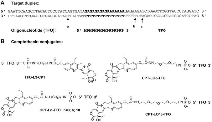
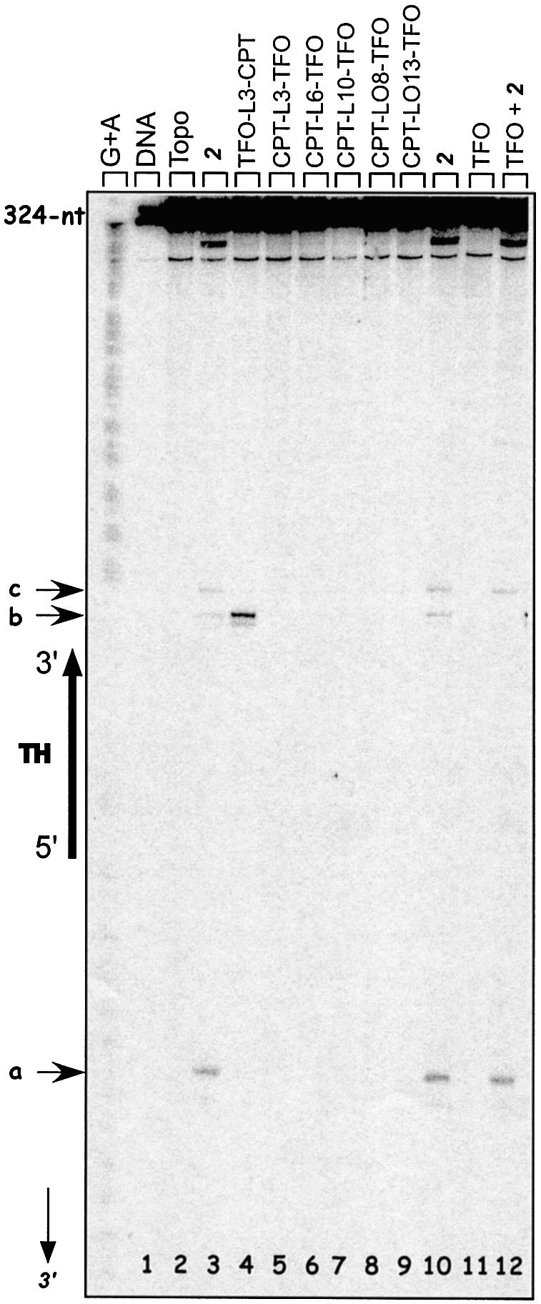
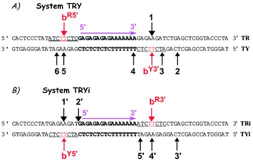
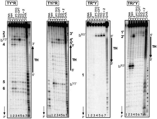
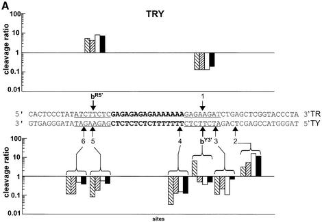
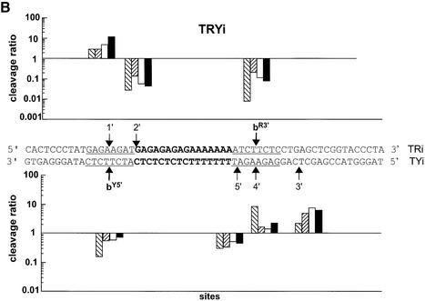
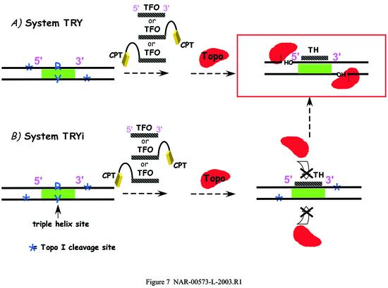
Triple helix-forming oligonucleotides covalently linked to topoisomerase I inhibitors, in particular the antitumor agent camptothecin, trigger topoisomerase I-mediated DNA cleavage selectively in the proximity of the binding site of the oligonucleotide vector. In the present study, we have performed a systematic analysis of the DNA cleavage efficiency as a function of the positioning of the camptothecin derivative, either on the 3' or the 5' side of the triplex, and the location of the cleavage site. A previously identified cleavage site was inserted at different positions within two triplex site-containing 59 bp duplexes. Sequence-specific DNA cleavage by topoisomerase I occurs only with triplex conjugates bearing the inhibitor at the 3'-end of the oligonucleotide and on the oligopyrimidine strand of the duplex. The lack of targeted cleavage on the 5' side is attributed to the structural differences of the 3' and 5' duplex-triplex DNA junctions. The changes induced in the double helix by the triple-helical structure interfere with the action of the enzyme according to a preferred spatial organization. Camptothecin conjugates of oligonucleotides provide efficient tools to probe the organization of the topoisomerase I-DNA complex and will be useful to understand the functioning of topoisomerase I in living cells.


Figures

Figure 1
Figure 1
Synthetic route used for the preparation of 7-ethyl-10-hydroxycamptothecin acetic acid (2).
Figure 2
Figure 2
(A) Sequence of the TFOs and the 77 bp DNA target used in this study. The TFO binds in the major groove parallel to the oligopurine strand of the duplex. The orientation of the triple helix is defined by the orientation of the oligopurine strand of the duplex. The triplex site is in bold. The 77 bp duplex target sequence was inserted between the BamHI and EcoRI sites of plasmid pbSK(+/–). M, 5-methyl-2′-deoxycytidine; P, 5-propynyl-2′-deoxyuridine. (B) Structure of the camptothecin conjugates used in this study. 7-Ethyl-10-hydroxycamptothecin acetic acid was attached through N-succinimide activation to the terminal amino group of the linker attached to the extremity of the TFO or MGB as described previously (18). L3, L6 and L10, propyldiamine, hexyldiamine and decyldiamine; LO8, 1,8-diaminodioxaoctane; LO13, 4,7,10-trioxa-1,13-tridecandiamine.
Figure 3
Figure 3
Sequence analysis of the Topo I-mediated cleavage products of the 324 bp target duplex (50 nM) 3′-end radiolabeled on the oligopyrimidine-containing strand. Adenine/guanine-specific Maxam–Gilbert chemical cleavage reactions were used as markers. The positions of the cleavage sites are indicated (sites ac), as well as the triple helix region and the nucleotide positions along the radiolabeled oligopyrimidine strand (arrow). Lane 1, duplex; lane 2, duplex incubated with Topo I; lanes 3–12, duplex incubated with Topo I in the presence of 5 µM 2 (lane 3 and 10), 0.5 µM TFO-L3-CPT (lane 4), 0.5 µM CPT-L3-TFO (lane 5), 0.5 µM CPT-L6-TFO (lane 6), 0.5 µM CPT-L10-TFO (lane 7), 0.5 µM CPT-LO8-TFO (lane 8), 0.5 µM CPT-LO13-TFO (lane 9), 5 µM TFO (lane 11) or 5 µM TFO + 5 µM 2 (lane 12).
Figure 4
Figure 4
Model 59 bp duplexes used in this study. (A) System TRY and (B) System TRYi. Sequence of the duplexes. The triple helix site is in bold and the same cleavage site b is indicated in red and underlined. The nomenclature of site b is as follows. The strand of the duplex, R or Y, is indicated as superscript followed by the side of the triple helix, 5′ or 3′, on which the site is located. For example, bR5′ stands for a site b positioned on the R strand of the duplex and on the 5′ side of the triplex end. Numbers indicate the other cleavage sites.
Figure 5
Figure 5
Positioning of Topo I in the presence of the triple-helical structure. Sequence analysis of the Topo I-mediated cleavage products on each 3′-end radiolabeled (50 nM) strand of the duplexes. Adenine/guanine-specific Maxam–Gilbert chemical cleavage reactions were used as markers. The positions of the cleavage sites are indicated on each strand according to the nomenclature defined in Figure 4. The orientation of the triple helix region is indicated, as well as the duplex system analyzed; radiolabeled strands of the duplexes are indicated by asterisks for each system. Lane 1, target duplex; lane 2, duplex incubated with Topo I; lanes 3–7, duplex incubated with Topo I and in the presence of 5 µM 2 (lane 3), 0.5 µM CPT-L3-TFO (lane 4), 0.5 µM TFO-L3-CPT (lane 5), 5 µM TFO (lane 6) or 5 µM TFO + 5 µM 2 (lane 7).
Figure 6
Figure 6
Comparison of the cleavage positions and intensities on each target duplex strand. The ratios between the intensity of cleavage in the presence of the triple helices and the intensity in the presence of CPT alone are presented, on a logarithmic scale, for duplexes TRY (A) and TRYi (B) at each cleavage site according to the position along the target. Data were compiled from eight independent experiments. TFO-L3-CPT, right-hatched bars; CPT-L3-TFO, left-hatched bars; TFO, white bars; TFO + CPT, black bars.
Figure 6
Figure 6
Comparison of the cleavage positions and intensities on each target duplex strand. The ratios between the intensity of cleavage in the presence of the triple helices and the intensity in the presence of CPT alone are presented, on a logarithmic scale, for duplexes TRY (A) and TRYi (B) at each cleavage site according to the position along the target. Data were compiled from eight independent experiments. TFO-L3-CPT, right-hatched bars; CPT-L3-TFO, left-hatched bars; TFO, white bars; TFO + CPT, black bars.
Figure 7
Figure 7
Schematic representation of the interaction between Topo I and DNA in the presence of the triple helices of the two systems studied. (A) TRY; (B) TRYi.

Similar articles

Cited by

References

    1. Champoux J.J. (2001) DNA topoisomerases: structure, function and mechanism. Annu. Rev. Biochem., 70, 369–413. - PubMed
    1. Wang J.C. (1996) DNA topoisomerases. Annu. Rev. Biochem., 65, 635–692. - PubMed
    1. Pommier Y., Pourquier,P., Fan,Y. and Strumberg,D. (1998) Mechanism of action of eukaryotic DNA topoisomerase I and drugs targeted to the enzyme. Biochim. Biophys. Acta, 1400, 83–105. - PubMed
    1. Redinbo M.R., Stewart,L., Kuhn,P., Champoux,J.J. and Hol,W.G. (1998) Crystal structures of human topoisomerase I in covalent and noncovalent complexes with DNA. Science, 279, 1504–1513. - PubMed
    1. Stewart L., Redinbo,M.R., Qiu,X., Hol,W.G.J. and Champoux,J.J. (1998) A model for the mechanism of human topoisomerase I. Science, 279, 1534–1541. - PubMed

Publication types