. 2003 Jul 15;31(14):4162-75.
doi: 10.1093/nar/gkg459.

Selective interactions of human kin17 and RPA proteins with chromatin and the nuclear matrix in a DNA damage- and cell cycle-regulated manner

Affiliations

Selective interactions of human kin17 and RPA proteins with chromatin and the nuclear matrix in a DNA damage- and cell cycle-regulated manner

Laurent Miccoli et al. Nucleic Acids Res. .

Abstract

Several proteins involved in DNA synthesis are part of the so-called 'replication factories' that are anchored on non-chromatin nuclear structures. We report here that human kin17, a nuclear stress-activated protein, associates with both chromatin and non-chromatin nuclear structures in a cell cycle- and DNA damage-dependent manner. After L-mimosine block and withdrawal we observed that kin17 protein was recruited in the nucleus during re-entry and progression through S phase. These results are consistent with a role of kin17 protein in DNA replication. About 50% of the total amount of kin17 protein was detected on nuclear structures and could not be released by detergents. Furthermore, the amount of kin17 protein greatly increased in both G(1)/S and S phase-arrested cells in fractions containing proteins anchored to nuclear structures. The detection of kin17 protein showed for the first time its preferential assembly within non-chromatin nuclear structures in G(1)/S and S phase-arrested cells, while the association with these structures was found to be less stable in the G(2)/M phase, as judged by fractionation of human cells and immunostaining. In asynchronous growing cells, kin17 protein interacted with both chromatin DNA and non-chromatin nuclear structures, while in S phase-arrested cells it interacted mostly with non-chromatin nuclear structures, as judged by DNase I treatment and in vivo UV cross-linking. In the presence of DNA damage in S phase cells, the distribution of kin17 protein became mainly associated with chromosomal DNA, as judged by limited formaldehyde cross-linking of living cells. The physical interaction of kin17 protein with components of the nuclear matrix was confirmed and visualized by indirect immunofluorescence and immunoelectron microscopy. Our results indicate that, during S phase, a fraction of the human kin17 protein preferentially associates with the nuclear matrix, a fundamentally non-chromatin higher order nuclear structure, and to chromatin DNA in the presence of DNA damage.


Figures
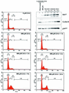
Figure 1
Figure 1
kin17 protein levels increased during the re-initiation of DNA synthesis and the progression into S phase. HeLa cells were treated with l-mimosine (400 µM) for 24 h. Cells were then released in fresh medium and recovered at the indicated times for cell cycle determination and immunoblot analysis of kin17, cyclin B and p34cdc2 protein contents.
Figure 2
Figure 2
Increased association of human kin17 protein with nuclear structures in G1/S and S phase-arrested cells. Asynchronous (mock-treated, lanes 1 and 10), l-mimosine-treated (lanes 2–5 and 11–14) or hydroxyurea- treated (lanes 6–9 and 15–18) RKO and HCT116 cells were lysed in buffer N (containing 1% Igepal CA-630) as indicated in Materials and Methods. The detergent-extractable and nuclei-bound fractions were collected by centrifugation, immunoblotted with anti-kin17 protein, anti-RPA or anti-PCNA monoclonal antibodies and revealed by chemiluminescence. A representative pattern of three different experiments is shown.
Figure 3
Figure 3
Endogenous kin17 protein associates with both DNA and nuclear structures as judged by selective extraction and immunostaining detection. (A) Triton X-100-extractable supernatants (TX-100 S, lane 1) and extracted nuclear pellets (TX-100 P, lane 2) were prepared from asynchronous HeLa cells. The extracted nuclear pellets were further treated with 1000 U/ml DNase I at 25°C for 30 min to obtain DNase I-solubilized supernatants [DNase(+) S, lane 5] and remaining DNase-resistant nuclear pellets [DNase(+) P, lane 6] or without DNase I at 25°C for 30 min [DNase(–) S, lane 3; DNase(–) P, lane 4]. These fractions were immunoblotted with anti-kin17 protein, anti-RPA or anti-PCNA monoclonal antibodies. (B) Indirect immunofluorescence detection of kin17 protein localization in in situ replication foci attached to the nuclear matrix prepared as described in Materials and Methods. Asynchronous (mock-treated) and hydroxyurea- or aphidicolin-arrested HeLa cells were fixed with paraformaldehyde (PAF) after Triton X-100 extraction (0.1% Triton-X100, panels a and e), DNase I treatment and ammonium sulfate extraction [0.1% Triton X-100 + (NH4)2SO4 + DNase I, panels b–d and f–h]. The extracted cells were then immunostained with anti-kin17 protein antibody (red). The samples were further treated with DAPI for DNA staining (blue). Digitized images of representative cells are shown at a magnification of 500×. Living asynchronous (C) or early S phase-arrested (D) HeLa cells on dishes were irradiated with UV at 50, 100, 150, 200 and 400 mJ/cm2 and subjected to successive extractions with Triton X-100 (Triton Ext., lanes 1–5) and 1 M NaCl (NaCl Ext., lanes 6–10) as described in Materials and Methods. Extracted kin17 and RPA proteins were detected by immunoblotting and revealed by chemiluminescence. A typical pattern is shown.
Figure 3
Figure 3
Endogenous kin17 protein associates with both DNA and nuclear structures as judged by selective extraction and immunostaining detection. (A) Triton X-100-extractable supernatants (TX-100 S, lane 1) and extracted nuclear pellets (TX-100 P, lane 2) were prepared from asynchronous HeLa cells. The extracted nuclear pellets were further treated with 1000 U/ml DNase I at 25°C for 30 min to obtain DNase I-solubilized supernatants [DNase(+) S, lane 5] and remaining DNase-resistant nuclear pellets [DNase(+) P, lane 6] or without DNase I at 25°C for 30 min [DNase(–) S, lane 3; DNase(–) P, lane 4]. These fractions were immunoblotted with anti-kin17 protein, anti-RPA or anti-PCNA monoclonal antibodies. (B) Indirect immunofluorescence detection of kin17 protein localization in in situ replication foci attached to the nuclear matrix prepared as described in Materials and Methods. Asynchronous (mock-treated) and hydroxyurea- or aphidicolin-arrested HeLa cells were fixed with paraformaldehyde (PAF) after Triton X-100 extraction (0.1% Triton-X100, panels a and e), DNase I treatment and ammonium sulfate extraction [0.1% Triton X-100 + (NH4)2SO4 + DNase I, panels b–d and f–h]. The extracted cells were then immunostained with anti-kin17 protein antibody (red). The samples were further treated with DAPI for DNA staining (blue). Digitized images of representative cells are shown at a magnification of 500×. Living asynchronous (C) or early S phase-arrested (D) HeLa cells on dishes were irradiated with UV at 50, 100, 150, 200 and 400 mJ/cm2 and subjected to successive extractions with Triton X-100 (Triton Ext., lanes 1–5) and 1 M NaCl (NaCl Ext., lanes 6–10) as described in Materials and Methods. Extracted kin17 and RPA proteins were detected by immunoblotting and revealed by chemiluminescence. A typical pattern is shown.
Figure 4
Figure 4
kin17 protein detection in purified protein–chromatin DNA and protein–protein complexes after in vivo cross-linking of living S phase HeLa cells with formaldehyde. After in vivo cross-linking with formaldehyde (1%, 4 min), the nuclei were isolated, lysed and both protein–chromatin DNA and protein–protein complexes were purified by equilibrium centrifugation in two consecutive cesium chloride gradients as described in Materials and Methods. After reversion of the cross-links by boiling, immunoblot analysis was performed on DNA–protein complexes obtained from untreated (A and B, lanes 1) and MNNG-treated (A and B, lanes 2) HeLa cells. (A) Treatment of cells for 3 h with 100 µM MNNG immediately after release from a double thymidine block. Detection of kin17 protein. (B) Detection of histone H1. (His)6-kin17, positive control [recombinant human (His)6-kin17 protein].
Figure 5
Figure 5
Localization of kin17 protein in HeLa cell extracts after in vivo cross-linking with DSP. Triton X-100-extractable supernatants (TX-100 S, lane 1) and extracted nuclei (TX-100 P, lane 2) were prepared from asynchronous HeLa cells before and after treatment with 25–200 µg/ml DSP. The nuclei were further extracted with buffer containing 0.5 M NaCl and the salt extracts (0.5 M NaCl S, lane 3) and the nuclear pellets (0.5 M NaCl P, lane 4) were separated by centrifugation. The nuclei extracted with detergent and salt were then treated with DNase I followed by salt extraction to obtain solubilized chromatin fractions [DNase(+) S, lane 7] and insoluble non-chromatin nuclear pellets [DNase(+) P, lane 8] or without DNase I [DNase(–) S, lane 5; DNase(–) P, lane 6]. These fractions were immunoblotted with monoclonal anti-kin17 antibodies and revealed by chemiluminescence (A). The samples were also subjected to SDS–PAGE followed by Coomassie blue staining for detection of core histones (B).
Figure 6
Figure 6
Subcellular localization of endogenous kin17 protein in HeLa cells during the cell cycle using in vivo cross-linking with DSP. Detection of kin17 protein, RPA or PCNA in asynchronous, early S phase- and G2/M-arrested cells, before and after treatment with DSP (200 µg/ml). Triton X-100-extractable supernatants (lanes 1 and 6) and extracted nuclei were prepared before (A) and after DSP cross-linking (B) of asynchronous or hydroxyurea-treated HeLa cells. The nuclei were further extracted with buffer containing 0.5 M NaCl and the salt extracts (lanes 2 and 7) and the nuclear pellets (lanes 3 and 8) were separated by centrifugation. The detergent- and salt-extracted nuclei were then treated with DNase I followed by salt extraction to obtain solubilized chromatin fractions (lanes 4 and 9) and insoluble non-chromatin nuclear pellets (lanes 5 and 10). (C) Triton X-100-extractable supernatants (0.1% TX-100 S) and extracted nuclei (0.1% TX-100 P) were prepared from nocodazole-treated cells with or without DSP. The nuclei were further extracted with buffer containing 0.5 M NaCl and the salt extracts and the nuclear pellets were separated by centrifugation. The detergent- and salt-extracted nuclei were then treated with DNase I followed by salt extraction to obtain solubilized chromatin fractions and insoluble non-chromatin nuclear pellets or without DNase I [DNase(–) S, DNase(–) P]. (D) Indirect immunofluorescence detection of kin17 protein localization in HeLa cells after DSP cross-linking and chromatin extraction. Asynchronous (left) or hydroxyurea-treated (early S phase, right) cells were fixed with formaldehyde directly (panels a and e), after extraction with Triton X-100 (panels b and f) or after chromatin extraction as described in Materials and Methods of cells treated with (panels c and g) or without DSP (w/o, panels d and h). The cells were then immunostained with anti-kin17 protein antibody (red) and treated with DAPI for DNA staining (blue). Digitized images of representative cells are shown at a magnification of 500×.
Figure 6
Figure 6
Subcellular localization of endogenous kin17 protein in HeLa cells during the cell cycle using in vivo cross-linking with DSP. Detection of kin17 protein, RPA or PCNA in asynchronous, early S phase- and G2/M-arrested cells, before and after treatment with DSP (200 µg/ml). Triton X-100-extractable supernatants (lanes 1 and 6) and extracted nuclei were prepared before (A) and after DSP cross-linking (B) of asynchronous or hydroxyurea-treated HeLa cells. The nuclei were further extracted with buffer containing 0.5 M NaCl and the salt extracts (lanes 2 and 7) and the nuclear pellets (lanes 3 and 8) were separated by centrifugation. The detergent- and salt-extracted nuclei were then treated with DNase I followed by salt extraction to obtain solubilized chromatin fractions (lanes 4 and 9) and insoluble non-chromatin nuclear pellets (lanes 5 and 10). (C) Triton X-100-extractable supernatants (0.1% TX-100 S) and extracted nuclei (0.1% TX-100 P) were prepared from nocodazole-treated cells with or without DSP. The nuclei were further extracted with buffer containing 0.5 M NaCl and the salt extracts and the nuclear pellets were separated by centrifugation. The detergent- and salt-extracted nuclei were then treated with DNase I followed by salt extraction to obtain solubilized chromatin fractions and insoluble non-chromatin nuclear pellets or without DNase I [DNase(–) S, DNase(–) P]. (D) Indirect immunofluorescence detection of kin17 protein localization in HeLa cells after DSP cross-linking and chromatin extraction. Asynchronous (left) or hydroxyurea-treated (early S phase, right) cells were fixed with formaldehyde directly (panels a and e), after extraction with Triton X-100 (panels b and f) or after chromatin extraction as described in Materials and Methods of cells treated with (panels c and g) or without DSP (w/o, panels d and h). The cells were then immunostained with anti-kin17 protein antibody (red) and treated with DAPI for DNA staining (blue). Digitized images of representative cells are shown at a magnification of 500×.
Figure 7
Figure 7
Immunogold electron microscopy localization of endogenous kin17 protein after in situ extraction of living HeLa cells. HeLa cells were extracted with Triton X-100, DNase I, and 2 M NaCl and embedded in Lowicryl. Immunolabeling was performed on sections using a mixture of anti-kin17 K36 and K58 monoclonal antibodies followed by goat anti-mouse antibody conjugated to colloidal gold particles 5 nm in diameter. Sections were stained with uranyl acetate. Gold particles were found either isolated along the fibers of the internal matrix (A and D) or in foci (arrows, A–D) close to the lamina (L). Interchromatin granules (Ig) and residual nucleoli (NU) were not labeled (A, C and D). Bars represent 0.5 µm.

Similar articles

Cited by

References

    1. Boulikas T. (1996) Common structural features of replication origins in all life forms. J. Cell. Biochem., 60, 297–388. - PubMed
    1. Boulikas T. (1995) Chromatin domains and the prediction of MAR sequences. Int. Rev. Cytol., 162A, 279–388. - PubMed
    1. Cook P.R. (1988) The nucleoskeleton: artefact, passive framework or active site? J. Cell Sci., 90, 1–6. - PubMed
    1. Berezney R., Mortillaro,M.J., Ma,H., Wei,X. and Samarabandu,J. (1995) The nuclear matrix: a structural milieu for genomic function. Int. Rev. Cytol., 162A, 1–65. - PubMed
    1. Nickerson J.A. (2001) Experimental observations of a nuclear matrix. J. Cell Sci., 114, 463–474. - PubMed

Publication types

MeSH terms