

Chemical shift changes provide evidence for overlapping single-stranded DNA- and XPA-binding sites on the 70 kDa subunit of human replication protein A
- PMID: 12853635
- PMCID: PMC165966
- DOI: 10.1093/nar/gkg451
Chemical shift changes provide evidence for overlapping single-stranded DNA- and XPA-binding sites on the 70 kDa subunit of human replication protein A
Abstract
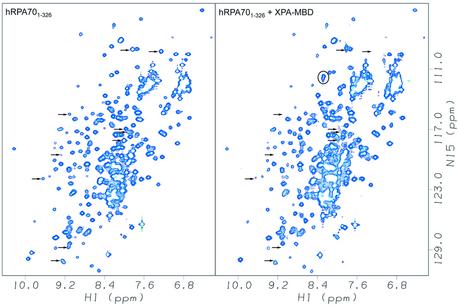
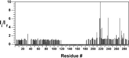
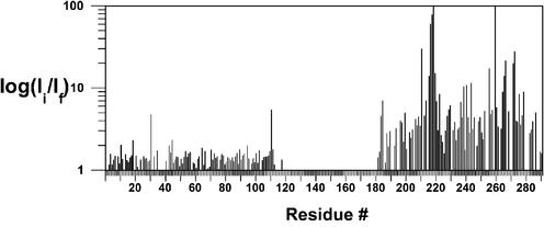
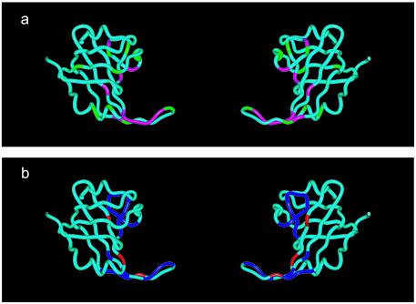
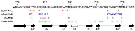
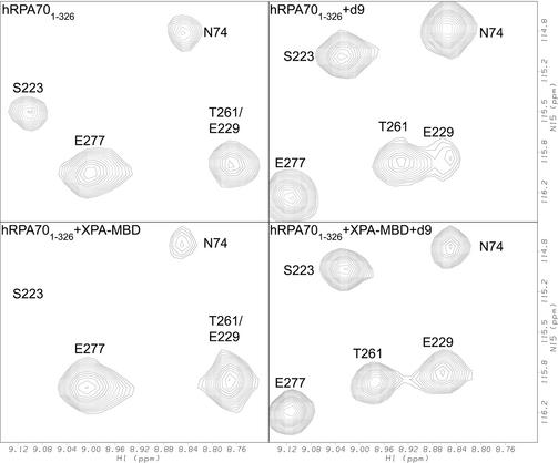
Replication protein A (RPA) is a heterotrimeric single-stranded DNA- (ssDNA) binding protein that can form a complex with the xeroderma pigmentosum group A protein (XPA). This complex can preferentially recognize UV-damaged DNA over undamaged DNA and has been implicated in the stabilization of open complex formation during nucleotide excision repair. In this report, nuclear magnetic resonance (NMR) spectroscopy was used to investigate the interaction between a fragment of the 70 kDa subunit of human RPA, residues 1-326 (hRPA70(1-326)), and a fragment of the human XPA protein, residues 98-219 (XPA-MBD). Intensity changes were observed for amide resonances in the (1)H-(15)N correlation spectrum of uniformly (15)N-labeled hRPA70(1-326) after the addition of unlabeled XPA-MBD. The intensity changes observed were restricted to an ssDNA-binding domain that is between residues 183 and 296 of the hRPA70(1-326) fragment. The hRPA70(1-326) residues with the largest resonance intensity reductions were mapped onto the structure of the ssDNA-binding domain to identify the binding surface with XPA-MBD. The XPA-MBD-binding surface showed significant overlap with an ssDNA-binding surface that was previously identified using NMR spectroscopy and X-ray crystallography. Overlapping XPA-MBD- and ssDNA-binding sites on hRPA70(1-326) suggests that a competitive binding mechanism mediates the formation of the RPA-XPA complex. To determine whether a ternary complex could form between hRPA70(1-326), XPA-MBD and ssDNA, a (1)H-(15)N correlation spectrum was acquired for uniformly (15)N-labeled hRPA70(1-326) after the simultaneous addition of unlabeled XPA-MBD and ssDNA. In this experiment, the same chemical shift perturbations were observed for hRPA70(1-326) in the presence of XPA-MBD and ssDNA as was previously observed in the presence of ssDNA alone. The ability of ssDNA to compete with XPA-MBD for an overlapping binding site on hRPA70(1-326) suggests that any complex formation between RPA and XPA that involves the interaction between XPA-MBD and hRPA70(1-326) may be modulated by ssDNA.
Figures
Similar articles
-
The weak interdomain coupling observed in the 70 kDa subunit of human replication protein A is unaffected by ssDNA binding.Nucleic Acids Res. 2001 Aug 1;29(15):3270-6. doi: 10.1093/nar/29.15.3270. Nucleic Acids Res. 2001. PMID: 11470885 Free PMC article.
-
DNA-XPA interactions: a (31)P NMR and molecular modeling study of dCCAATAACC association with the minimal DNA-binding domain (M98-F219) of the nucleotide excision repair protein XPA.Nucleic Acids Res. 2001 Jun 15;29(12):2635-43. doi: 10.1093/nar/29.12.2635. Nucleic Acids Res. 2001. PMID: 11410673 Free PMC article.
-
Interactions of human nucleotide excision repair protein XPA with DNA and RPA70 Delta C327: chemical shift mapping and 15N NMR relaxation studies.Biochemistry. 1999 Nov 16;38(46):15116-28. doi: 10.1021/bi991755p. Biochemistry. 1999. PMID: 10563794 Free PMC article.
-
DNA repair gets physical: mapping an XPA-binding site on ERCC1.DNA Repair (Amst). 2008 May 3;7(5):819-26. doi: 10.1016/j.dnarep.2008.01.018. Epub 2008 Mar 14. DNA Repair (Amst). 2008. PMID: 18343204 Free PMC article. Review.
-
The DNA damage-recognition problem in human and other eukaryotic cells: the XPA damage binding protein.Biochem J. 1997 Nov 15;328 ( Pt 1)(Pt 1):1-12. doi: 10.1042/bj3280001. Biochem J. 1997. PMID: 9359827 Free PMC article. Review.
Cited by
-
XPA: A key scaffold for human nucleotide excision repair.DNA Repair (Amst). 2016 Aug;44:123-135. doi: 10.1016/j.dnarep.2016.05.018. Epub 2016 May 20. DNA Repair (Amst). 2016. PMID: 27247238 Free PMC article. Review.
-
The Intriguing Mystery of RPA Phosphorylation in DNA Double-Strand Break Repair.Genes (Basel). 2024 Jan 27;15(2):167. doi: 10.3390/genes15020167. Genes (Basel). 2024. PMID: 38397158 Free PMC article. Review.
-
A new structural framework for integrating replication protein A into DNA processing machinery.Nucleic Acids Res. 2013 Feb 1;41(4):2313-27. doi: 10.1093/nar/gks1332. Epub 2013 Jan 8. Nucleic Acids Res. 2013. PMID: 23303776 Free PMC article.
-
Insights into hRPA32 C-terminal domain--mediated assembly of the simian virus 40 replisome.Nat Struct Mol Biol. 2005 Apr;12(4):332-9. doi: 10.1038/nsmb916. Epub 2005 Mar 27. Nat Struct Mol Biol. 2005. PMID: 15793585 Free PMC article.
-
A key interaction with RPA orients XPA in NER complexes.Nucleic Acids Res. 2020 Feb 28;48(4):2173-2188. doi: 10.1093/nar/gkz1231. Nucleic Acids Res. 2020. PMID: 31925419 Free PMC article.
References
-
- Friedberg E.C. (1997) Correcting the Blueprint of Life: An Historical Account of the Discovery of DNA Repair Mechanisms. Cold Springs Harbor Laboratory Press, Cold Spring Harbor, NY.
-
- Friedberg E.C., Walker,G.C. and Siede,W. (1995) DNA Repair and Mutagenesis. ASM Press, Washington, DC.
-
- deBoer J. and Hoeijmakers,J.H. (2000) Nucleotide excision repair and human syndromes. Carcinogenesis, 21, 453–460. - PubMed
-
- Lindahl T., Karran,P. and Wood,R.D. (1997) DNA excision repair pathways. Curr. Opin. Genet. Dev., 7, 158–169. - PubMed
-
- Wallace S.S., Van Houten,B. and Kow,Y.W. (1994) DNA damage: effect on DNA structure and protein recognition. Ann. NY Acad. Sci., 276, 236–292.
Publication types
MeSH terms
Substances
Grants and funding
LinkOut - more resources
Full Text Sources
Other Literature Sources
