. 2003 Jul 15;31(14):e76.
doi: 10.1093/nar/gng076.

Simultaneous on/off regulation of transgenes located on a mammalian chromosome with Cre-expressing adenovirus and a mutant loxP

Affiliations

Simultaneous on/off regulation of transgenes located on a mammalian chromosome with Cre-expressing adenovirus and a mutant loxP

Saki Kondo et al. Nucleic Acids Res. .

Abstract

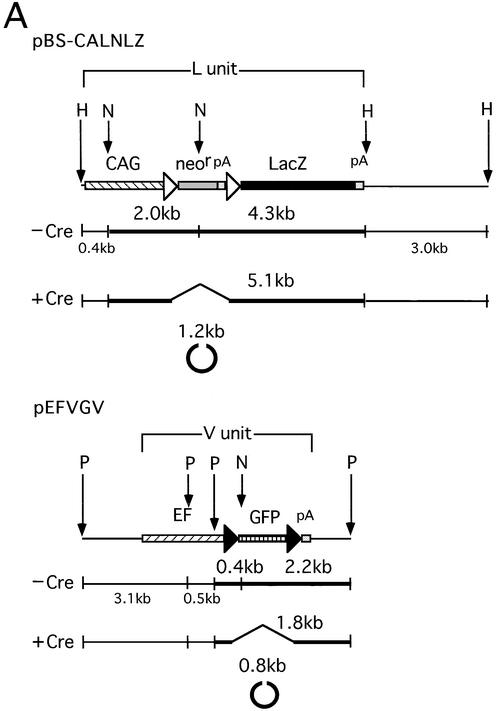
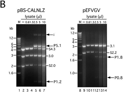
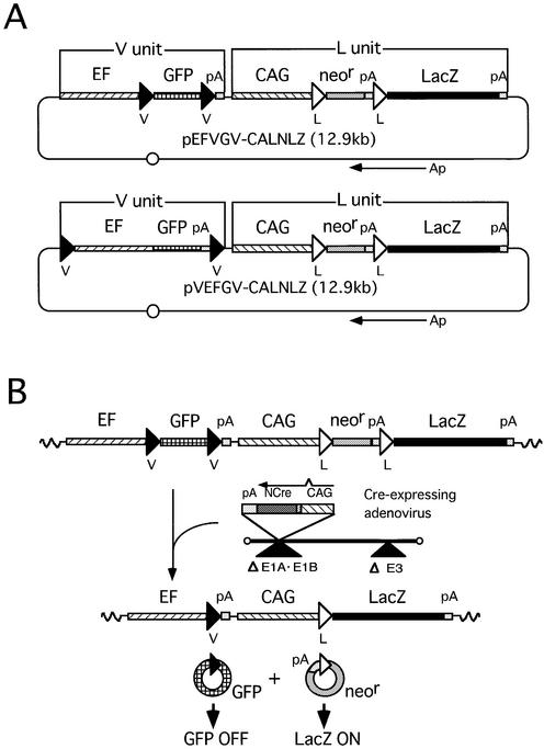
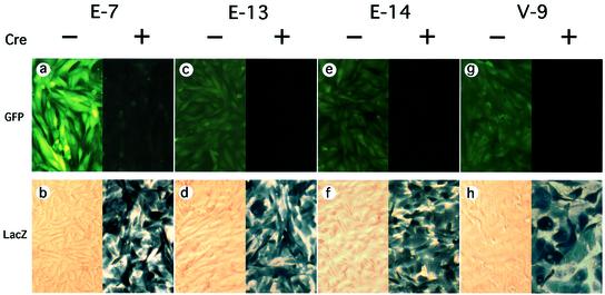
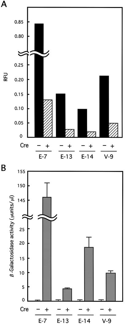
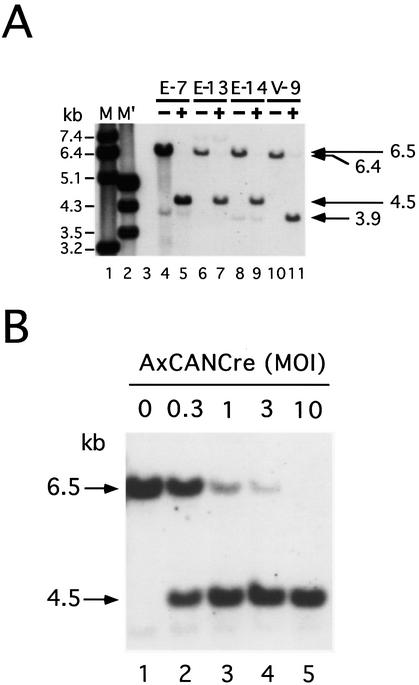
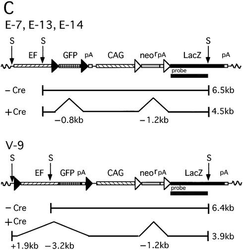
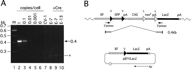
The site-specific recombinase Cre has often been used for on/off regulation of expression of transgenes introduced into the mammalian chromosome. However, this method is only applicable to the regulation of a single gene and cannot be used to simultaneously regulate two genes, because site-specific recombination occurs from the target loxP sequence of one regulating unit to the loxP sequence of any other unit and would eventually disrupt the structure of both regulating units. We previously reported a mutant loxP sequence with a two base substitution called loxP V (previously called loxP 2272), with which wild-type loxP cannot recombine but with which the identical mutant loxP recombines efficiently. In the present study we isolated cell lines bearing two regulating units on a chromosome containing a pair of wild-type loxP sequences or mutant loxP V sequences. After infection with Cre-expressing recombinant adenovirus AxCANCre, expression of a gene in each regulating unit was simultaneously turned on and off. Southern analyses showed that both regulating units were processed simultaneously and independently, even after infection with a limited amount of AxCANCre. The results showed that simultaneous regulation of gene expression on a mammalian chromosome mediated by Cre can be achieved by using mutant loxP V and wild-type loxP. This method may be a useful approach for conditional transgenic/knockout animals and investigation of gene function involving two genes simultaneously. Another possible application is for preparation of a new packaging cell line of viral vectors through simultaneous production of toxic viral genes.


Figures

Figure 1
Figure 1
In vitro recombination assay for comparison between the L target unit and V target unit. (A) Structure of substrate plasmids. (Top) Plasmid pBS-CALNLZ, substrate for L-L recombination. The Cre-mediated excisional deletion between a pair of L sequences removes both a neo resistance gene (neor) and SV40 early region poly(A) signal (pA) in the form of a circular DNA molecule. As a result of Cre recombination, a 5.1 kb band derived from outside the pair of L sequences and a 1.2 kb band derived from the NcoI-linearized circular molecule were detected. (Bottom) Plasmid pEFVGV, substrate for V-V recombination. The Cre-mediated excisional deletion between a pair of V sequences removes GFP cDNA in the form of a circular DNA molecule. As a result of Cre recombination, a 1.8 kb band derived from outside the pair of V sequences and a 0.8 kb band derived from the NcoI-linearized circular molecule were detected. N, NcoI; H, HindIII; P, PstI. See Figure 2 for the other abbreviations on the line of plasmid DNA. (B) Quantitation of in vitro recombination products. M (lanes 1 and 8), 1 kb Ladder Marker (Gibco BRL). Above the gel, amounts of Cre lysate are shown, and – indicates the negative control. After adding 10 µl of the Cre lysate to the reaction mixture, phenol/chloroform extraction was carried out to inactivate the Cre and the substrate DNA was added. S, substrate; i, intermediate; P, product.
Figure 1
Figure 1
In vitro recombination assay for comparison between the L target unit and V target unit. (A) Structure of substrate plasmids. (Top) Plasmid pBS-CALNLZ, substrate for L-L recombination. The Cre-mediated excisional deletion between a pair of L sequences removes both a neo resistance gene (neor) and SV40 early region poly(A) signal (pA) in the form of a circular DNA molecule. As a result of Cre recombination, a 5.1 kb band derived from outside the pair of L sequences and a 1.2 kb band derived from the NcoI-linearized circular molecule were detected. (Bottom) Plasmid pEFVGV, substrate for V-V recombination. The Cre-mediated excisional deletion between a pair of V sequences removes GFP cDNA in the form of a circular DNA molecule. As a result of Cre recombination, a 1.8 kb band derived from outside the pair of V sequences and a 0.8 kb band derived from the NcoI-linearized circular molecule were detected. N, NcoI; H, HindIII; P, PstI. See Figure 2 for the other abbreviations on the line of plasmid DNA. (B) Quantitation of in vitro recombination products. M (lanes 1 and 8), 1 kb Ladder Marker (Gibco BRL). Above the gel, amounts of Cre lysate are shown, and – indicates the negative control. After adding 10 µl of the Cre lysate to the reaction mixture, phenol/chloroform extraction was carried out to inactivate the Cre and the substrate DNA was added. S, substrate; i, intermediate; P, product.
Figure 2
Figure 2
Strategy for simultaneous gene regulation of the GFP gene and LacZ gene on mammalian chromosomes. EF, EF1α promoter; GFP, GFP cDNA; pA, polyadenylation signal; CAG, CAG promoter; neor, neo resistance gene; LacZ, β-galactosidase gene of E.coli; V, mutant loxP V; L, wild-type loxP; Ap, ampicillin resistance gene. (A) Structure of plasmids for simultaneous gene regulation. Each plasmid contains two Cre recombinase target units, the V unit and the L unit, respectively. Two different plasmids contain the identical L target unit containing the CAG promoter, a stuffer sequence flanked by a pair of L sequences, the LacZ gene and pA, in that order, while pEFVGV-CALNLZ (top) has a V target unit containing the EF1α promoter, GFP cDNA flanked by a pair of V sequences and pA, in that order, pVEFGV-CALNLZ (bottom) has the other V target unit containing a GFP expression unit flanked by a pair of V sequences. (B) Structure of a cell line containing the EFVGV-CALNLZ unit and the strategy of simultaneous gene regulation on mammalian chromosomes mediated by AxCANCre. Wavy line, cell chromosomal DNA.
Figure 3
Figure 3
Cre-mediated simultaneous GFP and LacZ gene regulation on mammalian chromosomes. Analysis of cell lines E-7 (a and b), E-13 (c and d), E-14 (e and f) and V-9 (g and h). (Top) (a, c, e and g) GFP portraits obtained by fluorescence microscopy. (Bottom) (b, d, f and h) X-gal staining. Cre – and Cre + refer to pre- and post-infection with AxCANCre, respectively. GFP and X-gal staining was performed 7 and 3 days after infection, respectively.
Figure 4
Figure 4
Quantitation of the expressed GFP fluorescence and β-galactosidase activity. (A) GFP expressed in each cell line was quantified before infection (–Cre) and 7 days post-infection with AxCANCre (+Cre). The fluorescence intensity (relative fluorescence units, RFU) of GFP was measured with Fluoroskan Ascent FL (Labsystems). (B) Expressed β-galactosidase activity was quantified before infection (–Cre) and 3 days post-infection with AxCANCre (+Cre). β-Galactosidase activity was quantified using CPRG and averages of three wells in a 24-well plate are shown. The lines on the graph are standard deviations.
Figure 5
Figure 5
Simultaneous recombination at gene regulation units on mammalian chromosomes. (A) Southern blot analysis of four cell lines before infection (–) and 3 days post-infection with AxCANCre at a MOI of 3 (+). M (lane 1) and M′ (lane 2) represent λ phage DNA digested with MscI and EcoRI + HindIII, respectively, as size markers. A faint extra band below the 6.5 kb band in lane 4 was derived from the region outside the detection area of the transfected plasmid DNA and was attributable to DNA contamination when preparing the probe. (B) Autoradiogram of the E-7 genome containing about five copies of regulation unit infected with AxCANCre at the MOIs indicated at the top of the gel. (C) A map of simultaneous Cre-mediated recombination at gene regulation units. The 6.5 kb band in cell lines E-7, E-13 and E-14 (top) before infection (–Cre) had shifted to a 4.5 kb band after infection (+Cre) because of the 0.8 and 1.2 kb deletion by V-V and L-L recombination, respectively. In cell line V-9 (lower), the 6.4 kb band before infection (–Cre) had shifted to a 3.9 kb band after infection (+Cre), because of a 1.2 kb deletion as a result of L-L recombination, a 3.2 kb deletion as a result of V-V recombination and 1.9 kb annexation due to the skipped SacI site. The position of the probe is shown as a closed bar. S, SacI.
Figure 5
Figure 5
Simultaneous recombination at gene regulation units on mammalian chromosomes. (A) Southern blot analysis of four cell lines before infection (–) and 3 days post-infection with AxCANCre at a MOI of 3 (+). M (lane 1) and M′ (lane 2) represent λ phage DNA digested with MscI and EcoRI + HindIII, respectively, as size markers. A faint extra band below the 6.5 kb band in lane 4 was derived from the region outside the detection area of the transfected plasmid DNA and was attributable to DNA contamination when preparing the probe. (B) Autoradiogram of the E-7 genome containing about five copies of regulation unit infected with AxCANCre at the MOIs indicated at the top of the gel. (C) A map of simultaneous Cre-mediated recombination at gene regulation units. The 6.5 kb band in cell lines E-7, E-13 and E-14 (top) before infection (–Cre) had shifted to a 4.5 kb band after infection (+Cre) because of the 0.8 and 1.2 kb deletion by V-V and L-L recombination, respectively. In cell line V-9 (lower), the 6.4 kb band before infection (–Cre) had shifted to a 3.9 kb band after infection (+Cre), because of a 1.2 kb deletion as a result of L-L recombination, a 3.2 kb deletion as a result of V-V recombination and 1.9 kb annexation due to the skipped SacI site. The position of the probe is shown as a closed bar. S, SacI.
Figure 6
Figure 6
Analysis of L-V recombination on mammalian chromosomes. (A) Ethidium bromide staining after PCR using chromosomal DNA before infection and 3 days post-infection with AxCANCre at a MOI of 3. The copy marker as a positive control was derived from plasmid pEFVLacZ, which contains the V sequence between the EF1α promoter and the LacZ gene. Copy markers for 1, 0.1, 0.01, 0.001 and 0 copies/cell were also included (lanes 2–6) to measure numbers of copies of EFVGV-CALNLZ per cell. CV1 (lane 7) and uninfected E-7 (lane 8) are also included. m (lane 1), 100 bp Ladder Marker (Gibco BRL). The asterisk shows the bands derived from CV1. (B) Structure of a putative recombination between L and V. Arrows show the positions of the 5′ and 3′ primers. Wavy line, cell chromosomal DNA. If the putative recombination between L and V occurred, recombinant structure will be the EF1α promoter sequence, loxP sequence and LacZ gene, in that order, as a result of recombination between the 5′ end of L and the 3′ end of V. The structure would be detected as a 0.4 kb band with these primers.

Similar articles

Cited by

References

    1. Sternberg N. and Hamilton,D. (1981) Bacteriophage P1 site-specific recombination. I. Recombination between loxP sites. J. Mol. Biol., 150, 467–486. - PubMed
    1. Barinaga M. (1994) Knockout mice: round two. Science, 265, 26–28. - PubMed
    1. Rossant J. and Nagy,A. (1995) Genome engineering: the new mouse genetics. Nature Med., 1, 592–594. - PubMed
    1. Rajewsky K., Gu,H., Kuhn,R., Betz,U.A., Muller,W., Roes,J. and Schwenk,F. (1996) Conditional gene targeting. J. Clin. Invest., 98, 600–603. - PMC - PubMed
    1. Lewandoski M. (2001) Conditional control of gene expression in the mouse. Nature Rev. Genet., 2, 743–755. - PubMed

Publication types

MeSH terms