

Fly and mammalian lipid phosphate phosphatase isoforms differ in activity both in vitro and in vivo
- PMID: 12856002
- PMCID: PMC1326340
- DOI: 10.1038/sj.embor.embor900
Fly and mammalian lipid phosphate phosphatase isoforms differ in activity both in vitro and in vivo
Erratum in
- EMBO Rep. 2005 Mar;6(3):289
Abstract
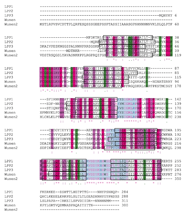
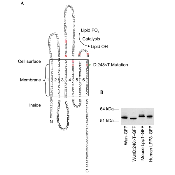
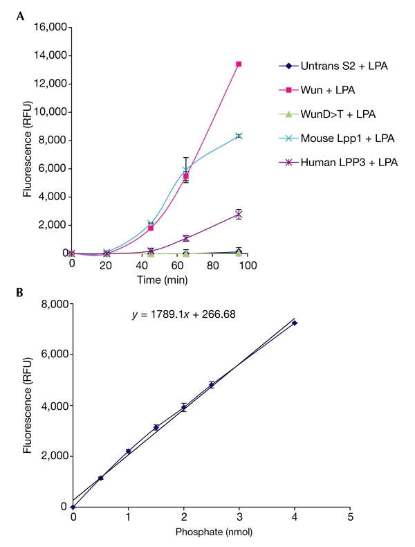
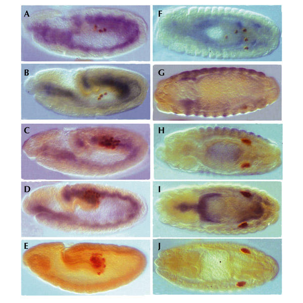
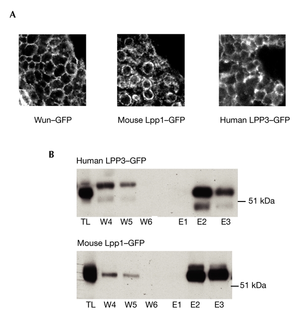
Wunen (Wun), a homologue of a lipid phosphate phosphatase (LPP), has a crucial function in the migration and survival of primordial germ cells (PGCs) during Drosophila embryogenesis. Past work has indicated that the LPP isoforms may show functional redundancy in certain systems, and that they have broad-range lipid phosphatase activities in vitro, with little apparent specificity between them. We show here that there are marked differences in biochemical activity between fly Wun and mammalian LPPs, with Wun having a narrower activity range than has been reported for the mammalian LPPs. Furthermore, although it is active on a range of substrates in vitro, mouse Lpp1 has no activity on an endogenous Drosophila germ-cell-specific factor in vivo. Conversely, human LPP3 is active, resulting in aberrant migration and PGC death. These results show an absolute difference in bioactivity among LPP isoforms for the first time in a model organism and may point towards an underlying signalling system that is conserved between flies and humans.
Figures

Similar articles
-
Lipid phosphate phosphatases dimerise, but this interaction is not required for in vivo activity.BMC Biochem. 2004 Jan 16;5:2. doi: 10.1186/1471-2091-5-2. BMC Biochem. 2004. PMID: 14725715 Free PMC article.
-
Lysophosphatidic acid and sphingosine 1-phosphate biology: the role of lipid phosphate phosphatases.Semin Cell Dev Biol. 2004 Oct;15(5):491-501. doi: 10.1016/j.semcdb.2004.05.007. Semin Cell Dev Biol. 2004. PMID: 15271294 Review.
-
Wunen, a Drosophila lipid phosphate phosphatase, is required for septate junction-mediated barrier function.Development. 2012 Jul;139(14):2535-46. doi: 10.1242/dev.077289. Epub 2012 Jun 6. Development. 2012. PMID: 22675212
-
Compartmentalizing the embryo: lipids and septate junction mediated barrier function.Fly (Austin). 2013 Jan-Mar;7(1):18-22. doi: 10.4161/fly.22938. Epub 2012 Dec 6. Fly (Austin). 2013. PMID: 23221483 Free PMC article.
-
Structural organization of mammalian lipid phosphate phosphatases: implications for signal transduction.Biochim Biophys Acta. 1999 Jul 30;1439(2):299-316. doi: 10.1016/s1388-1981(99)00102-x. Biochim Biophys Acta. 1999. PMID: 10425403 Review.
Cited by
-
Diverse roles of phosphatidate phosphatases in insect development and metabolism.Insect Biochem Mol Biol. 2021 Jun;133:103469. doi: 10.1016/j.ibmb.2020.103469. Epub 2020 Sep 12. Insect Biochem Mol Biol. 2021. PMID: 32931938 Free PMC article. Review.
-
Lipid phosphate phosphatase 3 stabilization of beta-catenin induces endothelial cell migration and formation of branching point structures.Mol Cell Biol. 2010 Apr;30(7):1593-606. doi: 10.1128/MCB.00038-09. Epub 2010 Feb 1. Mol Cell Biol. 2010. PMID: 20123964 Free PMC article.
-
Sex-specific DoublesexM expression in subsets of Drosophila somatic gonad cells.BMC Dev Biol. 2007 Oct 12;7:113. doi: 10.1186/1471-213X-7-113. BMC Dev Biol. 2007. PMID: 17935627 Free PMC article.
-
Origin and establishment of the germline in Drosophila melanogaster.Genetics. 2025 Apr 17;229(4):iyae217. doi: 10.1093/genetics/iyae217. Genetics. 2025. PMID: 40180587 Free PMC article. Review.
-
Lipid Phosphate Phosphatases and Cancer.Biomolecules. 2020 Sep 2;10(9):1263. doi: 10.3390/biom10091263. Biomolecules. 2020. PMID: 32887262 Free PMC article. Review.
References
-
- Barila D. et al. . ( 1996) The Dri 42 gene, whose expression is up-regulated during epithelial differentiation, encodes a novel endoplasmic reticulum resident transmembrane protein. J. Biol. Chem., 271, 29928–29936. - PubMed
-
- Brindley D. & Waggoner W. ( 1998) Mammalian lipid phosphate phosphohydrolases. J. Biol. Chem., 273, 24281–24284. - PubMed
-
- Dillon D.A. et al. . ( 1997) Mammalian Mg2+-independent phosphatidate phosphatase (PAP2) displays diacylglycerol pyrophosphate phosphatase activity. J. Biol. Chem., 272, 10361–10366. - PubMed
-
- Greig S. & Akam M. ( 1993) Homeotic genes autonomously specify one aspect of pattern in the Drosophila mesoderm. Nature, 362, 630–632. - PubMed
-
- Hofmann K. & Stoffel W. ( 1993) TMbase—a database of membrane spanning proteins segments. Biol. Chem. Hoppe Seyler, 374, 166.
Publication types
MeSH terms
Substances
Grants and funding
LinkOut - more resources
Full Text Sources
Molecular Biology Databases
Miscellaneous
