. 2003 Oct 10;278(41):39330-6.
doi: 10.1074/jbc.M305580200. Epub 2003 Jul 11.

A single internal ribosome entry site containing a G quartet RNA structure drives fibroblast growth factor 2 gene expression at four alternative translation initiation codons

Affiliations

A single internal ribosome entry site containing a G quartet RNA structure drives fibroblast growth factor 2 gene expression at four alternative translation initiation codons

Sophie Bonnal et al. J Biol Chem. .

Abstract

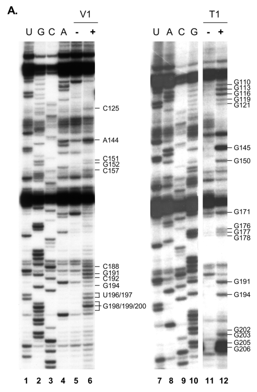
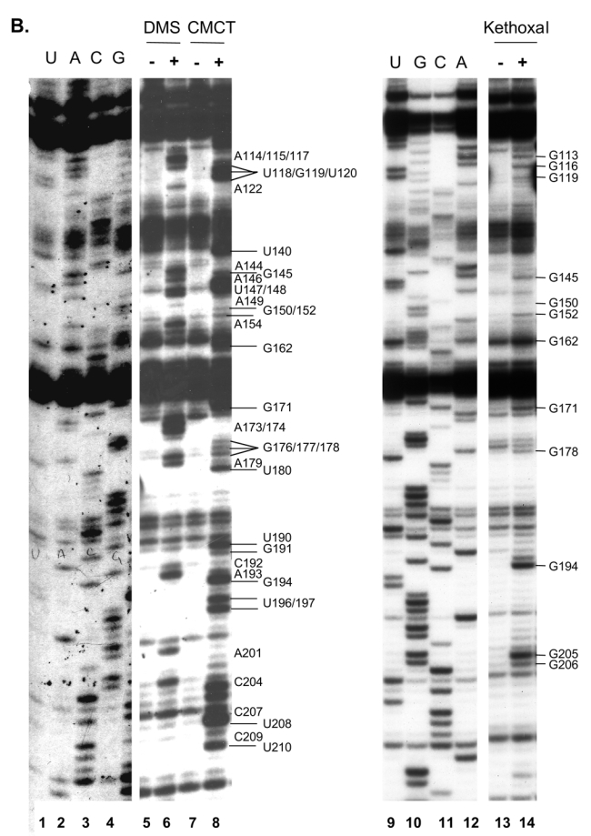
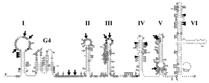
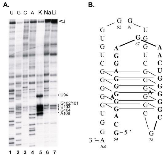
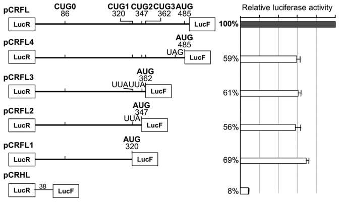
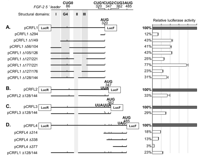
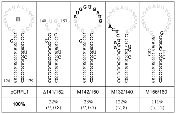
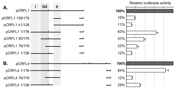
The 484-nucleotide (nt) alternatively translated region (ATR) of the human fibroblast growth factor 2 (FGF-2) mRNA contains four CUG and one AUG translation initiation codons. Although the 5'-end proximal CUG codon is initiated by a cap-dependent translation process, the other four initiation codons are initiated by a mechanism of internal entry of ribosomes. We undertook here a detailed analysis of the cis-acting elements defining the FGF-2 internal ribosome entry site (IRES). A thorough deletion analysis study within the 5'-ATR led us to define a 176-nt region as being necessary and sufficient for IRES function at four codons present in a downstream 308-nt RNA segment. Unexpectedly, a single IRES module is therefore responsible for translation initiation at four distantly localized codons. The determination of the FGF-2 5'-ATR RNA secondary structure by enzymatic and chemical probing experiments showed that the FGF-2 IRES contained two stem-loop regions and a G quartet motif that constitute novel structural determinants of IRES function.


Figures

Figure 1
Figure 1
Enzymatic (panel A) and chemical (panel B) probing of in vitro transcribed FGF-2 5′ ATR. Cleavage and modification sites were detected by primer extension using the 32P-5′ end-labelled RT285 oligonucleotide that hybridizes at position 285 to 303 of the FGF-2 RNA. The resulting cDNA was separated on 8% polyacrylamide/8 M urea sequencing gel and analyzed by autoradiography. RNA sequencing reactions were run in parallel. The nature and positions of cleaved (panel A) and modified (panel B) bases are indicated on the right side of each panel. RNAse V1 (V1), RNAse T1 (T1), or the three chemical agents (DMS, CMCT or Kethoxal) were added (lane +) or not (lane −) prior to the reverse transcription step.
Figure 1
Figure 1
Enzymatic (panel A) and chemical (panel B) probing of in vitro transcribed FGF-2 5′ ATR. Cleavage and modification sites were detected by primer extension using the 32P-5′ end-labelled RT285 oligonucleotide that hybridizes at position 285 to 303 of the FGF-2 RNA. The resulting cDNA was separated on 8% polyacrylamide/8 M urea sequencing gel and analyzed by autoradiography. RNA sequencing reactions were run in parallel. The nature and positions of cleaved (panel A) and modified (panel B) bases are indicated on the right side of each panel. RNAse V1 (V1), RNAse T1 (T1), or the three chemical agents (DMS, CMCT or Kethoxal) were added (lane +) or not (lane −) prior to the reverse transcription step.
Figure 2
Figure 2
RNA secondary structure model of the FGF-2 5′ ATR showing results from enzymatic cleavage and chemical modifications experiments. Domains I to VI are indicated as well as the G quartet motif (G4). White and black arrows represent weak-moderate and strong RNAse T1 cleavage sites respectively. White and black triangles represent weak-moderate and strong RNAse VI cleavage sites respectively. Dashed, white and black circles represent weak, moderate and strong modifications by DMS, CMCT or kethoxal respectively, x marks represent reverse transcriptase pauses, with those that are cation-dependent boxed. Lower case letters are positions with non determined reactivity. Translation initiation codons are boxed.
Figure 3
Figure 3
The FGF-2 G quartet motif. (A) Cation-dependent termination of reverse transcriptase at positions 94 to 106 of the FGF-2 5′ ATR. Reverse transcriptase reactions were performed in presence of 100mM KCl (lane 5), 100 mM NaCl (lane 6) or 100 mM LiCl (lane 7) and were run together with RNA sequencing reactions (lanes 1 to 4). Strong pauses are indicated by asteriks whereas the full-length extension product is marked with an open triangle. (B) Schematic model of the FGF-2 G-quartet motif. Lanes between guanine bases represent the Hoogsteen interactions that occur in a square-planar symmetric array
Figure 4
Figure 4
Schematic representation of bicistronic FGF-2 RNAs and their efficiency in directing internal entry of ribosome. The five translation initiation codons, their mutation to non-initiation UUA codons in the pCRFL2 and pCRFL3 constructs and the insertion of a UAG stop codon in the pCRFL 4 construct are shown. Relative luciferase activity is the LucF/LucR ratio in transiently transfected SK-Hep1 cells. The average relative luciferase activity was calculated from at least six experiments. An arbitrary value of 100 was set for the pCRFL construct. The pCRHL construct contains a 38 nts-long intercistronic spacer, as described in (10).
Figure 5
Figure 5
Mapping the domains required for IRES-mediated translation at each of the FGF-2 translation initiation codons. A schematic representation of the FGF-2 5′ leader RNA and the position of the structural domains I to III and G4 (see figure 4) are shown. Relative luciferase activities were calculated for each of deletion constructs transiently transfected in SK-Hep1 cells from at least four independent experiments performed in duplicate. An arbitrary value of 100 was set for the pCRFL1 construct in panel A, for pCRFL2 in panel B, for pCRFL3 in panel C and for pCRFL4 in panel D.
Figure 6
Figure 6
Analyzing the requirements for FGF-2 IRES-mediated translation in stem-loop II. Schematic representation of the wild type stem-loop II and of the different mutants. Wild type nucleotides in the loop are in gray characters whereas mutated nucleotides are in bold characters. The mutants were constructed in the pCRFL1 plasmid. The values for the average relative luciferase activities in transiently transfected SK-Hep 1 cells are shown at the bottom and were calculated from at least four experiments performed in duplicate. An arbitrary value of 100 was set for the parental pCRFL1 plasmid.
Figure 7
Figure 7
Mapping the domains sufficient for IRES-mediated translation. The various constructs were transfected in SK-Hep1 cells and the average relative luciferase activities were calculated from three independent experiments performed in duplicate.

Similar articles

Cited by

References

    1. Bikfalvi A, Klein S, Pintucci G, Rifkin DB. Endocr Rev. 1997;18:26–45. - PubMed
    1. Florkiewicz RZ, Sommer A. Proc Natl Acad Sci U S A. 1989;86:3978–3981. - PMC - PubMed
    1. Prats H, Kaghad M, Prats AC, Klagsbrun M, Lelias JM, Liauzun P, Chalon P, Tauber JP, Amalric F, Smith JA, et al. Proc Natl Acad Sci U S A. 1989;86:1836–1840. - PMC - PubMed
    1. Arnaud E, Touriol C, Boutonnet C, Gensac MC, Vagner S, Prats H, Prats AC. Mol Cell Biol. 1999;19:505–514. - PMC - PubMed
    1. Vagner S, Touriol C, Galy B, Audigier S, Gensac MC, Amalric F, Bayard F, Prats H, Prats AC. J Cell Biol. 1996;135:1391–1402. - PMC - PubMed

Publication types