. 2003 Jul;132(3):1228-40.
doi: 10.1104/pp.102.017707.

Cytokinin oxidase gene expression in maize is localized to the vasculature, and is induced by cytokinins, abscisic acid, and abiotic stress

Affiliations

Cytokinin oxidase gene expression in maize is localized to the vasculature, and is induced by cytokinins, abscisic acid, and abiotic stress

Norbert Brugière et al. Plant Physiol. 2003 Jul.

Abstract

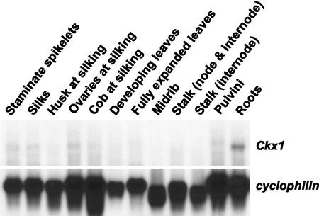
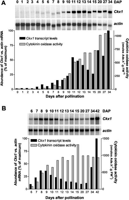
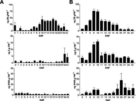
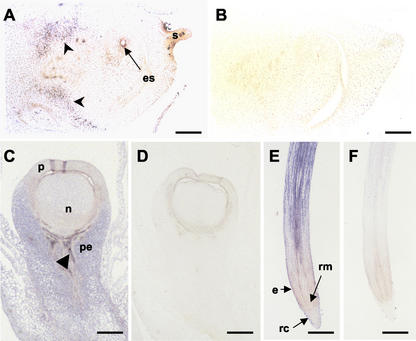
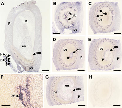
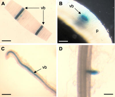
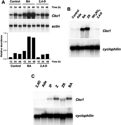
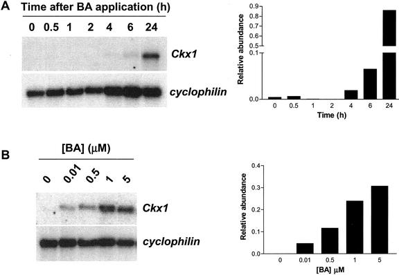
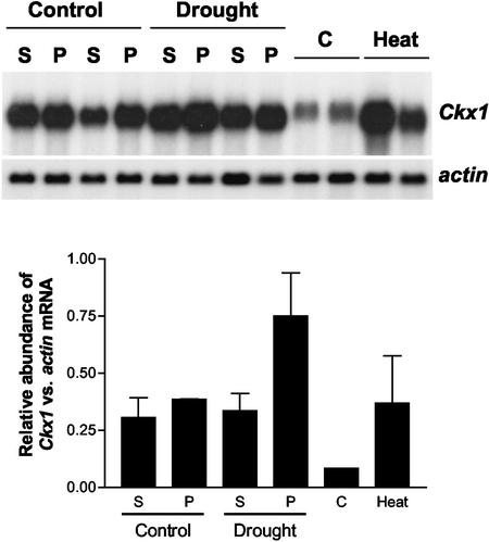
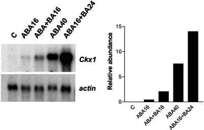
Cytokinins are hormones that play an essential role in plant growth and development. The irreversible degradation of cytokinins, catalyzed by cytokinin oxidase, is an important mechanism by which plants modulate their cytokinin levels. Cytokinin oxidase has been well characterized biochemically, but its regulation at the molecular level is not well understood. We isolated a cytokinin oxidase open reading frame from maize (Zea mays), called Ckx1, and we used it as a probe in northern and in situ hybridization experiments. We found that the gene is expressed in a developmental manner in the kernel, which correlates with cytokinin levels and cytokinin oxidase activity. In situ hybridization with Ckx1 and transgenic expression of a transcriptional fusion of the Ckx1 promoter to the Escherichia coli beta-glucuronidase reporter gene revealed that the gene is expressed in the vascular bundles of kernels, seedling roots, and coleoptiles. We show that Ckx1 gene expression is inducible in various organs by synthetic and natural cytokinins. Ckx1 is also induced by abscisic acid, which may control cytokinin oxidase expression in the kernel under abiotic stress. We hypothesize that under non-stress conditions, cytokinin oxidase in maize plays a role in controlling growth and development via regulation of cytokinin levels transiting in the xylem. In addition, we suggest that under environmental stress conditions, cytokinin oxidase gene induction by abscisic acid results in aberrant degradation of cytokinins therefore impairing normal development.


Figures

Figure 1.
Figure 1.
Expression of Ckx1 in different maize organs. Northern-blot analysis of Ckx1 transcript levels in different organs of maize (B73). Poly(A) RNA (3 μg) was extracted from the organs indicated. The same blot was hybridized with cyclophilin, which served as a loading control.
Figure 2.
Figure 2.
Relative abundance of Ckx1 transcripts and cytokinin oxidase activity during B73 kernel development. A, Ckx1 transcript levels were measured in 0- to 5-DAP kernels with pedicels and 6- to 34-DAP kernels without pedicels using [α-32P]dCTP labeled Ckx1 DNA probe. The same nylon membrane was stripped and probed with actin1-9. Black bars indicate the relative abundance of Ckx1 versus actin transcripts. Gray bars indicate cytokinin oxidase activity measured in the same samples. B, Ckx1 transcript levels were measured in pedicel samples of 6- to 42-DAP kernels. Transcripts (black bars) were detected, quantified, and normalized to actin transcript levels using the same procedure as in A. Cytokinin oxidase activity (gray bars) was measured as indicated in A. Poly(A) RNA (3 μg) were used for each northern-blot experiment.
Figure 3.
Figure 3.
Cytokinin levels measured in developing B73 kernels. Levels of ZR, Z, and iPAR were measured in the same samples used for analysis in Figure 2. A, Cytokinin levels in developing kernels from 0 to 5 DAP and in kernels of which the pedicel (pedicel/placental-chalazal region) was separated from the rest of the kernel starting at 6 DAP until 42 DAP. B, Cytokinin levels in pedicels samples collected from 6- to 42-DAP kernels.
Figure 4.
Figure 4.
In situ localization of Ckx1 transcripts in developing ovules and roots. A, An antisense RNA probe was used to detect Ckx1 sense transcripts on tissue sections of developing ovules 7 weeks after planting in genotype An1Bz2. B, Negative control probed with a sense Ckx1 probe. Bars = 350 μm. C, Detection of Ckx1 transcripts in developing ovules at silking (9 weeks after planting) in genotype An1Bz2. D, Negative controls probed with Ckx1 sense probe. Bars = 500 μm. E, Localization of Ckx1 transcripts in roots of plants at the V6 stage of inbred N46. F, Negative control probed with Ckx1 sense probe. Bars = 750 μm. Arrowheads (A and C) indicate signal detected in the vasculature. es, Embryo sac; n, nucellus; P, pericarp; pe, pedicel; e, epidermis; rc, root cap; rm, root meristem; and s, silk.
Figure 5.
Figure 5.
In situ localization of Ckx1 transcripts in 8-DAP kernels. A, A longitudinal section of an 8-DAP kernel (inbred N46) probed with an antisense Ckx1 probe. B through G, Transverse sections as indicated in A probed with an antisense Ckx1 probe. F, Close-up of C identified by the square; scale bar = 50 μm. H, Transverse section using sense probe as a control. en, Endosperm; em, embryo; n, nucellus; p, pericarp; pe, pedicel; vb, vascular bundles; and xy, xylem. Scale bar = 500 μm, except for F.
Figure 6.
Figure 6.
Characterization of transgenic maize plants expressing a fusion of the Ckx1 promoter and the GUS gene. A, GUS staining of a coleoptile cross-section showing strong labeling in the vascular bundles (vb). Bar = 3 mm. B, Close-up of a transverse coleoptile section showing intense labeling in the vascular bundles. Bar = 500 μm. C, Staining of lateral root showing GUS staining in the vascular bundle. Bar = 250 μm. D, Staining of a primary root showing activity in emerging lateral root. Bar = 250 μm. vb, Vascular bundle; and P, parenchyma.
Figure 7.
Figure 7.
Effect of exogenous application of cytokinins on Ckx1 transcript levels in different organs. A, Expression of Ckx1 in B73 4-DAP kernels after incubation for 12, 24, or 48 h in a solution containing water (control) or in water containing 10 μm BA or 2,4-D and quantification of transcript abundance compared with actin transcript levels (arbitrary units). Five micrograms of poly(A) was used for this experiment. B, Effect of exogenous application of cytokinins on Ckx1 transcript levels in roots. The root systems of 2-week-old seedlings were wrapped in germ paper soaked with deionized water (Control), 10 μm Ade, BA, ZR, 10 mm ammonium nitrate (NH4NO3), or 10 μm 2,4-D. Root systems were collected after 48 h of incubation, and 3 μg of poly(A) RNA was used for northern-blot quantification of Ckx1 transcripts. C, Ckx1 transcript levels in leaf discs incubated in the presence of 2,4-D, Ade, isopentenyladenine (iP), Z, ZR, or BA at 10 μm. Three micrograms of poly(A) RNA was used. Cyclophilin was used as a loading control.
Figure 8.
Figure 8.
Time course and dose responsiveness of Ckx1 transcript accumulation to BA application. A, Ckx1 transcript levels in leaf discs (B73) incubated in the presence of 10 μm BA for the indicated time period. B, Ckx1 transcript levels in leaf discs (B73) incubated with different concentrations of BA for 24 h. Three micrograms of poly(A) RNA was used. Cyclophilin was used as a loading control. The relative abundance of transcripts (arbitrary units) is indicated in the graph.
Figure 9.
Figure 9.
Effect of abiotic stress on Ckx1 expression in developing kernels. A drought stress was applied to growth chamber-grown plants (hybrid 3732) at 4 DAP by withholding water until 8 DAP. At that point, glumes were removed from developing seeds, and the pedicel region (P) was separated from the rest of the kernel (S). For the heat stress experiment, field-grown Mo17 plants ears were heat stressed, and transcript levels were measured in kernels of which the pedicel had been removed. Transcripts levels were measured using Ckx1 or actin as a probe and the relative abundance of transcripts is indicated in the graph (arbitrary units). Error bars represent the se. Four micrograms of poly(A) RNA was used.
Figure 10.
Figure 10.
Effect of ABA on accumulation of Ckx1 in leaf discs. Leaf discs were floated either for 40 h on distilled water (control) or for 16 h (ABA16) or 40 h (ABA40) on 10 μm ABA. Effect of ABA and BA cotreatment was studied by incubating leaf discs with ABA + BA (10 μm each) for 16 h (ABA+BA16) or with 10 μm BA for 24 h after pre-incubation with ABA for 16 h (ABA16+BA24). The graph to the right shows the relative abundance of Ckx1 and actin transcripts (arbitrary units). Three micrograms of poly(A) RNA was used.

References

    1. Armstrong DJ (1994) Cytokinin oxidase and the regulation of cytokinin degradation. In DWS Mok, MC Mok, eds, Cytokinins, Chemistry, Activity and Function. CRC Press, Boca Raton, FL, pp 139–154
    1. Bertell G, Eliasson L (1992) Cytokinin effects on root growth and possible interactions with ethylene and indol-3-acetic acid. Physiol Plant 84: 255–261
    1. Beveridge CA, Murfet IC, Kerhoas L, Sotta B, Miginiac E, Rameau C (1997) The shoot controls zeatin riboside export from pea roots: evidence from the branching mutant rms4. Plant J 11: 339–345
    1. Bilyeu KD, Cole JL, Laskey JG, Riekhof WR, Esparza TJ, Kramer MD, Morris RO (2001) Molecular and biochemical characterization of a cytokinin oxidase from maize. Plant Physiol 125: 378–386 - PMC - PubMed
    1. Bradley D, Carpenter R, Sommer H, Hartley N, Coen E (1993) Complementary floral homeoetic phenotypes result from opposite orientation of a transposon at the plena locus of Antirrhinum. Cell 72: 85–95 - PubMed

MeSH terms

LinkOut - more resources