. 2003 Jul;14(7):2728-43.
doi: 10.1091/mbc.e02-11-0767. Epub 2003 Mar 20.

Myo6 facilitates the translocation of endocytic vesicles from cell peripheries

Affiliations

Myo6 facilitates the translocation of endocytic vesicles from cell peripheries

Laura Aschenbrenner et al. Mol Biol Cell. 2003 Jul.

Abstract

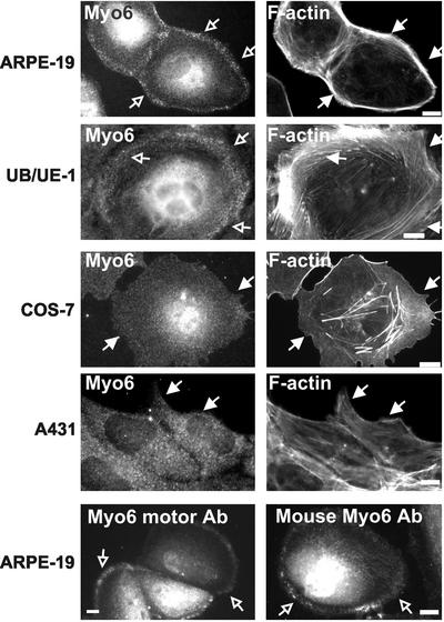
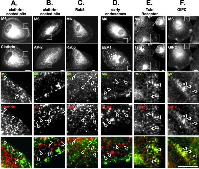
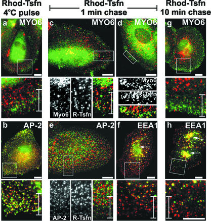
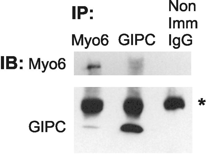
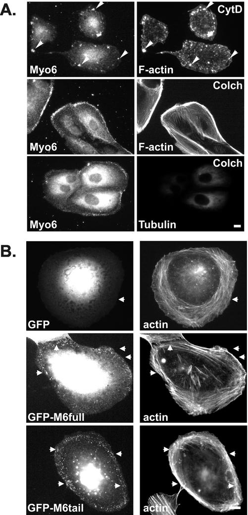
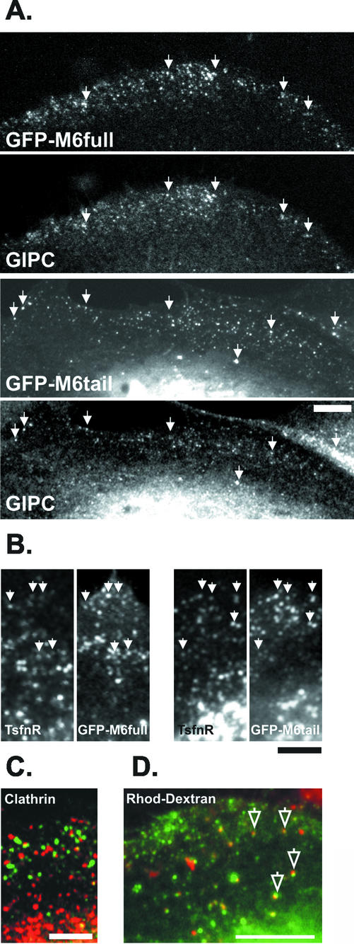
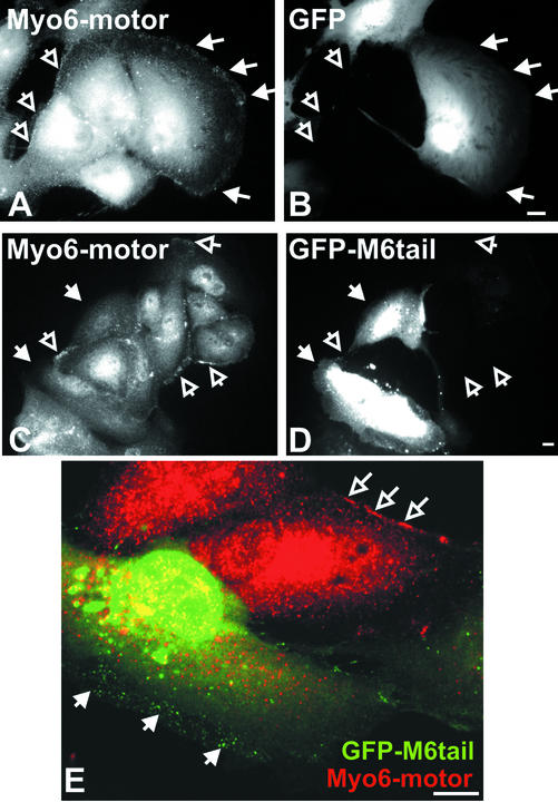
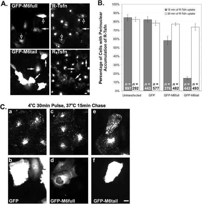
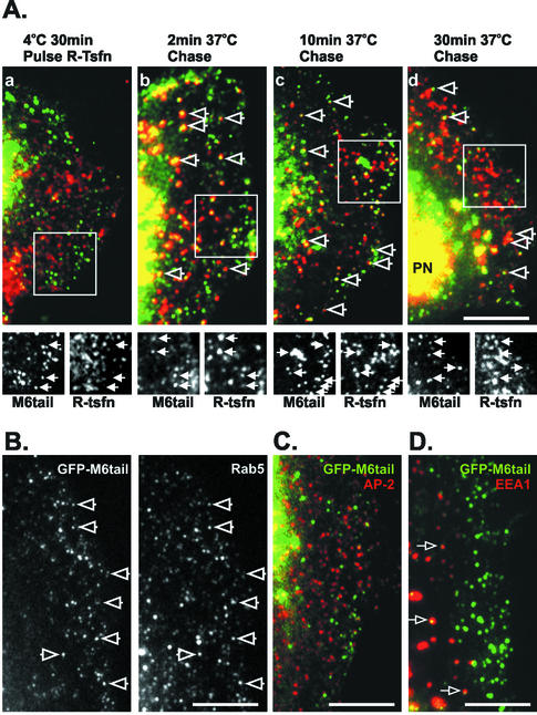
Immunolocalization studies in epithelial cells revealed myo6 was associated with peripherally located vesicles that contained the transferrin receptor. Pulse-chase experiments after transferrin uptake showed that these vesicles were newly uncoated endocytic vesicles and that myo6 was recruited to these vesicles immediately after uncoating. GIPC, a putative myo6 tail binding protein, was also present. Myo6 was not present on early endosomes, suggesting that myo6 has a transient association with endocytic vesicles and is released upon early endosome fusion. Green fluorescent protein (GFP) fused to myo6 as well as the cargo-binding tail (M6tail) alone targeted to the nascent endocytic vesicles. Overexpression of GFP-M6tail had no effect on a variety of organelle markers; however, GFP-M6tail displaced the endogenous myo6 from nascent vesicles and resulted in a significant delay in transferrin uptake. Pulse-chase experiments revealed that transferrin accumulated in uncoated vesicles within the peripheries of transfected cells and that Rab5 was recruited to the surface of these vesicles. Given sufficient time, the transferrin did traffic to the perinuclear sorting endosome. These data suggest that myo6 is an accessory protein required for the efficient transportation of nascent endocytic vesicles from the actin-rich peripheries of epithelial cells, allowing for timely fusion of endocytic vesicles with the early endosome.


Figures

Figure 1.
Figure 1.
Localization of myo6 in cultured cell lines. Indirect immunofluorescence staining of cells by using affinity-purified rabbit antibodies directed to the tail domain of myo6 (Myo6), the motor domain of myo6 (Myo6 motor Ab) or affinity-purified mouse antibodies directed to the tail domain (Mouse Myo6 Ab). Cells were counterstained for F-actin by using rhodamine-conjugated phalloidin. The cortical actin is demarcated by filled arrows. Myo6 is enriched in a peripheral punctate compartment (open arrows) that overlaps with the cortical actin in both ARPE-19 and UB/UE-1 cell lines. Bar, 10 μm.
Figure 2.
Figure 2.
Myo6 overlaps with GIPC, transferrin receptor, and Rab5 on vesicles distinct from clathrin-coated pits and EEA1-positive early endosomes. (A–F) ARPE-19 cells were double labeled with antibodies directed to the tail domain of myo6 (M6; detected with a fluorescein isothiocyanate-conjugated secondary antibody) and to components of the endocytic pathway (detected with a rhodamine-conjugated secondary antibody). The top panels show an example of the whole-cell staining pattern seen for myo6 and the markers. The boxed area in the top panels is enlarged in the bottom panels. In the overlay, myo6 is shown in green, the markers are shown in red, and overlap between the two markers is yellow. Staining was for clathrin heavy chain (A) the clathrin adaptor AP-2 (B), Rab5 (C), EEA1 (D), TsfnR (E), and GIPC (F). Bars (A–F), 10 μm.
Figure 3.
Figure 3.
Myo6 is recruited to uncoated endocytic vesicles. ARPE-19 cells were incubated with R-Tsfn at 4°C for 30 min. After surface labeling, the cells were either formaldehyde fixed (a and b) or warmed to 37°C for a 1-min chase (c–f) or a 10-min chase (g and h) before fixation. Cells were stained using antibodies to the tail domain of myo6 (a, c, d, and g), the clathrin-adapter AP-2 (b and e), or the early endosome protein EEA1 (f and h). Antibodies were visualized with a fluorescein-conjugated secondary antibody (green) and compared with the endocytosed rhodamine-conjugated transferrin (red) by immunofluorescence microscopy. Overlap between the R-Tsfn and either Myo6, AP-2, or EEA1 is shown in yellow. Cell peripheries of flat cells were chosen for analysis to allow clear visualization of the R-Tsfn as it moved through the endocytic pathway. Boxed areas in each top panel are enlarged in the bottom panel. The perinuclear early endosome is shown with arrows in f and h. Bars and brackets found in enlarged bottom panels are 10 μm.
Figure 4.
Figure 4.
Coimmunoprecipiation of myo6 with GIPC. Extracts from ARPE-19 cells were immunoprecipitated (IP) with antibodies to Myo6, GIPC, or nonimmune IgG. Immunoprecipitated proteins were detected by immunoblot (IB) analysis with antibodies to myo6 (top) and GIPC (bottom). The position of the IgG is marked with an asterisk.
Figure 5.
Figure 5.
Myo6 localization is dependent on F-actin and its C-terminal tail domain. (A) ARPE-19 cells were treated for 30 min with cytochalasin D (CytD) or colchicine (Colch) before staining with antibodies to myo6 and rhodamine-conjugated phalloidin or antitubulin antibody. Cytochalasin D treatment resulted in a collapse of the myo6-associated endocytic vesicles into peripheral clumps (arrowheads), whereas colchicine treatment had no effect on the location of the myo6-associated vesicles. (B) Fluorescence microscopy of ARPE-19 cells expressing GFP, GFP fused to full-length myo6 (GFP-M6full), or GFP fused to the C-terminal globular tail domain of myo6 (GFP-M6tail). Cells were costained with rhodamine-conjugated phalloidin to stain F-actin. Arrowheads point out actin-rich regions. Bars, 10 μm.
Figure 6.
Figure 6.
Myo6-GFP fusion proteins colocate with GIPC and Ts-fnR on an endocytic compartment in peripheries of ARPE-19 cells. Cells expressing GFP-M6full or GFP-M6tail were double stained with antibodies to GIPC (A) or the transferrin receptor (TsfnR) (B) and viewed by confocal microscopy. Both the full-length myo6 and the tail domain of myo6 targeted to the GIPC-associated, TsfnR-containing endocytic vesicles (arrowheads). (C) Cells expressing GFP-M6full (green) were stained with antibodies to clathrin (red). (D) Cells expressing GFP-M6tail (green) were incubated for 10 min with Rhod-dextran (red) to label endocytic compartments before fixation and visualization the confocal microscope. Myo6-tail-associated endocytic vesicles contain Rhod-dextran (open arrows; yellow color denotes overlap). Bars, 10 μm.
Figure 7.
Figure 7.
GFP-M6tail displaces myo6 from peripheral endocytic vesicles. Immunofluorescence microscopy of ARPE-19 cells transiently transfected with GFP (A and B) or GFP-M6tail (C–E) and stained with antibodies to the motor domain of myo6. GFP fluorescence is shown in B, D, and E (green), and myo6-motor antibodies staining is shown in A, C, and E (red). The region of the cell where peripheral vesicles are found is demarcated by filled arrows in transfected cells and by open arrows in untransfected cells. Bar, 10 μm.
Figure 8.
Figure 8.
Overexpression of the tail domain of myo6 delays endocytosis of transferrin. GFP, GFP-M6full, and GFP-M6tail–transfected cultures were incubated with R-Tsfn for either 15 or 30 min. (A) Overview of GFP-M6full and GFP-M6tail–transfected ARPE-19 cells after 15-min incubation with R-Tsfn. GFP-M6tail–expressing cells (demarcated by closed arrows) exhibited a decrease in net transferrin uptake, as is evidenced by a lack of perinuclear staining. In contrast, the majority of GFP-M6full–expressing cells accumulate transferrin normally (open arrows) compared with untransfected cells in the field. (B) Percentage of transfected and untransfected cells showing labeling of perinuclear endosomes after 15 min (gray bars) or 30 min (white bars) of R-Tsfn uptake. The total number of cells counted for each experiment is denoted as n. (C) Higher magnification view of GFP- (a and b), GFP-M6full–(c and d), and GFP-M6tail (e and f)–transfected cells after 15 min of incubation with R-Tsfn. Transferrin is shown in the top panels (a, c, and e); GFP in the bottom panels (b, d, and f). GFP-M6tail–transfected cells exhibit an increase in peripheral R-Tsfn staining, not seen in other cells. Bars, 10 μm.
Figure 9.
Figure 9.
GFP-M6tail is recruited to transferrin-containing uncoated endocytic vesicles, slowing traffic of transferrin to the early endosome. A portion of a transfected cell is shown in each panel, oriented with the nucleus positioned to the left and the cell periphery to the right. (A) Pulse-chase experiments after R-Tsfn in GFP-M6tail–transfected ARPE-19 cells. Transfected cells were incubated with R-Tsfn at 4°C for 30 min (a) and then warmed to 37°C for 2 min (b), 10 min (c), or 30 min (d). Open arrows point to overlap between the R-Tsfn (red) and the GFP-M6tail (green) staining at the periphery of each cell. The boxed region is presented at the same scale as separate M6tail and R-Tsfn images below each panel. The position of GFP-M6tail–associated vesicles is denoted with arrows in both lower panels. PN in d demarcates the position of the perinuclear early endosome. (B) Localization of rab5 in GFP-M6tail–transfected ARPE-19 cells. Open arrows mark the position of GFP-M6tail–associated endocytic vesicles that also contain rab5. (C) Localization of AP-2 (red) in GFP-M6tail (green)–transfected ARPE-19 cells reveals no overlap in CCPs. (D) Localization of EEA1 (red) in GFP-M6tail (green)–transfected ARPE-19 cells reveals that a subset of the GFP-M6tail is associated with peripherally located endosomes. Overlap in location is seen as yellow and is demarcated with an arrow. Bars, 10 μm.

References

    1. Apodaca, G. (2001). Endocytic traffic in polarized epithelial cells: role of the actin and microtubule cytoskeleton. Traffic 2, 149–159. - PubMed
    1. Avraham, K.B., Hasson, T., Sobe, T., Balsara, B., Testa, J., Copeland, N.G., and Jenkins, N.A. (1997). Characterization of human unconventional myosin-VI, a gene responsible for deafness in Snell's waltzer mice. Hum. Mol. Genet. 6, 1225–1231. - PubMed
    1. Avraham, K.B., Hasson, T., Steel, K.P., Kingsley, D.M., Russell, L.B., Mooseker, M.S., Copeland, N.G., and Jenkins, N.A. (1995). The mouse Snell's waltzer deafness gene encodes an unconventional myosin required for structural integrity of inner hair cells. Nat. Genet. 11, 369–374. - PubMed
    1. Biemesderfer, D., Mentone, S.A., Mooseker, M., and Hasson, T. (2002). Expression of myosin-VI within the endocytic pathway in the adult and developing proximal tubule. Am. J. Physiol. 282, F785–F794. - PubMed
    1. Breckler, J., Au, K., Cheng, J., Hasson, T., and Burnside, B. (2000). Novel myosin VI isoform is abundantly expressed in retina. Exp. Eye Res. 70, 121–134. - PubMed

Publication types

MeSH terms

LinkOut - more resources