. 2003 Jul;14(7):2744-55.
doi: 10.1091/mbc.e02-11-0740.

Rpb4p, a subunit of RNA polymerase II, mediates mRNA export during stress

Affiliations

Rpb4p, a subunit of RNA polymerase II, mediates mRNA export during stress

Marganit Farago et al. Mol Biol Cell. 2003 Jul.

Abstract

Changes in gene expression represent a major mechanism by which cells respond to stress. We and other investigators have previously shown that the yeast RNA polymerase II subunit Rpb4p is required for transcription under various stress conditions, but not under optimal growth conditions. Here we show that, in addition to its role in transcription, Rpb4p is also required for mRNA export, but only when cells are exposed to stress conditions. The roles of Rpb4p in transcription and in mRNA export can be uncoupled genetically by specific mutations in Rpb4p. Both functions of Rpb4p are required to maintain cell viability during stress. We propose that Rpb4p participates in the cellular responses to stress at the interface of the transcription and the export machineries.


Figures

Figure 4.
Figure 4.
Sequence alignment of Rpb4p and its mutant forms. The predicted amino acid sequence of WT Rpb4p and the Rpb4p moiety of the GFP-rpb4 mutants that conferred the ability to cope with stress upon rpb4Δ cells is presented here. Residues that underwent mutations are underlined. Star represents a stop codon. The four mutant forms that we classified as class II are depicted at the top. The rest are class I mutants.
Figure 1.
Figure 1.
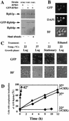
Expression level and localization of GFP-Rpb4p during optimal growth conditions and in response to various stresses. (A) Expression of GFP-RPB4 fusion gene in YMF1 strain is similar to the expression of RPB4 in WT cells and is not affected by HS. Proteins were extracted from YMF1 cells that had been growing exponentially at 22°C, or exposed to 42°C for 1 h, as indicated at the bottom. Samples (80 μg) were analyzed by Western analysis using anti-Rpb4p antibodies (Choder and Young, 1993). To demonstrate equal loading, the same filter was also reacted with anti-Rpb1p antibodies (8WG16 MAb). The GFP-Rpb4p bans also interacted with anti-GFP antibodies (our unpublished results). (B) GFP-Rpb4p localizes to the nucleus during growth under optimal environmental conditions. YMF1 cells were grown in rich medium (YPD) at 22°C and harvested in midlog for inspection using fluorescence microscopy. Top panel: localization of GFP-Rpb4p; middle and bottom panels show DAPI-stained and bright field images (BF), respectively, of the same field shown in the top panel. (C) GFP-Rpb4 is relocalized to the cytoplasm in response to various stresses. Cells were grown in rich liquid medium at 22°C till midlog and inspected by fluorescent microscope (designated Log). The remaining of the culture was then divided into three samples. One was allowed to grow at 22°C till cells entered stationary phase (stationary), and the others were shifted either to 37°C or to 6% ethanol (at 22°C) for an additional 10 h, as indicated at the top of each panel. Cell samples were then inspected using microscopy as above. (D) Kinetics of stress-induced relocation of GFP-Rpb4p: effect of temperature and cycloheximide. MF1 cells were grown in rich medium at 22°C until midlog phase. The culture was then split into two samples. Cycloheximide (25 μg/ml) was added to one sample, and the sample was further divided so that half remained at 22°C (designated 22°C + CHX) and the other half was shifted to 37°C (designated 37°C + CHX). The control culture to which no drug was added was divided into three portions and incubated at 22, 37, or 42°C, as indicated. To determine export kinetics, samples of each culture were examined by microscopy at the indicated times. Each sample was photographed and then ∼200 cells were randomly chosen and classified into those exhibiting nuclear localization of GFP-Rpb4p and those whose fluorescence was distributed all over the cell (cytoplasmic localization). The proportion of cells exhibiting cytoplasmic localization was plotted as a function of time. The control sample incubated at 22°C showed no change in the percentage of cells with cytoplasmic localization (∼10%) throughout the experiment (our unpublished results).
Figure 2.
Figure 2.
Immunolocalization of Rpb4p-(Myc)2, Rpb2p-HA, and Rpb3p-HA. Indicated strains were immunostained as described in MATERIALS AND METHODS. The bottom panels show the merge of the FITC and DAPI signals. When the two signals colocalize, the resulting color is yellow.
Figure 3.
Figure 3.
Growth and viability of cells carrying GFP-RPB4 or various mutant derivatives thereof under stress. (A–C) A plasmid expressing GFP-RPB4 (pMC143) restores the functions of Rpb4p. (A) Expression of pMC143 (see Table 1) recovers growth defects of Δrpb4 cells. WT Cells (SUB62) and Δrpb4 cells (MC11-1) were cultured in a synthetic complete medium, whereas Δrpb4 cells carrying pMC143 (YTN1) were cultured in a selective medium. Cells were allowed to grow at 22°C till midlogarithmic phase. Equivalent amounts of cells (diluted in fivefold steps) were spotted onto YPD plates supplemented with the indicated amounts of ethanol and were allowed to grown at the indicated temperatures for either 4 d (37°C) or 7 d (22°C). (B) Expression of GFP-RPB4 fusion gene in YTN1 strain is similar to the expression of GFP-RPB4 in YMF1 cells. Proteins were extracted from the indicated cells that had been grown logarithmically at 22°C till midlogarithmic growth phase. PT1 is isogenic to SUB62 strain and carrying a high-copy plasmid expressing GFP under ACT1 promoter. Samples (80 μg) were analyzed by Western analysis using anti-GFP antibodies. A band that was cross-reacted with the antibodies, marked by asterisk, demonstrates equal loading. (C) Expression of pMC143 recovers the transcriptional defect of Δrpb4 cells (MC11-1). Cells were grown in a selective medium at 22°C till midlogarithmic phase. The cultures were then split. One sample was left at 22°C, and the other was shifted rapidly to 42°C and incubated for 30 min at this temperature. Cell samples were collected and RNA was extracted as described previously (Choder, 1991; Sheffer et al., 1999). RNA samples (5 μg) were analyzed by Northern blot hybridization (Choder, 1991; Sheffer et al., 1999). The filter was hybridized with SSA4 DNA probe. rRNAs were detected by 0.5 μg/ml EtBr staining and shown at the bottom panel. (D and E) Both class I and class II mutants are sensitive to ethanol and temperature stresses. (D) Growth on YPD containing ethanol. Cells were grown in selective medium at 22°C till midlogarithmic phase. Equivalent amounts of cells (diluted in fivefold steps) were spotted onto YPD plates supplemented with the indicated amounts of ethanol. Plates were allowed to grown at 22°C for 7 d. (E) Cell viability as a function of time at 42°C. Experiment was done as described previously (Choder and Young, 1993).
Figure 5.
Figure 5.
Unlike class I (YTN75), class II mutants exhibit transcriptional capacity comparably to wild type. Temperature-sensitive mutants were selected as described in MATERIALS AND METHODS. (A) Levels of global mRNA. Cells were grown in selective medium at 22°C till midlog and then shifted rapidly to 37°C. Cell samples were collected either before HS (time 0) or at the indicated time points after temperature shift. After all samples had been collected, RNA was extracted. RNA samples (2 μg) from the indicated strains were dot-blotted onto nitrocellulose filter, in two sets of triplicates. One set was hybridized with 32P-poly(dT), to detect the global mRNA, and the other set with 32P-rDNA to detect rRNA. Only one dot of each set is shown. The intensities of the radioactive dots was quantified by phosphoimager. The average of the mRNA triplicate was divided by the average of the rRNA triplicate. The ratio obtained at time 0 of YTN1 was arbitrarily defined as 1, whereas the other columns are plotted relative to this ratio. Variation was <12%. This procedure was described previously (Choder, 1991). (B) Levels of specific mRNAs. Samples (5 μg) of RNA from the indicated strains were analyzed by Northern blot hybridization (Choder, 1991; Sheffer et al., 1999). The filter was sequentially hybridized with the indicated DNA probes. rRNA was detected by EtBr staining.
Figure 6.
Figure 6.
During incubation at 37°C, class II mutant cells synthesize poly(A)+ RNA that accumulates in the nucleus. The indicated strains were grown at 22°C till midlog. Cultures were then split into two samples. One was maintained at 22°C, and the other was shifted to 37°C for 1 h. Cells were then harvested and poly(A)+ RNA was detected by in situ hybridization using an oligo-dT50 probe (see MATERIALS AND METHODS). Preliminary test were made to make sure that the GFP fluorescence was not detected after cell fixation and permeabilization. See, for example, the 22°C panels in this figure. Nuclei were detected by DAPI staining.
Figure 7.
Figure 7.
During incubation at 42°C, class II mutant cells synthesize SSA4 mRNA that accumulates in the nucleus. (A) Quantitative analysis of SSA4 mRNA expression and localization. Indicated strains were grown in a selective medium at 22°C until midlog. Cultures were then divided into three samples. One was maintained at 22°C, and the others were shifted to 42°C for the indicated time. Cells were then harvested and the level of SSA4 mRNA and its localization was detected by in situ hybridization technique as described in MATERIALS AND METHODS. After taking pictures of a fluorescent microscope, cells were classified into those where SSA4 mRNA was detected in the cytoplasm, in the nucleus, or not detected at all. The code for the different expression and localization classes is depicted at the bottom right side of panel A. (B) Images of selected samples of cells that were visualized either before (22°C panels) or 15 min after a temperature shiftup (42°C panels), as described in Figure 1.
Figure 8.
Figure 8.
Effect of class I and class II mutations in GFP-RPB4 on the pattern of protein synthesis and localization of GFP-Rpb4p. (A) Pattern of protein synthesis. Strains, indicated at the top, were grown in a selective medium at 22°C till midlog and then incubated at 42°C for the indicated time periods, as detailed in MATERIALS AND METHODS. Cells were radiolabeled for 15 min before cell harvest as described in MATERIALS AND METHODS. Proteins were extracted and equal amounts were analyzed by SDS-PAGE (see MATERIALS AND METHODS), followed by fluorography and autoradiography. (C) Localization of class II GFP-Rpb4p mutants. Strains, indicated at the top, were grown in a selective medium at 22°C till midlog. The culture was then divided into two samples; one was further incubated at 22°C and the other was shifted to 42°C. One hour after the temperature shift, cell samples were inspected using microscopy as in Figure 1.

Similar articles

Cited by

References

    1. Amberg, D.C., Goldstein, A.L., and Cole, C.N. (1992). Isolation and characterization of RAT1: an essential gene of Saccharomyces cerevisiae required for the efficient nucleocytoplasmic trafficking of mRNA. Genes Dev. 6, 1173–1189. - PubMed
    1. Brodsky, A., and Silver, P. (2000). Pre-mRNA processing factors are required for nuclear export. RNA 6, 1737–1749. - PMC - PubMed
    1. Buratowski, S. (2000). Snapshots of RNA polymerase II transcription initiation. Curr. Opin. Cell Biol. 12, 320–325. - PubMed
    1. Choder, M. (1991). A general topoisomerase I-dependent transcriptional repression in the stationary phase in yeast. Genes Dev. 5, 2315–2326. - PubMed
    1. Choder, M. (1993). A growth rate-limiting process in the last growth phase of the yeast life cycle involves RPB4, a subunit of RNA polymerase II. J. Bacteriol. 175, 6358–6363. - PMC - PubMed

Publication types

MeSH terms