. 2003 Jul;14(7):2768-80.
doi: 10.1091/mbc.e02-10-0647. Epub 2003 Apr 4.

Intercellular organelle traffic through cytoplasmic bridges in early spermatids of the rat: mechanisms of haploid gene product sharing

Affiliations

Intercellular organelle traffic through cytoplasmic bridges in early spermatids of the rat: mechanisms of haploid gene product sharing

Sami Ventelä et al. Mol Biol Cell. 2003 Jul.

Abstract

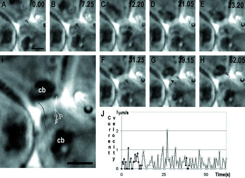
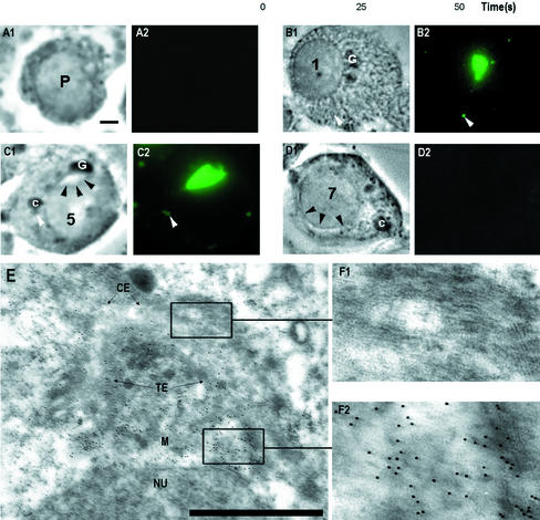
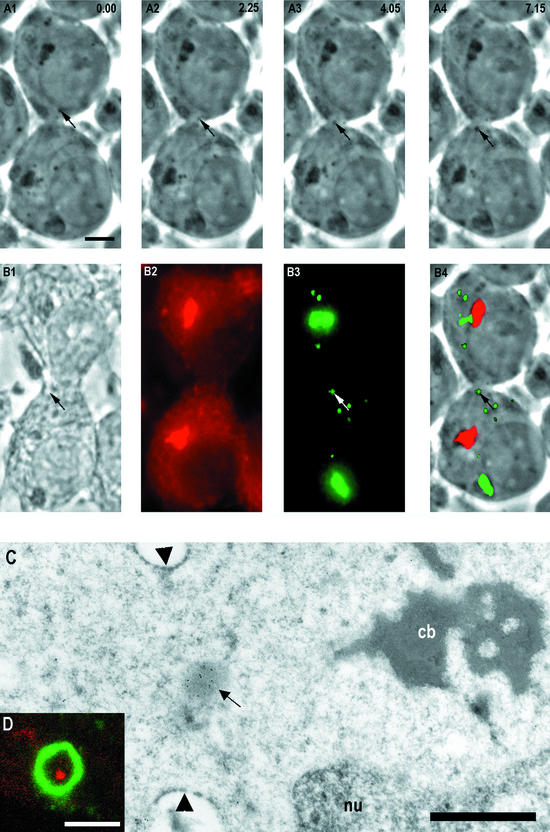
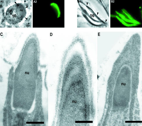
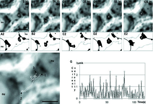
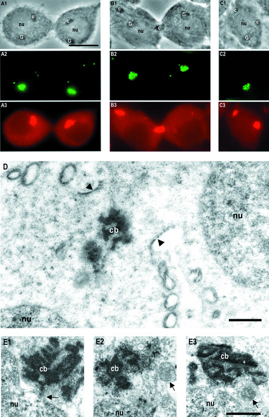
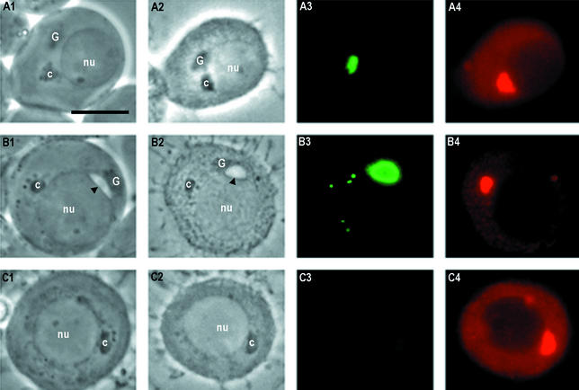
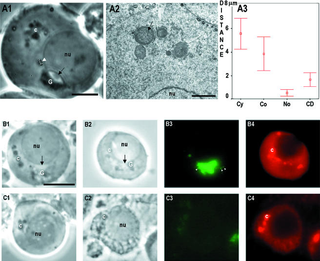
Stable cytoplasmic bridges (or ring canals) connecting the clone of spermatids are assumed to facilitate the sharing of haploid gene products and synchronous development of the cells. We have visualized these cytoplasmic bridges under phase-contrast optics and recorded the sharing of cytoplasmic material between the spermatids by a digital time-lapse imaging system ex vivo. A multitude of small (ca. 0.5 microm) granules were seen to move continuously over the bridges, but only 28% of those entering the bridge were actually transported into other cell. The average speed of the granules decreased significantly during the passage. Immunocytochemistry revealed that some of the shared granules contained haploid cell-specific gene product TRA54. We also demonstrate the novel function for the Golgi complex in acrosome system formation by showing that TRA54 is processed in Golgi complex and is transported into acrosome system of neighboring spermatid. In addition, we propose an intercellular transport function for the male germ cell-specific organelle chromatoid body. This mRNA containing organelle, ca. 1.8 microm in diameter, was demonstrated to go over the cytoplasmic bridge from one spermatid to another. Microtubule inhibitors prevented all organelle movements through the bridges and caused a disintegration of the chromatoid body. This is the first direct demonstration of an organelle traffic through cytoplasmic bridges in mammalian spermatogenesis. Golgi-derived haploid gene products are shared between spermatids, and an active involvement of the chromatoid body in intercellular material transport between round spermatids is proposed.


Figures

Figure 1.
Figure 1.
A successive series of time-scaled (in seconds, upper right corner) of images (A–H) of step 2 spermatid ex vivo, showing a two-directional movement of granules through the cytoplasmic bridge. Representative positions of the granule are shown by arrows. The path of movement is shown by white line at I. Another granule (arrowhead in G), traced by black line in I shows how it dissociate from the vicinity of chromatoid body and moves uni-directionally over the bridge. The changing velocity of granule at various time points is summarized in J; the movement inside the bridge is indicated with solid triangles. cb, chromatoid body, Bars, 2 μm.
Figure 2.
Figure 2.
TRA54 localization in early spermatids. Squash preparation from different stages were fixed and immunostained with TRA54 (A–D). Phase contrast (1), immunofluorescence of TRA54 (2). Pachytene spermatocytes (P) were negative (A). Translation of TRA54 started in the early spermatids (B). Numbers indicate the differentiation steps of spermatids. Golgi complex (G) was highly TRA54 positive. Small granules inside the cytoplasm were TRA54 positive (white arrowhead). At step 5 spermatids acorosome system (black arrowheads, C) is TRA54 positive. Also small granules in the vicinity of chromatoid body (c) were positive (white arrowhead). Golgi complex was TRA54 negative at step 5 spermatids. (D) A control slide from step 7 spermatid incubated with rat nonimmunized serum. Acrosome system is marked with black arrowheads. Bar, 2 μm. Immunoelectron microscopy (E) of early spermatids revealed strongest TRA54 labeling in trans-element (TE) of the Golgi complex. cis-element (CE) contains no TRA54 labeling (enlargement, F1). The strongest labeling is seen at the trans-saccular element (TE) and in the medulla (M), where proacrosomic granules are formed (enlargement, F2). Bar, 1 μm.
Figure 3.
Figure 3.
A successive time-scaled series of phasecontrast images ex vivo showing granule (arrows) transport between neighbor spermatids (A1–4). The time points in seconds are indicated at top right corner. (B1) Phase-contrast image from the same cells as in A after quick fixation, immunoreaction of anti-Mvh (mouse VASA-homologue; B2), TRA54 (B3), and superimposed image (B4) where granules and Golgi complex were TRA54 positive (green) and chromatoid body anti-Mvh positive (red). The same granule passing through the cytoplasmic bridge ex vivo (A) was TRA54 positive (arrows in B3 and B4). Bar, 5 μm. (C) Immunoelectron microscopy revealed a TRA54-positive granule (arrow) inside the cytoplasmic bridge (arrowheads). The chromatoid body (cb) was seen close to the cytoplasmic bridge. nu, nucleus. Bar, 1 μm. (D) Close-up view of a cytoplasmic bridge from a whole-mounted step 1–4 spermatid under confocal microscope double-immunostained with heat shock factor 2 (HSF-2) and TRA54 antibodies. HSF-2 (green) labeled the cytoplasmic bridge. TRA54-positive granule (red) was seen inside the cytoplasmic bridge. Bar, 2 μm.
Figure 4.
Figure 4.
TRA54 location during later spermatid development. In step 10 spermatids only acrosome system (arrowheads in A1) was TRA54 positive (A2). In step 16 spermatids acrosome systems (arrowheads in B1) were highly TRA54 positive (B2). Bar, 2 μm. Electron microscopy of step 15–17 spermatids (C–E) revealed TRA54 labeling in acrosome system (C) specifically in the middle part of the acrosomal membranes (D). Controls incubated with nonimmune normal rat serum were negative (E). Bar, 1 μm.
Figure 5.
Figure 5.
Time-scaled (top right corner in seconds) series of phase-contrast images showing two-directional movement of the chromatoid body (c) between two step 1 spermatids (A1–E1) through the cytoplasmic bridge. The same series is thresholded in A2–E2 to show in detail the chromatoid body and associated granules at the nuclear (nu) envelope. Moreover, in threshold image series the edges of the cells (gray) and nucleus (black) are presented. The movement path of the centroid of the chromatoid body is shown in F. Arrow 1 shows the starting point of the movement and arrow 2 the point where chromatoid body was close to the nucleus of the lower cell. Arrow 3 indicates the end point of chromatoid body movement during the 124.2-s recording period. The velocity of chromatoid body at various time points is shown in G. Bars, 2 μm.
Figure 6.
Figure 6.
Different localizations of the chromatoid body in early spermatids in fixed squash preparations (A–C) seen under phase-contrast optics (1) and after double-immunostaining with TRA54 (2) and Mvh (3) antibodies. Chromatoid body (c) located next to the nucleus (nu; A1–A3), inside the cytoplasmic bridge (B1–B3) and C1-C3 shows two chromatoid bodies inside the same spermatid. Bar, 10 μm. Electronmicrograph of the chromatoid body (cb) in the cytoplasmic bridge (arrowheads) of step 1–4 spermatids (D). nu, nucleus. (E1–E3) Electron micrographs showing the interrelationships between chromatoid body and the nuclear envelope in snap-frozen preparations (see MATERIALS AND METHODS). A large contact area was seen between nucleus and chromatoid body, and a nuclear pore complex with material continuities to chromatoid body (arrow in E1). (E2) A membranous structure (arrow) closely associated with the chromatoid body. (E3) A chromatoid body close to the nucleus and a multivesicular body (arrow). Bars, 1 μm.
Figure 7.
Figure 7.
Phase contrast (1), fixed (2), TRA54 (3), and VASA (4) immunostained images showing an early step 1 spermatids at a starting point of incubation (A) and after a 48-h incubation in control conditions (B) and with 1.0 μg/ml Golgi complex-disturbing agent brefeldin A (C). In early step 1 spermatids (A) only faint labeling of TRA54 was seen in the Golgi complex (A3). The chromatoid body was stained with Mvh antibody (A4). After 48 h of incubation in control conditions (B) acrosome system is developed (arrowheads in B1 and B2). After TRA54 immunostaining the acrosome system and some cytoplasmic granules were positive (B3). When step 1 spermatids were incubated for 48 h with brefeldin A, no Golgi complex was seen (C1 and C2). Neither the acrosome system nor TRA54-positive granules were seen in spermatids (C3). The chromatoid body was intact after treatment with brefeldin A (C4). Bar, 10 μm.
Figure 8.
Figure 8.
Effect of microtubule inhibitors. Stage I seminiferous tubules segments were incubated for 48 h in the presence of 10 μg/ml nocodazole or vincristine. (A1) Step 3 spermatid after a 48-h incubation in 10 μg/ml vincristine. The chromatoid body (c) was disintegrated into several separate granules. Normal cytoplasmic granule movement was changed into random Brownian motion. Movement path of one granule is indicated at the white arrowhead. The morphology of the Golgi complex (G) remained intact under phase-contrast optics. Th acrosome system is indicated with black arrow. Bar, 5 μm. Electron microscopic appearance of the chromatoid body after 48-h incubation with 10 μg/ml vincristine. Several spheres of chromatoid material containing ribosome-like granules were seen (arrow). Bar, 2 μm. Phase contrast ex vivo (B1 and C1), phase contrast after fixation (B2 and C2), TRA54 immunofluorescence (B3 and C3), and Mvh immunostaining (B4 and C4) after incubation step 1 spermatids for 48 h in the presence of 10 μg/ml nocodazole. The chromatoid body was converted into several spheres (B1). Also the morphology of the Golgi complex (G) was altered. After TRA54 immunostaining two types of spermatids were found: spermatids that contained the TRA54-positive acrosome system (B3) and small TRA54-positive granules in the vicinity of the Golgi complex (white arrowheads) and spermatids where the acrosome system was absent (C3). Disintegrated morphology of the chromatoid body was seen after immunostaining with Mvh (B4 and C4). Bar, 10 μm.

Similar articles

Cited by

References

    1. Alastalo, T.P., Lönnström, M., Leppä, S., Kaarniranta, K., Pelto-Huikko, M., Sistonen, L., and Parvinen, M. (1998). Stage-specific expression and cellular localization of the heat shock factor 2 isoforms in the rat seminiferous epithelium. Exp. Cell Res. 240, 16–27. - PubMed
    1. Biggiogera, M., Fakan, S., Leser, G., Martin, T.E., and Gordon, J. (1990). Immunoelectron microscopical visualization of ribonucleoproteins in the chromatoid body of mouse spermatids. Mol. Reprod. Dev. 26, 150–158. - PubMed
    1. Bohrmann, J., and Biber, K. (1994). Cytoskeleton-dependent transport of cytoplasmic particles in pre-vitellogenic to mid-vitellogenic ovarian follicles of Drosophila: time-lapse analysis using video-enhanced contrast microscopy. J. Cell Sci. 107, 849–858. - PubMed
    1. Braun, R.E., Behringer, R.R., Peschon, J.J., Brinster, R.L., and Palmiter, R.D. (1989). Genetically haploid spermatids are phenotypically diploid. Nature 337, 373–376. - PubMed
    1. Burgos, M.H., and Fawcett, D.W. (1955). Studies on the fine structure of the mammalian testis. J. Biophys. Biochem. Cytol. 1, 287–300. - PMC - PubMed

Publication types

MeSH terms

LinkOut - more resources