. 2003 Jul;14(7):2876-89.
doi: 10.1091/mbc.e03-02-0108. Epub 2003 Apr 17.

Nek2A kinase stimulates centrosome disjunction and is required for formation of bipolar mitotic spindles

Affiliations

Nek2A kinase stimulates centrosome disjunction and is required for formation of bipolar mitotic spindles

Alison J Faragher et al. Mol Biol Cell. 2003 Jul.

Abstract

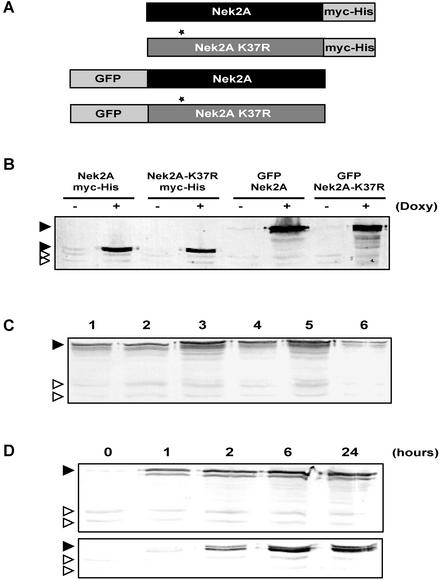
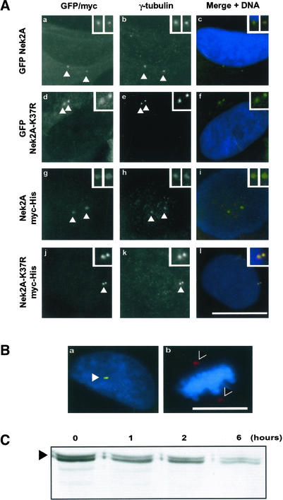
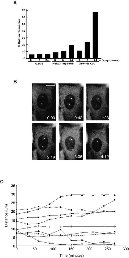
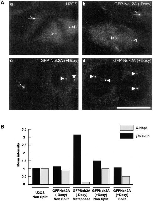
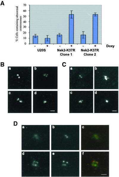
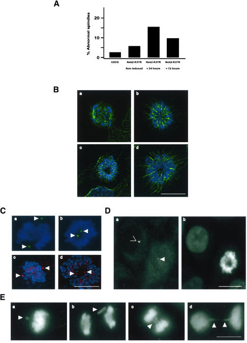
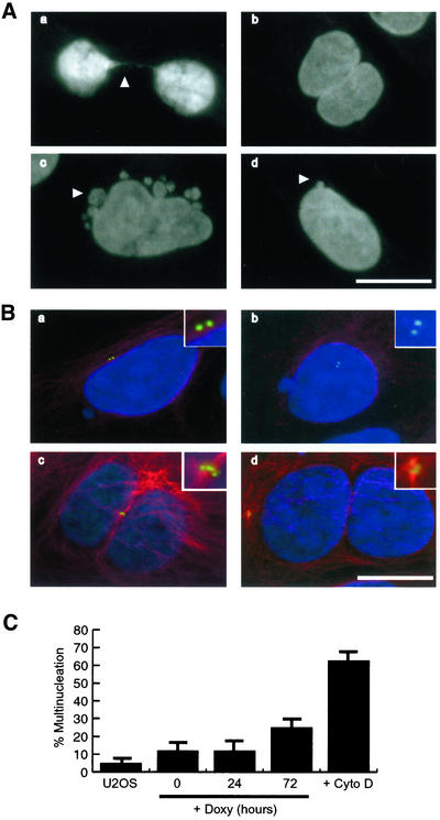
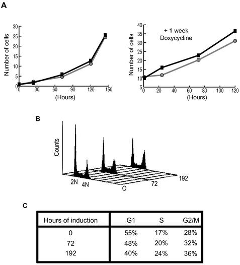
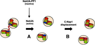
Nek2A is a cell cycle-regulated kinase of the never in mitosis A (NIMA) family that is highly enriched at the centrosome. One model for Nek2A function proposes that it regulates cohesion between the mother and daughter centriole through phosphorylation of C-Nap1, a large coiled-coil protein that localizes to centriolar ends. Phosphorylation of C-Nap1 at the G2/M transition may trigger its displacement from centrioles, promoting their separation and subsequent bipolar spindle formation. To test this model, we generated tetracycline-inducible cell lines overexpressing wild-type and kinase-dead versions of Nek2A. Live cell imaging revealed that active Nek2A stimulates the sustained splitting of interphase centrioles indicative of loss of cohesion. However, this splitting is accompanied by only a partial reduction in centriolar C-Nap1. Strikingly, induction of kinase-dead Nek2A led to formation of monopolar spindles with unseparated spindle poles that lack C-Nap1. Furthermore, kinase-dead Nek2A interfered with chromosome segregation and cytokinesis and led to an overall change in the DNA content of the cell population. These results provide the first direct evidence in human cells that Nek2A function is required for the correct execution of mitosis, most likely through promotion of centrosome disjunction. However, they suggest that loss of centriole cohesion and C-Nap1 displacement may be distinct mitotic events.


Figures

Figure 1.
Figure 1.
Generation of tetracycline-inducible Nek2A cell lines. (A) Schematic representation of the four Nek2A fusion proteins used for generation of tetracycline-inducible U2OS cell lines. Nek2A-K37R represents the catalytically inactive mutant kinase with a point mutation (asterisk) in the kinase domain. GFP, enhanced green fluorescent protein tag; Myc-His, myc epitope, and hexahistidine tag. (B) Western blot with α-Nek2 antibodies of extracts from each cell line after 24 h induction with (+) or without (–) doxycycline. (C) Western blot with α-Nek2 antibodies of six independent clones of the GFP-Nek2A-K37R cell line after 24-h doxycycline induction, indicating moderate clone to clone variation in recombinant protein expression level. (D) Western blot with α-Nek2 antibodies showing a time course of expression from a GFP-Nek2A (top) and Nek2A-K37R-myc-His (bottom) cell line after tetracycline induction (hours). In B, C, and D, filled arrowheads indicate recombinant Nek2A proteins; open arrowheads indicate migration of endogenous Nek2A (top band) and Nek2B (bottom band) proteins.
Figure 2.
Figure 2.
Recombinant Nek2A localizes to centrosomes but not spindle poles. Cell lines were induced with doxycycline for 24 h before processing for fluorescence microscopy. GFP-Nek2A (a–c), GFP-Nek2A-K37R (d–f), Nek2A-myc-His (g–i), and Nek2A-K37R-myc-His (j–l). GFP signal (a and d), α-myc antibodies (g and j), anti-γ-tubulin antibodies (b, e, h, and k), and merged image of GFP or myc (green), γ-tubulin (red), and Hoechst 33258 (blue) (c, f, i, and l). Bar, 15 μm. Centrosomes (arrowheads) are shown at increased magnification in insets. (B) Merged images showing Nek2A-K37R-myc-His–induced cells in interphase (a) or metaphase (b) stained with antibodies against γ-tubulin (red) and myc (green). DNA is stained in blue. Recombinant Nek2A is detected at interphase centrosomes (arrowhead) but not mitotic spindle poles (>). Bar, 15 μm. (C) Western blot with anti-Nek2 antibodies of extracts from cells induced to express Nek2A-K37R-myc-His (arrowhead) for 24 h before addition of cycloheximide for the times indicated (hours).
Figure 3.
Figure 3.
Active Nek2A kinase induces loss of centrosome cohesion. (A) Percentage of cells in which γ-tubulin–stained centrosomes were separated by >2 μm is indicated for U2OS-T-REx, Nek2A-myc-His, or GFP-Nek2A cells treated with doxycycline for 0, 6, or 24 h. (B) Selected images from a time-lapse movie of a cell induced to express GFP-Nek2A for 24 h showing that centrosomes remain split for more than 4 h (times in hours:minutes indicated on each panel). Bar, 15 μm. (C) Intercentriolar distances (micrometers) were measured over time (minutes) on captured images from single cell time-lapse experiments as shown in B. Cells were selected for imaging if the intercentriolar distance was initially >5 μm. Results from nine experiments are shown.
Figure 4.
Figure 4.
C-Nap1 is still present on Nek2A-induced split centrosomes. (A) U2OS T-REx cells (a) or GFP-Nek2A cells without (b) or with 24-h doxycycline induction (c and d) were fixed and immunostained with anti-C-Nap1 antibodies. Images were captured under identical conditions to allow comparison of C-Nap1 signal intensity. Unsplit (>) and split (filled arrowheads) centrosomes in interphase centrosomes, and mitotic spindle poles (open arrowheads) are indicated. Bar, 15 μm. (B) Abundance of C-Nap1 and γ-tubulin at centrosomes was calculated by measuring mean pixel intensities as indicated in MATERIALS AND METHODS. Centrosomes in 20 cells for each condition were imaged, quantitated, and normalized to the intensity of unsplit interphase centrosomes in the parental U2OS cell line (given an arbitrary value of 1.0). Results show the mean of three independent experiments in which all cells were fixed at the same time and immunostained with C-Nap1 antibodies under identical conditions.
Figure 5.
Figure 5.
Induction of centrosome abnormalities in cells expressing kinase-dead Nek2A. (A) Percentage of cells with abnormal centrosomes was calculated in U2OS T-REx cells or in two clones of the Nek2A-K37R-myc-His cell line treated with (+) or without (–) doxycycline for 24 h. (B and C) Representative digital deconvolution microscopy images of normal (a) or abnormal (b–d) centrosomes in cells induced to express Nek2A-K37R-myc-His for 24 h before processing for immunofluorescence microscopy with α-Nek2 (B) or anti-γ-tubulin (C) antibodies. In some cells, the centrosome look like a tight cluster of more than two dots (b and c); in other cells, abnormal centrosomes look like a single aggregate that is significantly larger than a normal centrosome (d). (D) Nek2A-K37R-myc-His cells were induced for 24 h before detergent extraction, fixation, and immunostaining with antibodies against γ-tubulin (a and d) and the centriolar marker GT335 (b and e). In cells containing clusters of centrosomes, each dot seems to contain a single centriole. Merged images (c and f) are shown of γ-tubulin (green) and GT335 (red). Bars (B–D), 2 μm.
Figure 6.
Figure 6.
Mitotic spindle and chromosome segregation defects in Nek2A-K37R cells. (A) Frequency of abnormal (monopolar or multipolar) spindles in mitotic cells was counted in U2OS T-REx cells or in Nek2A-K37R-myc-His cells with (+) or without (–) doxycycline induction for 24 and 72 h. (B) Digital deconvolution microscopy of spindles in Nek2A-K37R-myc-His mitotic cells stained for DNA (blue) and microtubules (green) comparing normal (a) with monopolar (b–d) spindles. (C) Bipolar (a and c) and monopolar (b and d) spindles stained for DNA (blue) and γ-tubulin (a and b; green) or Plk1 (c and d; red) in Nek2A-K37R-myc-His cells. Both Plk1 and γ-tubulin are recruited to poles of monopolar as well as bipolar spindles (arrowheads). Plk1 is also present on kinetochores. (D) C-Nap1 (a) and DNA (b) staining of an adjacent pair of interphase and mitotic cells expressing Nek2A-K37R-myc-His protein. Note that C-Nap1 is significantly less abundant at poles of a monopolar spindle (arrowhead) compared with at interphase centrosomes (>). (E) DNA staining of Nek2A-K37R-myc-His mitotic cells showing clear evidence of missegregation defects (arrowheads) with unaligned chromosomes in metaphase (a), lagging chromosomes in anaphase (b and c), and thin chromatin bridges in telophase (d). Bars (B–E), 15 μm.
Figure 7.
Figure 7.
Increased multinucleation in Nek2A-K37R interphase cells. (A) DNA staining of Nek2A-K37R-myc-His interphase cells after 24-h induction with doxycycline showing cells still attached through thin chromatin bridges (a), multinucleated cells (b), and micronucleated cells (c and d). Bar, 15 μm. (B) Immunofluorescence microscopy of micronucleated (a and b) and multinucleated (c and d) Nek2A-K37R-myc-His cells stained for centrosomes (C-Nap1, green), microtubules (α-tubulin, red), and DNA (blue), showing that multinucleated cells frequently contain multiple centrosomes suggestive of defects in cytokinesis. Bar, 15 μm. (C) Percentage of Nek2A-K37R-myc-His cells containing multinucleated cells after 0, 24, and 72 h of doxycycline induction compared with the frequency of multinucleation in parental U2OS cells that were either untreated (U2OS) or treated for 24 h with cytochalasin D (Cyto D).
Figure 8.
Figure 8.
Induction of kinase-dead Nek2A leads to an altered cell cycle distribution. (A) Counts (× 105) of Nek2A-K37R-myc-His cells in the absence (black line, ▪) or presence (grey line, •) of doxycycline. In the left-hand graph, doxycycline was added at T = 0, whereas in the right-hand graph, cells had been grown in the presence of doxycycline for 1 wk before T = 0. (B) Flow cytometry profiles for Nek2A-K37R-myc-His cells induced with tetracycline for 0, 72, and 192 h. The positions of 2N and 4N DNA are indicated. (C) Percentage of cells in each phase of the cell cycle was calculated from the flow cytometry profiles shown in B.
Figure 9.
Figure 9.
Centrosome disjunction and C-Nap1 displacement may be distinct events. The schematic model illustrates centrosome disjunction (A) and C-Nap1 displacement (B) as independent events that take place upon onset of mitosis. Disjunction of centrosomes, i.e., loss of cohesion between mother and daughter centrioles, is triggered by activation of Nek2A kinase. This itself is the result of a loss of the antagonistic activity of PP1, perhaps through phosphorylation of PP1 or binding of Inhibitor-2. The substrates of Nek2 involved in centrosome cohesion are likely to include C-Nap1 (blue sphere), although phosphorylation by Nek2 alone may not be sufficient to completely displace C-Nap1 from the centrosome. Nek2 may also have other substrates within the intercentriolar linkage. Likewise, full C-Nap1 displacement may require additional phosphorylation by other mitotic kinases.

References

    1. Blangy, A., Lane, H.A., d'Herin, P., Harper, M., Kress, M., and Nigg, E.A. (1995). Phosphorylation by p34cdc2 regulates spindle association of human Eg5, a kinesin-related motor essential for bipolar spindle formation in vivo. Cell 83, 1159–1169. - PubMed
    1. Bobinnec, Y., Moudjou, M., Fouquet, J.P., Desbruyeres, E., Edde, B., and Bornens, M. (1998). Glutamylation of centriole and cytoplasmic tubulin in proliferating non-neuronal cells. Cell Motil. Cytoskeleton 39, 223–232. - PubMed
    1. Bornens, M., and Moudjou, M. (1999). Studying the composition and function of centrosomes in vertebrates. Methods Cell Biol. 61, 13–34. - PubMed
    1. Bornens, M., Paintrand, M., Berges, J., Marty, M.C., and Karsenti, E. (1987). Structural and chemical characterization of isolated centrosomes. Cell Motil. Cytoskeleton 8, 238–249. - PubMed
    1. Chevrier, V., Piel, M., Collomb, N., Saoudi, Y., Frank, R., Paintrand, M., Narumiya, S., Bornens, M., and Job, D. (2002). The Rho-associated protein kinase p160ROCK is required for centrosome positioning. J. Cell Biol. 157, 807–817. - PMC - PubMed

Publication types

LinkOut - more resources