. 2003 Jul;14(7):3041-54.
doi: 10.1091/mbc.e03-01-0016.

ROCK and nuclear factor-kappaB-dependent activation of cyclooxygenase-2 by Rho GTPases: effects on tumor growth and therapeutic consequences

Affiliations

ROCK and nuclear factor-kappaB-dependent activation of cyclooxygenase-2 by Rho GTPases: effects on tumor growth and therapeutic consequences

Salvador Aznar Benitah et al. Mol Biol Cell. 2003 Jul.

Abstract

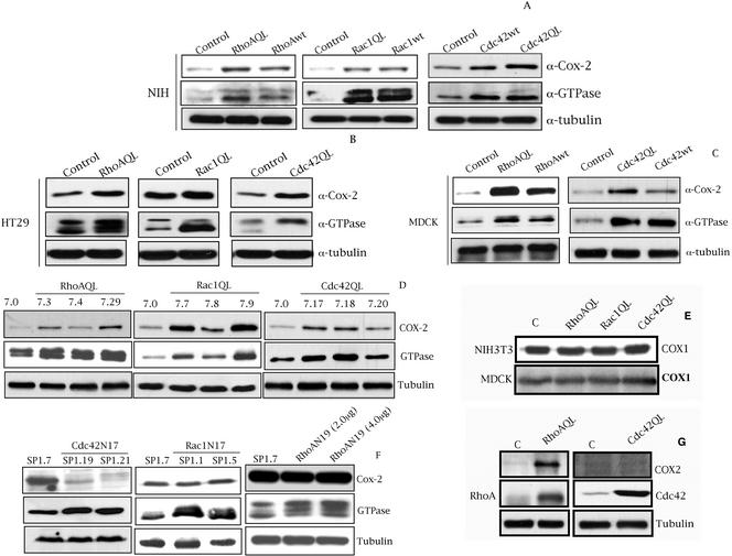
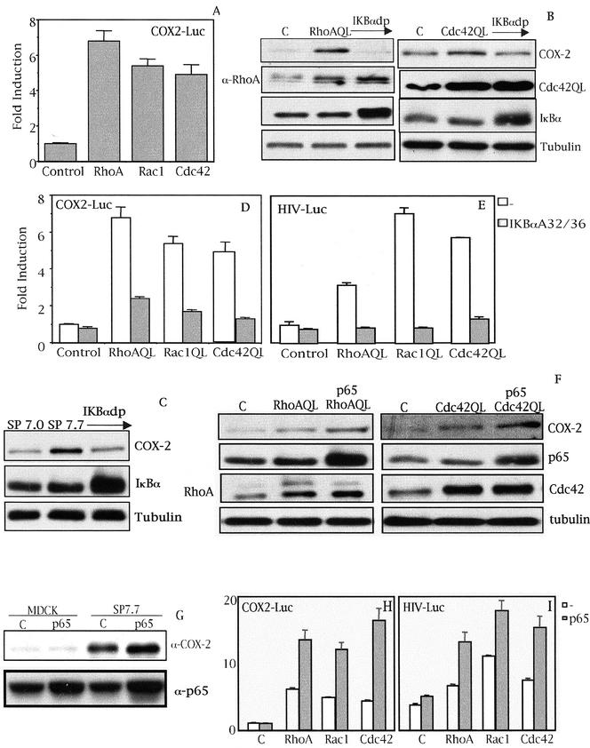
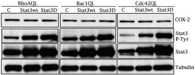
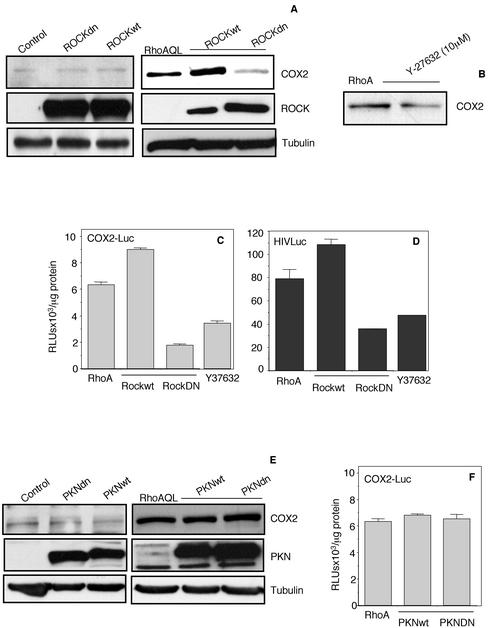
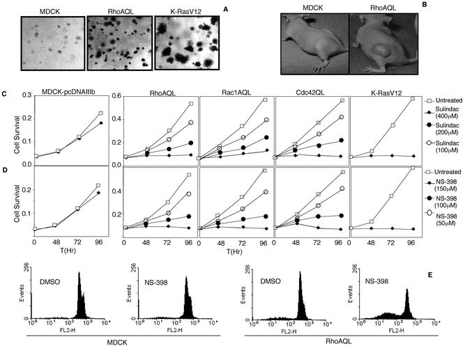
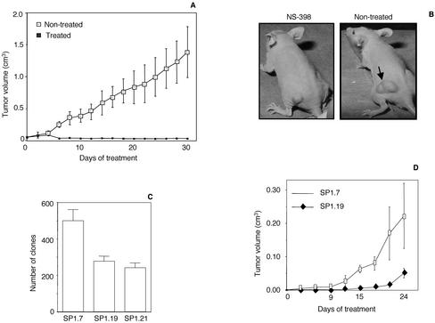
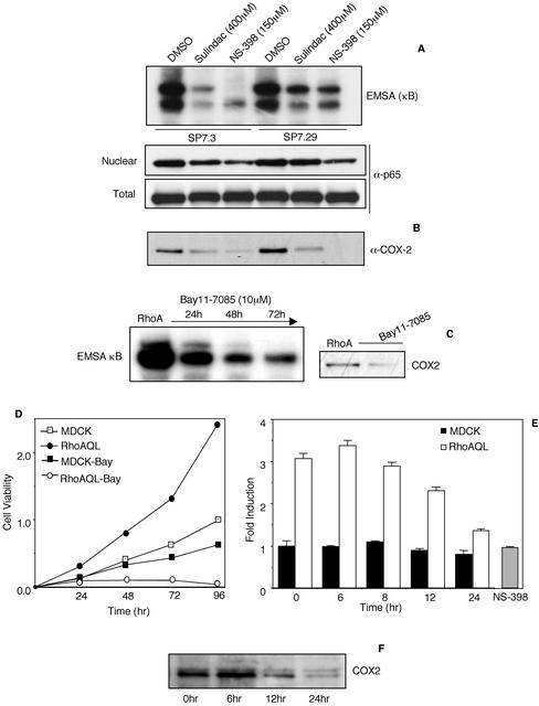
Rho GTPases are overexpressed in a variety of human tumors contributing to both tumor proliferation and metastasis. Recently, several studies demonstrate an essential role of transcriptional regulation in Rho GTPases-induced oncogenesis. Herein, we demonstrate that RhoA, Rac1, and Cdc42 promote the expression of cyclooxygenase-2 (COX-2) at the transcriptional level by a mechanism that is dependent on the transcription factor nuclear factor-kappaB (NF-kappaB), but not Stat3, a transcription factor required for RhoA-induced tumorigenesis. With respect to RhoA, this effect is dependent on ROCK, but not PKN. Treatment of RhoA-, Rac1-, and Cdc42-transformed epithelial cells with Sulindac and NS-398, two well-characterized nonsteroid antiinflammatory drugs (NSAIDs), results in growth inhibition as determined by cell proliferation assays. Accordingly, tumor growth of RhoA-expressing epithelial cells in syngeneic mice is strongly inhibited by NS-398 treatment. The effect of NSAIDs over RhoA-induced tumor growth is not exclusively dependent on COX-2 because DNA-binding of NF-kappaB is also abolished upon NSAIDs treatment, resulting in complete loss of COX-2 expression. Finally, treatment of RhoA-transformed cells with Bay11-7083, a specific NF-kappaB inhibitor, leads to inhibition of cell proliferation. We suggest that treatment of human tumors that overexpress Rho GTPases with NSAIDs and drugs that target NF-kappaB could constitute a valid antitumoral strategy.


Figures

Figure 1.
Figure 1.
Rho GTPases induce the expression of COX-2 in NIH3T3, HT29, DLD-1, and MDCK cells. (A) Transient expression of RhoA, Rac1, and Cdc42 (wt and QL) induce the expression of COX-2 in NIH3T3 cells compared with empty vector (pcDNAIIIb) transfected cells. (B) Transient expression of RhoAQL, Rac1QL, and Cdc42QL increases the expression of COX-2 in HT29 human colorectal cell line, compared with HT29-pcDNAIIIb transfected cells. (C) Transient expression of wild-type and oncogenic RhoA and Cdc42 induces the expression of COX-2 in MDCK epithelial cells compared with empty vector transfected cells. For A, B, and C, all cell lines were transfected as indicated in MATERIALS AND METHODS and whole cell lysates were obtained 48 h posttransfection. (D) Stable MDCK-RhoAQL, Rac1QL, and Cdc42QL cell lines exhibit high expression of COX-2 with respect to stable MDCK-pcDNAIIIB cells. (E) RhoAQL, Rac1QL, and Cdc42QL do not stimulate the expression of COX-1 in MDCK (same extracts as in D) or NIH3T3 cells (same extracts as in A). (F) Stable HT29 transfectants of dominant negative Cdc42 (N17) (left), but not Rac1N17 (middle), display a complete loss of COX-2 expression compared with control empty vector transfected parental cell line. Transient expression of dominant negative RhoA (N19) does not inhibit COX-2 expression in HT29 cells (right). (G) RhoAQL, but not Cdc42, induces the expression of COX-2 in human colorectal cell line DLD1, which lack endogenous expression of COX-2. Equal loading was verified with anti-tubulin in all blots. All experiments were performed at least three independent times.
Figure 2
Figure 2
(facing page). Rho GTPase-dependent expression of COX-2 is at the transcriptional level and dependent on NF-κB. (A) RhoA, Rac1, and Cdc42 (QL) induce the transcription of the proximal region of the cox-2 promoter (-1772 to +106) in MDCK cells. Stable cell lines of RhoAQL, Rac1QL, and Cdc42QL (7.3, 7.9, and 7.18, respectively) were transfected with COX2-Luc reporter vector (0.5 μg), and luciferase activity was measured 48 h posttransfection. Data shown represents a single experiment performed in triplicate ± SD. (B) IκBαS32/36A inhibits RhoA- and Cdc42QL-induced COX-2 expression. Transient transfection of 2 μg of IκBαS32/36A (IκBdp) together with 1 μg of pcDNAIIIb, RhoAQL, or Cdc42QL was carried out in MDCK cells and extracts for Western blot analysis were obtained 48 h posttransfection. (C) IκBαS32/36A inhibits Rac1QL-induced COX-2 expression in MDCK cells. MDCK-pcDNAIIIb or clone SP7.9 (MDCK-Rac1QL) were transiently transfected with IκBαS32/36A (2 μg), and Western blot analysis was carried out 48 h posttransfection. Equal loading was verified with an antitubulin antibody. (D) IκBαS32/36A inhibits transcription of the cox-2 promoter induced by RhoAQL, Rac1QL, and Cdc42QL. COX2-Luc (0.5 μg) and IκBαS32/36A (1.0 μg) were transiently cotransfected in 7.0 (MDCK-pcDNAIIIb), 7.3 (MDCK-RhoAQL), 7.9 (MDCK-Rac1QL), and 7.18 (MDCK-Cdc42QL), and luciferase activity was measured 48 h posttransfection (E) Expression of IκBαS32/36A leads to a functional inhibition of NF-κB transcriptional activity in MDCK cells. Same experiment as in D was carried out with 0.5 μg of HIV-luc reporter instead of COX-2-luc, and luciferase activity was measured 48 h posttransfection. Data shown in D and E represent a single experiment performed in triplicate ± SD. (F) Overexpression of p65 augments COX-2 expression in RhoAQL- and Cdc42QL-expressing cells. MDCK cells were transfected with 2.0 μg of p65 together with pcDNAIIIB (1.0 μg) (referred as control cells) RhoAQL (1.0 μg) or Cdc42QL (1.0 μg), and extracts were obtained 48 h posttransfection. (G) p65 potentiates COX-2 expression in SP7.7 (Rac1QL-MDCK) clone without any effect in parental MDCK cells. p65 (2.0 μg) was transfected in MDCK-pcDNAIIIB control cells, or in SP7.7 cells and extracts were obtained 48 h posttransfection. Expression of p65 was verified in F and G with an anti-p65 antibody. Equal loading was determined with anti-tubulin. (H) Expression of p65 produces a synergism in Rho-mediated transcription of the cox-2 promoter. (I) Expression of p65 in MDCK cells together with RhoAQL, Rac1QL, or Cdc42QL leads to a functional increase of NF-κB transcriptional activity. For parts H and I transfection was carried out as indicated in parts D and E, but cotransfecting p65 rather than IκBαS32/36A. Transfection efficiencies in D, E, H, and I were normalized using an RSV5-CAT reporter (0.5 μg) transfected along with the indicated plasmids. All experiments were performed four times with similar results.
Figure 3.
Figure 3.
Stat3 transcription factor is not involved in the expression of COX-2 under Rho GTPases signaling. Activation of Stat3 and Stat3 expression were determined using an anti-Stat3PY-694 and anti-Stat3 antibody respectively. Equal loading was verified with anti-tubulin. Results shown are representative of five independent experiments.
Figure 4.
Figure 4.
ROCK, but not PKN, is involved in RhoA-mediated COX-2 expression and NF-κB transcriptional activity. (A) ROCKwt and ROCKdn synergize and inhibit, respectively, RhoAQL-induced COX-2 expression (right) without any effect over COX-2 expression in parental MDKC-pcDNAIIIb cells (left). ROCKwt and ROCKdn (2.0 μg each) were cotransfected with pcDNAIIIb or RhoAQL in MDCK cells and whole cell extracts were collected 48 h posttransfection and subjected to Western blot analysis. Expression of ROCKwt and ROCKdn were verified with anti-myc antibody. (B) Y-27632 inhibits COX-2 expression induced by RhoAQL. MDCK-RhoAQL cells were treated with Y-27632 (10 μM) for 24 h, and whole cell extracts were then obtained and used to verify COX-2 expression. (C and D) ROCK is necessary for both RhoA-induced transcription of the cox2 proximal promoter region and NF-κB transcriptional activation. COX2-Luc or HIV-luc (0.5 μg) was cotransfected with ROCKwt or ROCKdn in MDCK cells along with empty vector or RhoAQL. Twenty-four hours posttransfection, luciferase activity was measured. As well, RhoAQL transfected cells were treated with Y-27632 (10 μM) for 24 h and luciferase activity was measured at this time. (E) PKN is not necessary for RhoAQL-induced COX-2 expression in MDCK cells. PKNwt or PKNdn (2.0 μg) were transfected into MDCK cells along with pcDNAIIIb or RhoAQL vectors, and 48 h posttransfection whole cell extracts were obtained. Expression of FLAG-PKNwt and FLAG-PKNdn was verified using an anti-FLAG antibody. (F) PKN has no effect overtranscription of the cox2 promoter under RhoA signaling. MDCK-RhoAQL cells were transfected either with empty vector or PKNwt and PKNdn along with the COX2-luc reporter vector. Twenty-four hours posttransfection, luciferase activity was measured as described in MATERIALS AND METHODS. Results shown in all parts are representative of three independent experiments.
Figure 5.
Figure 5.
Sulindac and NS-398 inhibit proliferation and induce apoptosis of RhoAQL-, Rac1QL-, Cdc42QL-, and KrasV12-expressing epithelial cells. (A) RhoAQL and KrasV12 promote anchorage independent growth in MDCK cells. (B) RhoAQL promotes tumor growth in vivo. MDCK or MDCK-RhoAQL cells (2 × 10⁶) were inoculated subcutaneously in athymic mice, and tumor volume was monitored at 2-d intervals. Pictures were taken 60 d upon inoculation. (C and D) Indicated concentrations of sulindac or NS-398 inhibit proliferation of MDCK cells transformed with RhoAQL, Rac1QL, Cdc42QL, and MDCK-RasV12 cells with no significant effect on MDCK parental cells. (E) NS-398 induces apoptosis of MDCK-RhoAQL but not MDCK parental cells. NS-398 (150 μM) was added to MDCK-pcDNAIIIb or MDCK-RhoAQL, and cells were treated for 96 h. At this point, cells were collected by trypsin treatment and were stained with propidium iodide for fluorescence-activated cell sorting analysis (see MATERIALS AND METHODS).
Figure 6.
Figure 6.
NS-398 inhibits tumor growth of RhoAQL-transformed MDCK cells. (A) NS-398 inhibits tumor growth promoted by oncogenic RhoA. Cells (2 × 10⁶) were inoculated subcutaneously in nu/nu mice and when tumors had reached a mean volume of 0.05–0.1 cm3, mice were treated intraperitoneally with NS-398 (3 mg/kg body weight) at 3-d intervals. Statistical significance was achieved at day 8 of treatment and was maintained throughout the rest of the treatment (p < 0.05). (B) Representative pictures of NS398-treated and vehicle-treated mice inoculated with MDCK-RhoAQL cells at 2 wk of treatment. (C) Inhibition of Cdc42 in HT29 cells results in a 50% reduction of anchorage-independent growth in soft agar. Clones were stained with crystal violet and were quantified 1 mo after seeding of cells. (D) Tumor growth of HT29-Cdc42N17 (SP1.19) in syngeneic mice is delayed with respect to empty vector transfected cells HT29 cells (SP1.7). Cells (2 × 10⁶; SP1.7 and SP1.19) were inoculated subcutaneously in immunosuppressed mice and tumor volume was monitored at 2-d intervals. All experiments shown were performed three independent times with similar results.
Figure 7.
Figure 7.
Sulindac and NS-398 inhibit both NF-κB DNA-binding and COX-2 expression induced by RhoAQL. (A) Sulindac and NS-398 inhibit NF-κB DNA-binding without affecting p65 expression. MDCK-RhoQL cells (clones SP7.3 and SP7.29) were treated with NS-398 and sulindac at the indicated concentrations for 72 h, and nuclear and whole cell extracts were obtained. EMSA analysis was carried out using a κB consensus sequence, and p65 expression was verified using an anti-p65 antibody. (B) Sulindac and NS-398 treatment inhibit COX-2 expression induced by RhoAQL. Same whole cell extracts as in A were used to verify COX-2 expression with an anti-COX-2 antibody. (C) MDCK-RhoAQL cells (SP7.3) were treated with Bay11-7083 (10 μM) for 72 h and nuclear and whole cell extracts were obtained. EMSA analysis and COX-2 expression were performed as in A and B. (D) Bay11-7083 inhibits MDCK-RhoAQL cell proliferation with a minor effect over empty vector transfected MDCK cells. MDCK-pcDNAIIIb and MDCK-RhoAQL cells were plated on 24-well dishes and were treated with Bay11-7083 (10 μM) at the indicated time points. Cell viability was determined by the crystal violet method. (E) Bay11-7083 does not affect the enzymatic activity of COX-2 (PGE2 production) in MDCK cells transformed with RhoAQL. MDKC-pcDNAIIIb and MDCK-RhoAQL (SP7.3) were plated in 24-well dishes and were treated with Bay11-7083 (10 μM) for the indicated period of time (0, 6, 8, and 24 h). Supernatant was collected and PGE2 concentration was measured as described in MATERIALS AND METHODS. As a positive control, NS-398 (100 μM) was added for 8 h and PGE2 synthesis was measured as described above. Ordinate indicate fold induction of PGE2 relative to control cells. (F) Bay11-7083 inhibits RhoAQL-induced COX-2 expression in MDCK cells at 12 h of treatment. The same experiment as in E was carried in parallel and whole cell extracts were obtained and subjected to Western blot analysis by using an anti-COX-2 antibody. All experiments were performed three times with similar results.

References

    1. Aznar, S., and Lacal, J.C. (2001a). Rho signals to cell growth and apoptosis. Cancer Lett. 165, 1-10. - PubMed
    1. Aznar, S., and Lacal, J.C. (2001b). Searching new targets for anticancer drug design: the families of Ras and RhoGTPases and their effectors. Prog. Nucleic Acid Res. Mol. Biol. 67, 193-234. - PubMed
    1. Aznar, S., and Lacal, J.C. (2003). Rho GTPases in human carcinogenesis: a tale of excess. Rev. Oncol. (in press).
    1. Aznar, S., Valeron, P.F., del Rincon, S.V., Perez, L.F., Perona, R., and Lacal, J.C. (2001). Simultaneous tyrosine and serine phosphorylation of stat3 transcription factor is involved in rho a GTPase oncogenic transformation. Mol. Biol. Cell 12, 3282-3294. - PMC - PubMed
    1. Bar-Sagi, D., and Hall, A. (2000). Ras and Rho GTPases: a family reunion. Cell 103, 227-238. - PubMed

Publication types

MeSH terms

Substances