. 2003 Aug;77(15):8196-206.
doi: 10.1128/jvi.77.15.8196-8206.2003.

Structural analyses of purified human immunodeficiency virus type 1 intracellular reverse transcription complexes

Affiliations

Structural analyses of purified human immunodeficiency virus type 1 intracellular reverse transcription complexes

Milan V Nermut et al. J Virol. 2003 Aug.

Abstract

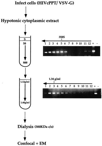
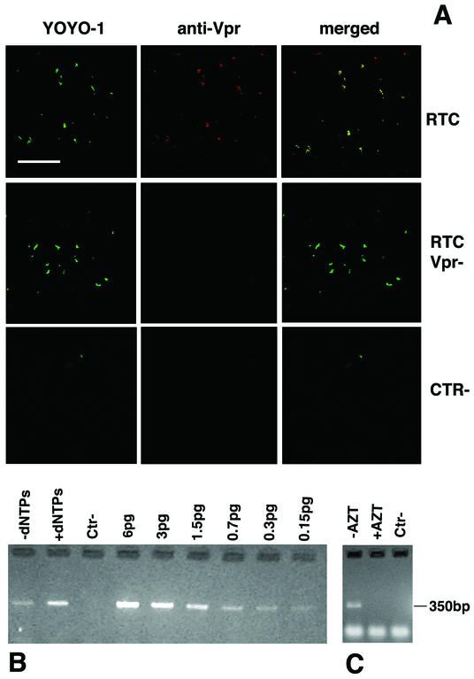
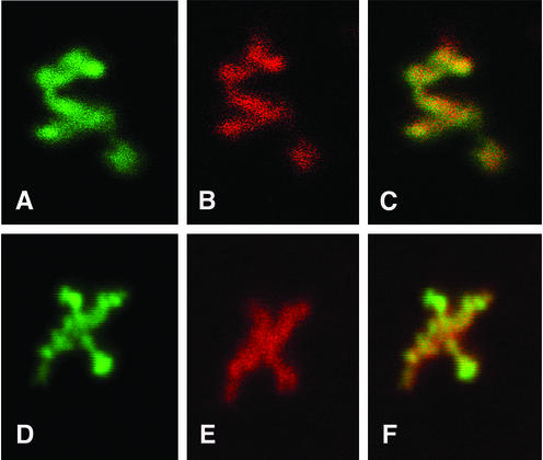
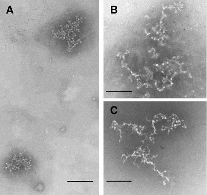
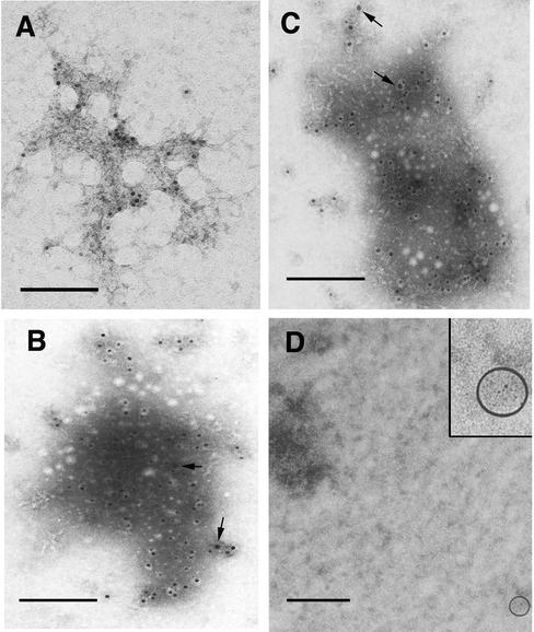
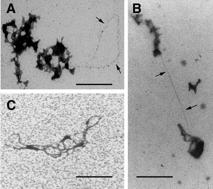
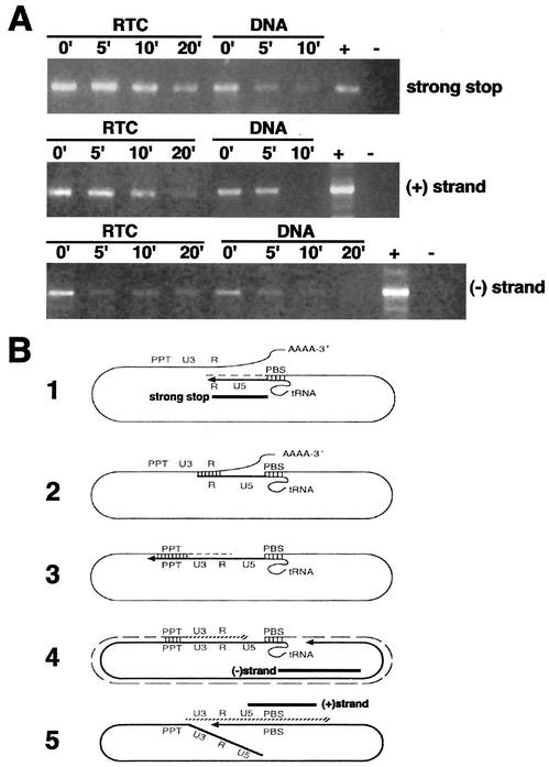
Retroviruses copy their RNA genome into a DNA molecule, but little is known of the structure of the complex mediating reverse transcription in vivo. We used confocal and electron microscopy to study the structure of human immunodeficiency virus type 1 (HIV-1) intracellular reverse transcription complexes (RTCs). Cytoplasmic extracts were prepared 3, 4, and 16 h after acute infection by Dounce homogenization in hypotonic buffer. RTCs were purified by velocity sedimentation, followed by density fractionation in linear sucrose gradients and dialysis in a large pore cellulose membrane. RTCs had a sedimentation velocity of approximately 350 S and a density of 1.34 g/ml and were active in an endogenous reverse transcription assay. Double labeling of nucleic acids and viral proteins allowed specific visualization of RTCs by confocal microscopy. Electron microscopy revealed that RTCs are large nucleoprotein structures of variable shape consisting of packed filaments ca. 6 nm thick. Integrase and Vpr are associated with discrete regions of the 6-nm filaments. The nucleic acids within the RTC are coated by small proteins distinct from nucleocapsid and are partially protected from nuclease digestion.


Figures

FIG. 1.
FIG. 1.
Scheme of the method used to purify intracellular HIV-1 RTCs. HeLa cells were infected at a high multiplicity of infection with an HIV-1-based vector pseudotyped with VSV-G. After 2 h of incubation at 4°C, cells were transferred at 37°C for 3, 4, or 16 h, and cytoplasmic extracts were prepared by Dounce homogenization in hypotonic buffer. Extracts were loaded onto a 5 to 20% linear sucrose gradient and subjected to velocity sedimentation. The position of the viral DNA in the gradient was detected by PCR with primers specific for the strong-stop DNA. The peak of the viral DNA was consistently found sedimenting at approximately 350 S. Fractions 4 and 5 were subjected to density fractionation into a 20 to 70% linear sucrose gradient, and the position of the viral DNA was detected as before by PCR. The fractions having a density of ca. 1.34 g/ml contained the peak of the viral DNA and were dialyzed in a large pore cellulose membrane (300,000-Da cutoff). Dialyzed samples were used for confocal and EM analyses. Arrows indicate the direction of the gradients.
FIG. 2.
FIG. 2.
Analysis of purified HIV-1 RTCs by confocal microscopy and endogenous reverse transcription assay. (A) Purified RTCs isolated 4 h postinfection were adsorbed onto plastic tissue culture dishes, fixed, and labeled with the nucleic acid dye YOYO-1 and an anti-Vpr polyclonal antibody. Images were acquired sequentially and merged by using the Confocal Assistant software. Mutant RTCs lacking Vpr (RTC Vpr−), samples from uninfected cells (CTR−), and nonimmune rabbit sera were used as controls. Scale bar, 15 μm. (B) Endogenous RT assay on the equilibrium density fractions containing the peak of the viral DNA (4 h postinfection). Samples were incubated for 6 h at 37°C in the presence or absence exogenous dNTPs and then subjected to PCR with primers specific for the positive-strand DNA (expected band size is 350 bp). Serial dilutions of the HIV-1 vector plasmid were amplified in parallel. Ctr−, uninfected cells. (C) RTCs were extracted from cells infected in the presence (+AZT) or absence (−AZT) of 200 μM AZT, purified, and subjected to an endogenous RT assay in the presence of dNTPs. The expected band size for the positive-strand DNA is 350 bp. Lower-molecular-weight bands are PCR artifacts. Ctr−, uninfected cells.
FIG. 3.
FIG. 3.
Analyses of purified RTC by confocal microscopy at ×6,300 magnification. Purified RTCs (4 h postinfection) were adsorbed onto plastic tissue culture dishes, fixed, and doubly labeled with YOYO-1 (A and D) and anti-Vpr (B) or anti-IN (E) antibodies. Images were acquired sequentially at ×6,300 magnification by using an optical zoom and merged by using Lasersharp Confocal Assistant software (C and F).
FIG. 4.
FIG. 4.
Visualization of purified RTCs (3 and 4 h postinfection) by negative stain EM. Samples were adsorbed onto carbon-coated grids and negatively stained with 4% STA (pH 8.2). (A) Two complexes in different orientations are visible after negative staining at ×88,000 magnification. Scale bar, 140 nm. (B) Higher-magnification image of a typical complex as detected at ×180,000 magnification. Scale bar, 85 nm. (C) Samples (20 μl) were mixed 1:1 with GNE buffer (pH 10) for 3 h, washed, and negatively stained with STA (pH 8.2). Note the indication of continuity of the filaments after relaxation of the complexes. Scale bar, 85 nm.
FIG. 5.
FIG. 5.
Gold immunolabeling of purified RTCs (4 h postinfection) with anti-Vpr polyclonal antibody (A), with anti-IN antibody (B and C) or nonimmune rabbit serum (D), followed by incubation with 5-nm colloidal gold-conjugated secondary antibody. Samples were stained with ethanolic UA in panel A or STA (pH 7.6) in panels B, C, and D. The arrows in panels B and C point to small clusters of gold particles. The round white dots in panels B and C are sublimation artifacts of the STA staining. The circled area in panel D contains a gold particle and is magnified in the inset. Scale bar, 100 nm.
FIG. 6.
FIG. 6.
Visualization of nucleic acids in purified RTCs. Samples (3 and 4 h postinfection) were treated with proteinase K for 1 h (A) or 2 h (B), ethanol precipitated, adsorbed onto carbon-coated 400-mesh grids, washed in distilled water, and stained with 1% ethanolic UA diluted 1:5 in acetone for 5 min. (C) Bundle of nucleic acid filaments from a preparation deproteinized with guanidine thiocyanate. The arrows point to nucleic acids. Scale bar, 150 nm.
FIG. 7.
FIG. 7.
The viral DNA within the RTC is partially protected from micrococcal nuclease digestion. (A) Equilibrium density fractions containing the peak of the viral DNA (4 h postinfection) were incubated in the presence of micrococcal nuclease and 1.6 mM CaCl2. The reactions were stopped by addition of 4 mM EGTA at the indicated time points and analyzed by PCR with primers specific for negative-strand, positive-strand, and strong-stop DNAs. Naked viral DNA was prepared from the same density fractions by proteinase K digestion, phenol-chloroform extraction, and ethanol precipitation and then incubated as described above in the presence of micrococcal nuclease. −, No DNA; +, HIV-1 plasmid DNA. (B) Schematic representations of reverse transcription. The regions of the genome amplified by PCR in panel A are indicated by black bars. Diagram 1 shows the synthesis of the negative-strand strong-stop DNA starts at the primer-binding site (PBS), where a partially unfolded tRNA is bound. RNase H degrades the positive-strand RNA template so that the first strand transfer can take place. In diagram 2, a bridge is formed between the two complementary R sequences, and RT can jump on either of the RNA strands to elongate the negative-strand DNA in diagram 3. The RNA template is degraded except for the PPT. In diagram 4, the synthesis of the negative strand is completed. In diagram 5, the synthesis of the positive-strand strong-stop DNA starts at the PPT. The tRNA primer is removed, and the two strong-stop strands pair, forming a circular molecule suitable for elongation of the positive strand. At the end of the elongation process, the circular intermediate is opened into a linear double-stranded DNA molecule.

References

    1. Allain, B., M. Lapadat-Tapolsky, C. Berlioz, and J.-L. Darlix. 1994. Transactivation of the minus-strand DNA transfer by nucleocapsid protein during reverse transcription of the retroviral genome. EMBO J. 13:973-981. - PMC - PubMed
    1. Besnier, C., Y. Takeuchi, and G. Towers. 1925. 2002. Restriction of lentivirus in monkeys. Proc. Natl. Acad. Sci. USA 99:11920-11921. - PMC - PubMed
    1. Boom, R., C. J. A. Sol, M. M. M. Salimans, C. L. Jansen, P. M. E. Wertheim-van Dillen, and J. van der Noordaa. 1990. Rapid and simple method for purification of nucleic acids. J. Clin. Microbiol. 28:495-503. - PMC - PubMed
    1. Bowerman, B., P. O. Brown, J. M. Bishop, and H. E. Varmus. 1989. A nucleoprotein complex mediated the integration of retroviral DNA. Genes Dev. 3:469-478. - PubMed
    1. Braaten, D., E. K. Franke, and J. Luban. 1996. Cyclophilin A is required for an early step in the life cycle of human immunodeficiency virus type 1 before initiation of reverse transcription. J. Virol. 70:3551-3560. - PMC - PubMed

Publication types

MeSH terms

LinkOut - more resources