. 2003 Aug;77(15):8394-407.
doi: 10.1128/jvi.77.15.8394-8407.2003.

Replication of modified vaccinia virus Ankara in primary chicken embryo fibroblasts requires expression of the interferon resistance gene E3L

Affiliations

Replication of modified vaccinia virus Ankara in primary chicken embryo fibroblasts requires expression of the interferon resistance gene E3L

Simone Hornemann et al. J Virol. 2003 Aug.

Abstract

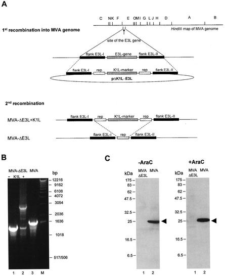
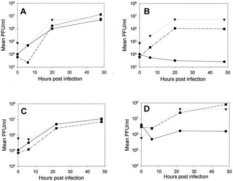
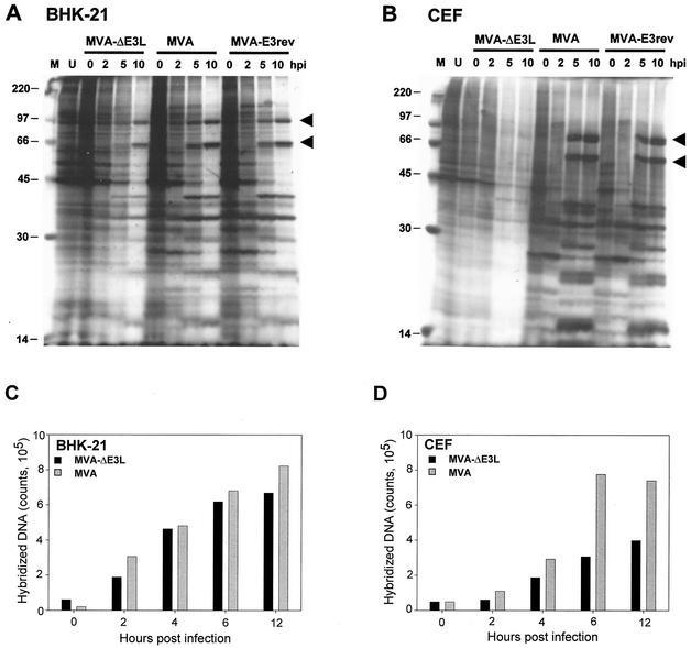
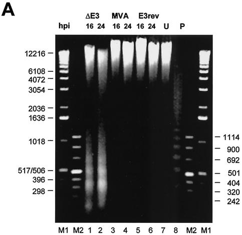
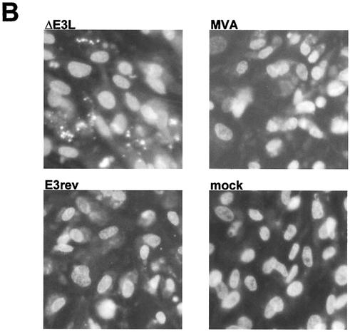
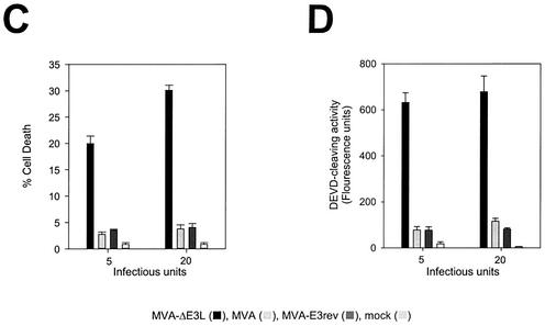
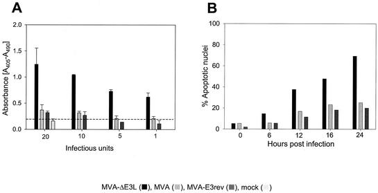
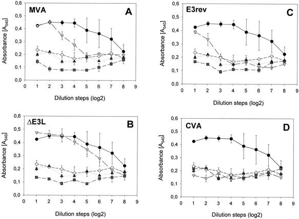
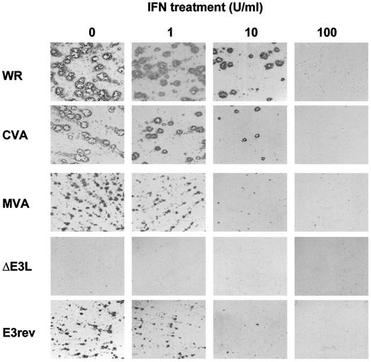
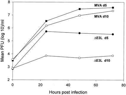
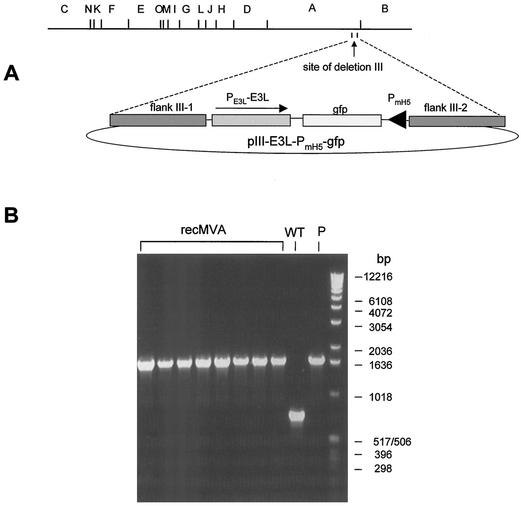
Highly attenuated modified vaccinia virus Ankara (MVA) serves as a candidate vaccine to immunize against infectious diseases and cancer. MVA was randomly obtained by serial growth in cultures of chicken embryo fibroblasts (CEF), resulting in the loss of substantial genomic information including many genes regulating virus-host interactions. The vaccinia virus interferon (IFN) resistance gene E3L is among the few conserved open reading frames encoding viral immune defense proteins. To investigate the relevance of E3L in the MVA life cycle, we generated the deletion mutant MVA-DeltaE3L. Surprisingly, we found that MVA-DeltaE3L had lost the ability to grow in CEF, which is the first finding of a vaccinia virus host range phenotype in this otherwise highly permissive cell culture. Reinsertion of E3L led to the generation of revertant virus MVA-E3rev and rescued productive replication in CEF. Nonproductive infection of CEF with MVA-DeltaE3L allowed viral DNA replication to occur but resulted in an abrupt inhibition of viral protein synthesis at late times. Under these nonpermissive conditions, CEF underwent apoptosis starting as early as 6 h after infection, as shown by DNA fragmentation, Hoechst staining, and caspase activation. Moreover, we detected high levels of active chicken alpha/beta IFN (IFN-alpha/beta) in supernatants of MVA-DeltaE3L-infected CEF, while moderate IFN quantities were found after MVA or MVA-E3rev infection and no IFN activity was present upon infection with wild-type vaccinia viruses. Interestingly, pretreatment of CEF with similar amounts of recombinant chicken IFN-alpha inhibited growth of vaccinia viruses, including MVA. We conclude that efficient propagation of MVA in CEF, the tissue culture system used for production of MVA-based vaccines, essentially requires conserved E3L gene function as an inhibitor of apoptosis and/or IFN induction.


Figures

FIG. 1.
FIG. 1.
Construction and characterization of MVA-ΔE3L. (A) Schematic maps of the MVA genome and the plasmid pΔK1L-E3L designed for deletion of the E3L gene sequences. The HindIII restriction endonuclease sites of the MVA genome are indicated at the top of the panel. MVA DNA sequences adjacent to the E3L gene (flank E3L-I and flank E3L-II) were cloned into pΔK1L to direct insertion of the K1L selectable marker by homologous recombination, resulting in the deletion of the E3L ORF from the MVA genome. K1L gene expression allows selective growth of the unstable intermediate virus MVA-ΔE3L+K1L in RK-13 cells. The final mutant virus MVA-ΔE3L resulted after the deletion of the K1L marker gene during a second homologous recombination involving additional repetitive sequences (rep). (B) PCR analysis of viral DNA. Genomic template DNA was prepared from MVA-ΔE3L without K1L (lane 1) or with K1L marker gene sequences (lane 2) or from MVA (lane 3). Oligonucleotides E3A and E3B from gene sequences adjacent to the E3L gene locus were used for amplification of specific DNA fragments. PCR products were separated by agarose gel electrophoresis. The 1-kb ladder (Roche Diagnostics) served as molecular weight marker (M). (C) Western blot analysis of lysates from CEF infected with MVA-ΔE3L or MVA in the presence (+AraC) or absence (−AraC) of AraC. The E3L protein bands detected by the E3L-specific mouse monoclonal antibody TW9 are marked by arrowheads.
FIG. 2.
FIG. 2.
Analysis of virus growth in BHK (A and C) and CEF (B and D) cells after infection with a low (A and B) or high (C and D) dose of MVA-ΔE3L (•), MVA (▪), or MVA-E3rev (▾).
FIG. 3.
FIG. 3.
Viral polypeptide synthesis. (A and B) BHK (A) and CEF (B) cells were infected with MVA-ΔE3L, MVA, or MVA-E3rev and labeled with [35S]methionine for 30 min at the indicated hour p.i. (hpi). Cell lysates were analyzed by gel electrophoresis on a 10% polyacrylamide gel and visualized by autoradiography. Protein standards (lanes M) are indicated by their molecular masses (in kilodaltons). Uninfected cells (lanes U) served as controls. Representative late virus-induced proteins are marked by arrowheads. (C and D) Viral DNA synthesis. DNA isolated from BHK-21 cells (C) or CEF (D) at 0, 2, 4, 6, and 12 h after infection by MVA-ΔE3L or MVA was immobilized on a Hybond N+ membrane and analyzed by hybridization of a 32P-labeled MVA DNA probe. Radioactivity was quantitated with a phosphorimager analyzer.
FIG. 4.
FIG. 4.
Induction of apoptosis by MVA-ΔE3L in CEF cells. (A) For DNA fragmentation analysis, semiconfluent monolayers of CEF cells were either left uninfected (lane 7) or infected with MVA-ΔE3L (lanes 1 and 2), MVA (lanes 3 and 4), or MVA-E3rev (lanes 5 and 6) at an MOI of 20 IU/cell. Total DNA extracts were obtained at 16 h p.i. (lanes 1, 3, and 5) and 24 h p.i. (lanes 2, 4, and 6), separated by gel electrophoresis through 1% agarose, and visualized by ethidium bromide staining. As a positive control, a sample from a DNA laddering kit (Roche Diagnostics) was applied (lane 8). Lanes M1 and M2, molecular weight standards. (B) The Hoechst 3343 staining was performed with mock-infected or MVA-infected CEF cells grown on coverslips. After 16 h, cells were stained for 30 min with Hoechst 3343. Micrographs were taken of CEF cells either mock infected (mock) or infected with MVA-ΔE3L (ΔE3L), nonrecombinant MVA (MVA), or MVA-E3rev (E3rev) at an MOI of 5 IU/cell. (C) Apoptotic cells stained with Hoechst 3343 were counted in CEF cells infected with MVA-ΔE3L, MVA, or MVA-E3rev at an MOI of 5 or 20 PFU/cell and in mock-infected cells. Results are given as means plus or minus 3× SEM. (D) Induction of DEVD-cleaving activity was determined in CEF cells infected with MVA-ΔE3L, MVA, and MVA-E3rev at an MOI of 5 or 20 IU/cell or in uninfected CEF cells. DEVD-cleaving activity levels were measured in triplicate with 10 μl from each mixture as described in Materials and Methods. Results are given as means plus or minus 3× SEM.
FIG. 4.
FIG. 4.
Induction of apoptosis by MVA-ΔE3L in CEF cells. (A) For DNA fragmentation analysis, semiconfluent monolayers of CEF cells were either left uninfected (lane 7) or infected with MVA-ΔE3L (lanes 1 and 2), MVA (lanes 3 and 4), or MVA-E3rev (lanes 5 and 6) at an MOI of 20 IU/cell. Total DNA extracts were obtained at 16 h p.i. (lanes 1, 3, and 5) and 24 h p.i. (lanes 2, 4, and 6), separated by gel electrophoresis through 1% agarose, and visualized by ethidium bromide staining. As a positive control, a sample from a DNA laddering kit (Roche Diagnostics) was applied (lane 8). Lanes M1 and M2, molecular weight standards. (B) The Hoechst 3343 staining was performed with mock-infected or MVA-infected CEF cells grown on coverslips. After 16 h, cells were stained for 30 min with Hoechst 3343. Micrographs were taken of CEF cells either mock infected (mock) or infected with MVA-ΔE3L (ΔE3L), nonrecombinant MVA (MVA), or MVA-E3rev (E3rev) at an MOI of 5 IU/cell. (C) Apoptotic cells stained with Hoechst 3343 were counted in CEF cells infected with MVA-ΔE3L, MVA, or MVA-E3rev at an MOI of 5 or 20 PFU/cell and in mock-infected cells. Results are given as means plus or minus 3× SEM. (D) Induction of DEVD-cleaving activity was determined in CEF cells infected with MVA-ΔE3L, MVA, and MVA-E3rev at an MOI of 5 or 20 IU/cell or in uninfected CEF cells. DEVD-cleaving activity levels were measured in triplicate with 10 μl from each mixture as described in Materials and Methods. Results are given as means plus or minus 3× SEM.
FIG. 5.
FIG. 5.
(A) Detection of apoptosis after infection with different doses of virus. The extent of apoptosis was measured by ELISA either in mock-infected CEF or in cells infected with MVA-ΔE3L, MVA, or MVA-E3rev. Cell death detection ELISAs were performed according to the manufacturer's instructions at 16 h p.i. Absorbance at 405 nm (reference, 490 nm) was measured. Results are given as means plus or minus 2× SEM. (B) Kinetics and extent of apoptosis were analyzed in CEF cells that were mock infected or infected with MVA-ΔE3L, MVA, or MVA-E3rev at an MOI of 20 IU/cell. Cells were harvested at indicated time points, fixed overnight, stained with propidium iodide, and analyzed by flow cytometry.
FIG. 6.
FIG. 6.
Synthesis of chicken IFN-α/β after infection of CEF with MVA-ΔE3L. CEF monolayers grown in 6-well tissue culture plates were inoculated with 10 IU/cell of MVA (A), MVA-ΔE3L (B), MVA-E3rev (C), or wild-type vaccinia virus CVA (D). Cell-free supernatants were collected at 0 h (▴, mock infected; ▪, virus infected) or 24 h (◊, mock infected; ▿, virus infected) after infection and tested for antiviral activities of chicken IFN-α/β in comparison to those of recIFN (•; 250 U/ml).
FIG. 7.
FIG. 7.
Effect of IFN treatment on MVA infection. CEF monolayers were incubated with increasing amounts of recIFN-α/β for 24 h before the cultures were infected at low MOIs with MVA, MVA-ΔE3L, or MVA-E3rev or with vaccinia virus CVA or WR. At 48 h after infection, cell monolayers were fixed and foci of virus infected cells were visualized using vaccinia virus-specific immunostaining.
FIG. 8.
FIG. 8.
Analysis of virus growth in primary fibroblast cultures prepared from 5-day-old (d5) chicken embryos (closed symbols) or 10-day-old (d10) chicken embryos (open symbols) after infection with a low-level dose of MVA-ΔE3L (ΔE3L) or MVA.
FIG. 9.
FIG. 9.
Generation of recombinant MVA by E3L rescue and growth selection in CEF cells. (A) Schematic maps of the MVA genome (HindIII restriction map) and the vector plasmid pIII-E3L-PmH5-gfp are shown. Flank III-1 and flank III-2 correspond to DNA sequences which target foreign genes as well as the selectable marker E3L in the site of deletion III within the MVA genome. The foreign gene is controlled by the modified vaccinia virus early-late promoter PmH5. (B) PCR analysis of viral DNA. Genomic DNA isolated from eight different clones of recombinant MVA-PmH5-gfp (recMVA) or from nonrecombinant MVA (WT) and plasmid pIII-E3L-PmH5-gfp DNAs (P) served as template DNAs for the amplification of characteristic DNA fragments. A 1-kb DNA ladder was used as a standard.

Similar articles

Cited by

References

    1. Afonso, C. L., E. R. Tulman, Z. Lu, L. Zsak, G. F. Kutish, and D. L. Rock. 2000. The genome of fowlpox virus. J. Virol. 74:3815-3831. - PMC - PubMed
    1. Amara, R. R., F. Villinger, J. D. Altman, S. L. Lydy, S. P. O'Neill, S. I. Staprans, D. C. Montefiori, Y. Xu, J. G. Herndon, L. S. Wyatt, M. A. Candido, N. L. Kozyr, P. L. Earl, J. M. Smith, H. L. Ma, B. D. Grimm, M. L. Hulsey, J. Miller, H. M. McClure, J. M. McNicholl, B. Moss, and H. L. Robinson. 2001. Control of a mucosal challenge and prevention of AIDS by a multiprotein DNA/MVA vaccine. Science 292:69-74. - PubMed
    1. Antoine, G., F. Scheiflinger, F. Dorner, and F. G. Falkner. 1998. The complete genomic sequence of the modified vaccinia Ankara strain: comparison with other orthopoxviruses. Virology 244:365-396. - PubMed
    1. Beattie, E., J. Tartaglia, and E. Paletti. 1991. Vaccinia virus-encoded eIF-2α homolog abrogates the antiviral effect of interferon. Virology 183:419-422. - PubMed
    1. Beattie, E., K. L. Denzler, J. Tartaglia, M. E. Perkus, E. Paoletti, and B. L. Jacobs. 1995. Reversal of the interferon-sensitive phenotype of a vaccinia virus lacking E3L by expression of the reovirus S4 gene. J. Virol. 69:499-505. - PMC - PubMed

Publication types

MeSH terms

LinkOut - more resources